Abdomen
 Call Kai
 Learn
Learn Practice Test
 Spaced Repetition
 Match
Match Flashcards
 Knowt Play
1/1858

There's no tags or description
Looks like no tags are added yet.
Name | Mastery | Learn | Test | Matching | Spaced | Call with Kai | Chat |
|---|
No analytics yet
Send a link to your students to track their progress
1859 Terms
A roughly cylindrical chamber extending from the inferior margin of the thorax to the superior margin of the pelvis and lower limb
Abdomen

Forms the superior opening to the abdomen, closed by the diaphragm
Inferior thoracic aperture

Inferiorly, the deep abdominal wall is continuous with the Pelvic wall at the ____.
Pelvic inlet

The cavity enclosed by the abdominal wall that freely communicates with the pelvic cavity
Peritoneal cavity

what is suspended in the peritoneal cavity by mesenteries or positioned between the cavity and musculoskeletal wall
Abdominal viscera

Functions of the Abdomen
Houses and protects major viscera and breathing
The abdominal viscera partly protected by the thoracic wall because they lie beneath the domes of the diaphragm
Much of the Liver, gallbladder, stomach, spleen, and parts of the colon

The urinary organs located deep to the lower ribs
Superior poles of the kidneys
The structures that predominantly support and protect abdominal viscera not located beneath the diaphragmatic domes
Muscular abdominal walls
When the abdominal wall relaxes to accommodate expansion of the thoracic cavity and the inferior displacement of abdominal viscera during contraction of the diaphragm?
Inspiration

When the abdominal wall contracts to assist in elevating the domes of the diaphragm, thus reducing thoracic volume?
Expiration

The type of expiration that can expel material from the airway using abdominal muscles
Forced expiration
Two examples of forced expiration using the abdominal muscles
Coughing and sneezing
The pressure within the abdominal cavity that can be dramatically increased by contraction of the abdominal wall when the diaphragm is in a fixed position
Intraabdominal pressure

The mechanism retaining air in the lungs during increased intraabdominal pressure
Closing valves in the larynx in the neck

The bodily processes aided by increased intraabdominal pressure
Voiding bladder contents(micturition), voiding rectal contents(defecation), and childbirth

Skeletal elements of the abdominal wall
L5 and their intervertebral discs, superior expanded parts of the pelvic bones, and bony components of the inferior thoracic wall(costal margin, rib XII, end of rib XI, and xiphoid process)
The three muscles reinforcing the posterior abdominal wall lateral to the vertebral column
Quadratus lumborum, psoas major, and iliacus

Muscles that pass into the thigh and are major flexors of the hip joint.
Distal ends of the psoas major and iliacus

The three muscle layers predominantly forming the lateral abdominal wall
Transversus abdominis, internal oblique, and external oblique
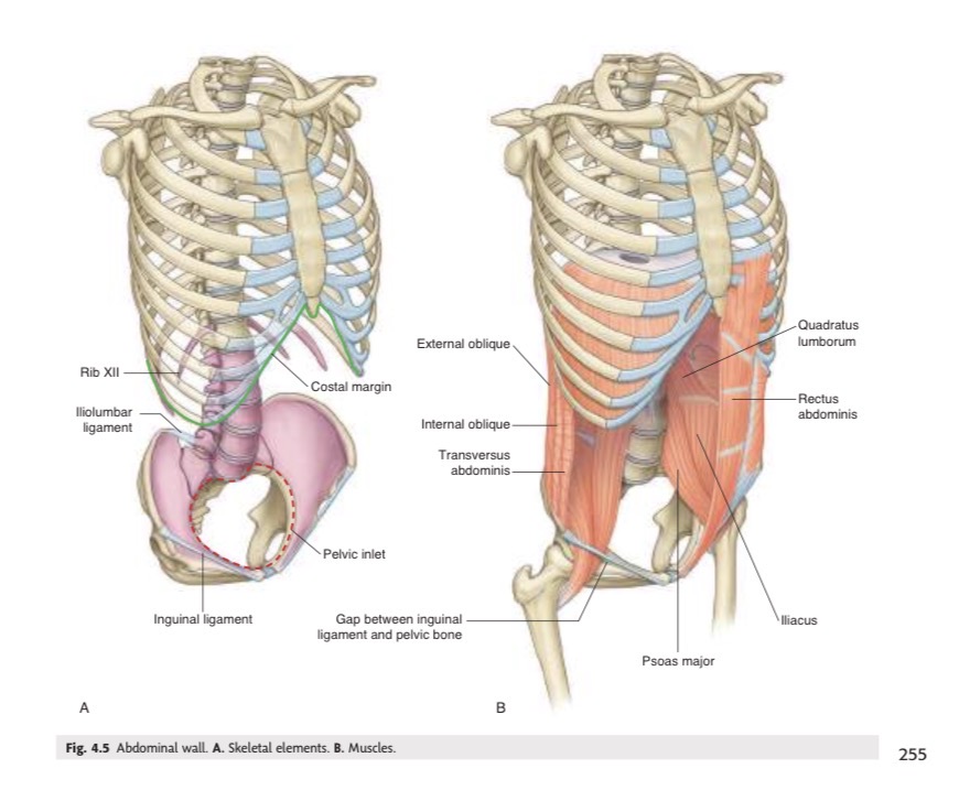
The orientation of the three lateral abdominal-wall muscle layers compared with thoracic muscles
Similar to the orientation of the intercostal muscles
The anterior paired segmented vertical muscle spanning between the inferior thoracic wall and pelvis
Rectus abdominis

The tissue providing structural continuity posteriorly in the abdominal wall
Thick fascia
The flat tendinous sheets providing structural continuity between lateral and anterior abdominal-wall regions; derived from muscles of the lateral wall.
Aponeuroses

The central digestive tube suspended from the abdominal wall by mesenteries
Gut tube (gastrointestinal system)

The thin sheets of tissue suspending the gastrointestinal tract from abdominal walls
Mesenteries

The mesentery associated with proximal regions of the gut tube
Ventral (anterior) mesentery

The mesentery extending along the entire length of the gastrointestinal system
Dorsal (posterior) mesentery

The basis for naming different portions of the ventral and dorsal mesenteries
According to the organs they suspend or with which they are associated
The membrane lining the abdominal cavity
Peritoneum

The epithelial-like single layer of cells forming part of the peritoneum together with a supportive layer of connective tissue.
Mesothelium
The thoracic membranes comparable to the peritoneum
Pleura and serous pericardium in the thorax
The part of the peritoneum lining the abdominal wall
Parietal peritoneum

The part of the peritoneum covering suspended organs
Visceral peritoneum

The normally potential space between visceral and parietal peritoneum
Peritoneal cavity
The two major positional categories of abdominal viscera
Intraperitoneal and retroperitoneal
An abdominal structure suspended by a mesentery within the peritoneal cavity such as the elements of the GI system.
Intraperitoneal structure

An abdominal structure positioned between the parietal peritoneum and abdominal wall without a mesentery
Retroperitoneal structure

The two primary urinary structures that develop and remain behind the peritoneum.
Kidneys and ureters
The specific gastrointestinal structures mentioned that transition from being suspended to being fixed against the posterior wall.
Parts of the small and large intestines
The structures associated with the posterior abdominal wall along the body's median axis
Large vessels, nerves, and lymphatics
The mesentery supporting the developing gut tube during development
Dorsal mesentery
1)The superior aperture of the abdomen; 2) the structure closing the 1st term
Inferior thoracic aperture; Diaphragm
The margin of the inferior thoracic aperture consists of:
vertebra TXII rib XII, distal end of rib XI, costal margin, and xiphoid process.
The musculotendinous structure separating the thorax from the abdomen
Diaphragm
The muscular extension anchoring each side of the diaphragm to the vertebral column
Crus

The right diaphragmatic crus extends inferiorly to approximately which vertebral level?
LIII

The left diaphragmatic crus extends inferiorly to approximately which vertebral level?
LII

The arch-shaped ligaments anchoring the diaphragm posteriorly
Arcuate ligaments

The arcuate ligament crossing the psoas major muscle
Medial arcuate ligament

The arcuate ligament crossing the quadratus lumborum muscle
Lateral arcuate ligament

The ligament that crosses the aorta and is continuous with both crura
Median arcuate ligament

The vertebral attachment of the medial arcuate ligament
Transverse process of LI

The bony attachment of the lateral arcuate ligament
Rib XII

The boundary where the abdominal wall becomes continuous with the pelvic wall and the opening through which the abdominal cavity communicates with the pelvic cavity
Pelvic inlet

The posterior bony boundary of the pelvic inlet
Sacrum

The anterior bony boundary of the pelvic inlet
Pubic symphysis

The lateral boundaries of the pelvic inlet
Distinct bony rims of the pelvic bones

The structure separating the abdomen from the thorax
Diaphragm

The opening that connects the abdomen directly to the pelvis, serving as the passageway for structures moving between the two cavities.
Pelvic inlet

The membrane continuous between the abdominal and pelvic cavities
Peritoneum

The pelvic organ that can expand superiorly into the abdominal cavity
Bladder

The reproductive organ that can expand superiorly into the abdominal cavity during pregnancy
Uterus

The structure marking the inferior abdominal-wall margin at the abdomen-thigh aperture
Inguinal ligament

The major vascular structures passing between the abdomen and thigh
Major artery and vein of the lower limb
The nerve passing through the abdomen-thigh aperture and innervating the quadriceps femoris
Femoral nerve

The major action of the quadriceps femoris muscle
Knee extension
The non-neural, nonvascular structures also passing between abdomen and thigh
Lymphatics and distal psoas major and iliacus
The action of psoas major and iliacus at the hip joint
Flexion of the thigh at the hip joint
The abdominal artery and vein that become the femoral artery and vein below the inguinal ligament
External iliac artery and vein of the abdomen

The thigh vessels formed when the external iliac vessels pass inferior to the inguinal ligament
Femoral artery and femoral vein of the thigh

The gastrointestinal tube suspended by both ventral and dorsal mesenteries
Foregut
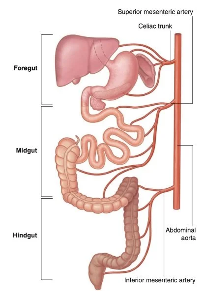
The primitive gut tube regions
Foregut, midgut, and hindgut
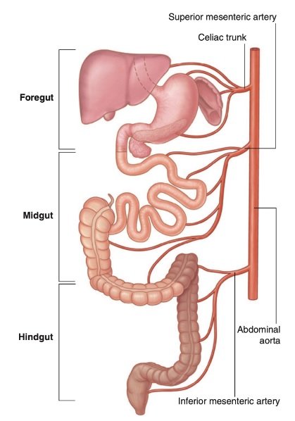
The abdominal derivatives of the foregut
Distal esophagus, stomach, and proximal duodenum

The only gut region suspended by both ventral and dorsal mesenteries
Foregut

The foregut diverticulum that grows into the ventral mesentery, giving rise to the liver and gallbladder, and, ultimately to the ventral part of the pancreas
Anterior foregut diverticulum
The direction in which the developing stomach rotates
Clockwise

The effect of stomach rotation on the dorsal mesentery
It moves left and greatly expands

The structures that swing right with the duodenum
Dorsal mesentery and appreciable part of the pancreas

The opening created by restriction of the space associated with the expanded dorsal mesentery
Omental/Epiploic foramen

The space posterior to the stomach enclosed by the expanded dorsal mesentery
Omental bursa (Lesser sac)

The larger part of the peritoneal cavity surrounding the lesser sac
Greater sac

The route providing access from the greater sac to the omental bursa
Omental foramen

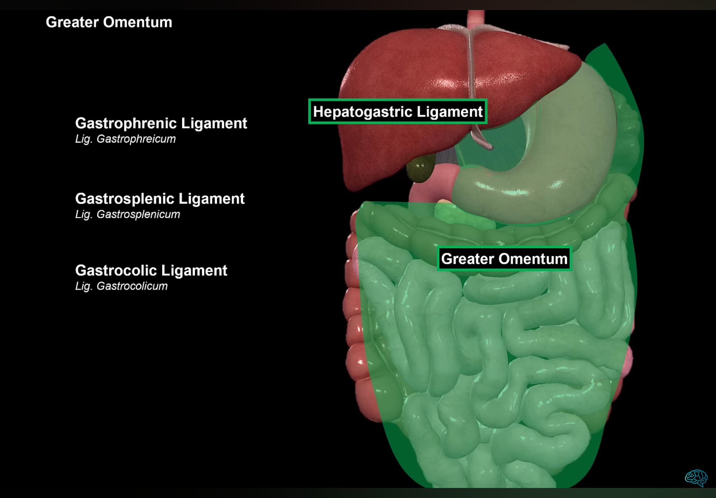
The dorsal mesentery portion that greatly enlarges inferiorly and forms an apron-like structure
Greater omentum

The stomach attachment of the greater omentum
Greater curvature of the stomach

The first structure observed when the abdominal cavity is opened anteriorly
Greater omentum
The gastrointestinal structures derived from the midgut
Distal duodenum, jejunum, ileum, ascending colon, and proximal two-thirds of transverse colon

The embryonic structure projecting anteriorly from the developing midgut into the umbilicus
Small Yolk sac
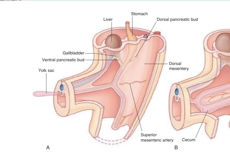
The event caused by rapid gastrointestinal growth in which the midgut loop leaves the abdominal cavity
Midgut herniation
The direction of rotation of the two limbs of the midgut loop
Counterclockwise

The portion of the midgut loop that descends into the inferior right abdomen
Cecum

The artery located at the center of the midgut rotation axis which supplies the midgut
Superior mesenteric artery

The mesentery suspending the transverse colon
Transverse mesocolon

The structure that hangs over the transverse colon and mesocolon
Greater omentum

The hindgut derivatives in the abdomen
Distal one-third of transverse colon, descending colon, sigmoid colon, and superior rectum

The structures formed as proximal hindgut parts swing left
Descending colon and sigmoid colon
The route taken by the sigmoid colon before becoming continuous with the rectum
Through the pelvic inlet
The vertebral level where the sigmoid colon becomes continuous with the rectum
Vertebra level SIII
The thoracic spinal nerves whose anterior rami enter the abdominal wall
T7 to T12

The intercostal nerves supplying abdominal-wall skin and muscle
T7 to T11
