neuro unit 2 lecture 3: hearing and olfaction
 Call Kai
 Learn
 Practice Test
 Spaced Repetition
 Match
 Flashcards
 Knowt Play
1/25

There's no tags or description
Looks like no tags are added yet.
26 Terms
How does the human auditory system detect sounds?
amplitude (intensity, measured as decibels (volume) and Frequency- (measured as cycles per second or hertz (hz))
parts of the external ear
pinna, ear canal
pinna
outer part of ear (where earrings are, funnels sound into the ear canal) shape of the ear modifies the character of sound frequencies that reach the middle ear
what does the middle ear do
concentrates sound energies
what are the ossicles
3 bones (malleus, incus, stapes) that connect the eardrum (tympanic membrane) to the oval window
What are the tensor tympani and the stapedius and what do they do
two muscles in the middle ear that control volume- they can stiffen and reduce sound’s effect on the eardrum (tympanic membrane) what a
what are the two muscles of the inner ear
tensor tympani and stapedius, control volume- tense and reduce sound’’ effects

what is the oval window
opening from the middle ear to the inner ear (connected to the eardrum by the ossicles- malleus, incus, stapes)

what is the ear drum and how does it work
eardrum= tympanic membrane, sound waves travel down ear canal and hit the tympanic membrane, causing it to vibrate which causes ossicles to vibrate which amplify the vibrations into the cochlea, hair cells in the cochlea then pick up on the vibrations and transduce the waves of fluid into electrical signals
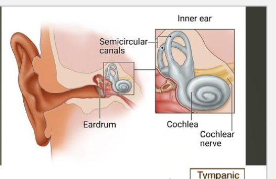
cochlea
part of the inner ear that converts vibrations into neural activity/ electrical impulses, has 3 canals- scala vestibuli (vestibular canal), scala media (middle canal)/ cochlear duct, scala tympani (tympanic canal)

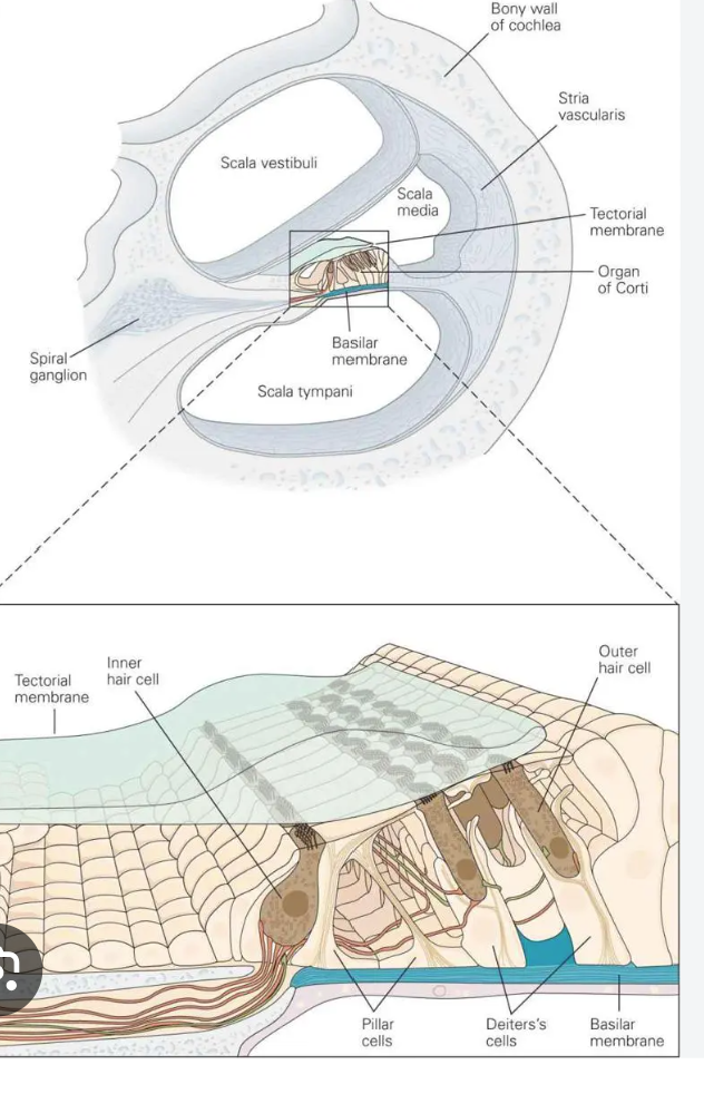
organ of corti
part of the cochlea (in the scala media/ cochlear duct) that converts sound to neural activity- contains hair cells and nerve fibers, hair cells are connected to the auditory nerve (nerve 8, vestibulocochlear) and rest on the basilar membrane

basilar membrane
organ of corti rests on it, rolled up in the cochlea, sound vibrations cause it to ripple, like shaking out a rug, different parts of it vibrate most at different frequencies- tip of basilar= thinner and stiffer, vibrates more at high frequencies and activates corresponding hair cells, end of basilar membrane= thick in diameter but thinner/ floppier, vibrates more at lower frequencies, and activates corresponding hair cells hai
hair cells
convert sound into neural activity, sloping group of stereocilia (microscopic hair cells) protrude from the cell, and sway when inner ear fluid moves, connected by tiplinks which are tiny fibers that open ion channels when stereocilia bend- these are mechanically gated ion channels, hairs sway bc of movement of basilar membrane and fluid,

tip links
stretch and open mechanically gated ion channels which let k+ and ca2+ rush into the cell, generated a graded potential that causes an action potential which will release NTS to the vestibulocochlear nerve

how are hair cells organized
two groups- inner hair cells, IHCs- single row near central axis/ inside, needed for audio detection, and other hair cells, OHC- three rows on outside, not rlly for detection, moreso for cochlear amplification- amplify dif frequencies of sound by increasing vibrations of the basilar membrane, vestibulocochlear nerve contacts the base of the hair cells
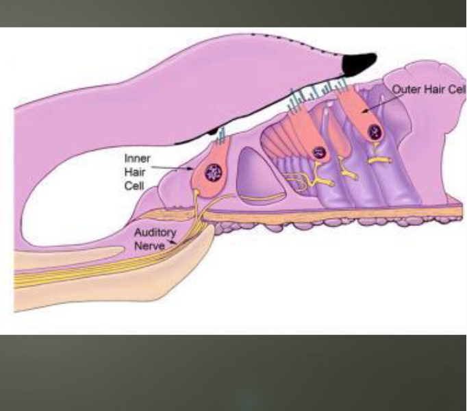
inner hair cells
one row, impt for audio detection ou
outer hair cells
3 rows, important cochlear amplifiers, amplify dif frequencies of sound by increasing the vibration of the basilar membrane, without cochlear amplification, 20-30 TIMES LESS HEARING
where do auditory nerve fibers from hair cells terminate
cochlear nuclei, which sends info to the superior olivary nucleus (first site of binaural (two ear) processing, info shared here contralaterally as well as processed from same side), which sends information to the inferior colliculi, auditory center of the midbrain, then to the medial geniculate nuclei in the thalamus, then to the auditory cortex

What is the pathway from ear to brain?
hair cells (tip links) to vestibulocochlear, to cochlear nucleus, to superior olivary nucleus, to inferior colliculi in midbrain, to medial geniculate nucleus in thalamus (forebrain) to auditory cortex)
where is the primary auditory cortex
temporal lobe ish, right by the sylvian fissure, mainly activated by sound, speech activates other, more specialized auditory areas

olfaction
ability to detect smells (odors)- every odor we detect has a specific chemical structure
where does the sense of smell start
receptor neurons in the nose- the olfactory epithelium (similar to the basilar membrane)
olfactory receptors (what’s special about them)
differ from other sensory neurons of the brain, so many dif types (350+, mostly metabotropic), die and are replaced in adulthood (bc of hazardous environment of nostril), each olfactory axon extends into an olfactory bulb of the brain, terminates on a specific glomerulus (r we serious) which processes info from one specific type of receptor
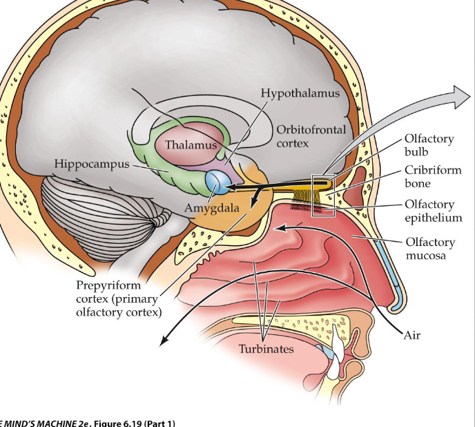
WHERE DO AXONS OF OLFACTORY RECEPTOR CELLS GO
directly to the olfactory bulb, where they extend onto a glomerulus, which processes info from one specific type of odorant receptors

from olfactory receptors to glomerulus inward
glomerulus recieve info from neurons w same type of receptors, organized like a map, glomeruli recieve info from receptors and by association chemicals, that are closely related, this organization is maintained throughout the brain,
Where does info go from nose into brain?
from the olfactory bulb, smell is the only sensory type that synapses directly onto the cortex without first passing thru the thalamus, olfactory neurons synapse directly onto areas involved in emotion and memory (amygdala and thalamus) (smell super important to many memories (Ppl’s perfumes, stories in Bible) smell very biologically impt
