55- Resp System Upper Resp Tract Conditions
 Call Kai
 Learn
 Practice Test
 Spaced Repetition
 Match
 Flashcards
 Knowt Play
1/65

There's no tags or description
Looks like no tags are added yet.
66 Terms
what are the body parts of the upper resp tract?
nasal cavity, pharynx, guttural pouch, larynx, trachea
dilation of mucosal vessels (congestion) indicates what
circulatory failure preceding death
epistaxis
blood flow from nose (nasal mucosa or from deep in lungs)
whats unilateral epistaxis from
upper resp tract
whats bilateral epistaxis from
lungs
causes of epistaxis
local trauma, submucosal erosions from inflammation, neoplasia
specific form of pulm hemorrhage in racehorses that occurs after exercise and is characterized by epistaxis in severe cases
exercise induced pulmonary hemorrhage

gross lesion: large areas of dark brown discoloration on caudal lung lobes
exercise induced pulmonary hemorrhage
common pathogens of the guttural pouch
fungus, bacteria
why is infection of the guttural pouch bad?
doesnt drain well, close to carotid arteries and cranial nerves
whats guttural pouch mycosis caused by
inhalation of moldy hay
ASPERGILLUS

horse presents with dorsal and lat walls of gut pouch mucosa covered by focal rounded raised plaques that coalesce into larger masses, unilateral
guttural pouch mycosis

histo: necrotic inflamm of the mucosa with vasculitis and intralesional fungal hyphae
gut pouch mycosis
empyema
accumulation of neutrophils within a potential space

what is gut pouch empyema a sequela to
suppurative inflamm of the nasal cavities from strep equi infection aka strangles !!
what is diff about the secondary lesions of gut pouch empyema
similar to gut pouch mycosis but no erosion of internal carotid artery
when does infectious rhinitis occur
secondary to changes in microbiota balance of the nasal cavity and normally resolves completely
chronic rhinitis
destruction of nasal conchae and deviation of the septum
sequela to infectious rhinitis
hemorrhage, ulcers, nasopharyngeal polyps arising from inflamed mucosa
infectious rhinitis in the facial bones could cause
osteomyelitis
infectious rhinitis in the cribiform plate could cause
meningitis
infectious rhinitis in the eustachian tubes could cause
otitis media or gut pouch empyema in horses
infectious rhinitis in the inner ear could cause
otitis interna and vestibular syndrome
gross lesion: mucosal hyperemia, increased prod of clear fluid manufactured by serous glands present in the nasal submucosa
serous rhinitis (milldest form)
causes of serous rhinitis
mild irritants, cold air, early viral infections, upper resp system infections, mild allergic rxns

gross lesion: serous and mucus discharge, mucus exudate is THICK translucent or kinda turbid viscous fluid
catarrhal rhinitis

histo: mucus gland hyperplasia (inc goblet cells)
catarrhal rhinitis
describe the acute inflamm process of suppurative rhinitis
neutrophilic exudate occurs when the nasal mucosa suffers a more severe injury
sometimes includes mucosal necrosis and secondary bacteria infection

gross lesion: thick and opaque exudate that is white/green/brown, nasal passages are obstructed by exudate, and possibly mucosuppurative if features of catarrhal inflamm
suppurative rhinitis

inflammation that causes increased vascular permeability, resulting from abundant plasma fibrinogen which forms FIBRIN
fibrinous rhinitis
gross lesion of fibrinous rhinitis
fibrin appears as a yellow tan or gray friable mat on the nasal mucosa
fibrinous rhinitis: if the fibrin mat is fibrinous =
thin film of exudate
fibrinous rhinitis: if the fibrin mat is pseudodiphtheritic rhinitis =
fibrinous exudate can be removed leaving an intact underlying mucosa
fibrinous rhinitis: if the fibrin mat is diphtheritic or pseudomembrane rhinitis =
pseudomembrane is difficult to remove and leaves an ulcerated mucosa

gross lesion: multifocal to coalescing tan granulomas, chronic inflamm can lead to the formation of polypoid nodules that can obstruct nasal passages
granulomatous rhinitis

histo of granulomatous rhinitis
infiltration of epitheloid macrophages mixed with a few lymphocytes and plasma cells
what is granulomatous rhinitis associated with
chronic allergic inflammation and infectious etiologies like fungi or tuberculosis
most common causes of tracheitis are ?
VIRAL infections like infectious bovine rhinotracheitis, equine viral rhinopneumonia, canine distemper, feline rhinotracheitis

with tracheitis, viral lesions are typically mild but become complicated with
secondary BACTERIAL infections and can lead to tracheal ulceration
what is seen after aspiration, inhalation of fumes during fire, aspiration of food or grass awns
tracheitis
what occurs freq with rhinitis
sinusitis
what can cause sinutitis
sequela to penetrating object or septic wounds of nasal, frontal, maxillary, palatine bones
improper dehorning
max tooth infection

whats this pic show
sinusitis
dog presents with acute onset coughing and has rhinitis/pharyngitis but also was normal prior
canine infectious resp disease complex
pathogenesis of CIRD involves mult pathogens like
CAdV-2, bordetella, strep zoo
severity increases with mult pathogens and poor ventilation
gross lesions: CATARRHAL TO MUCOPURULENT MATERIAL, pneumonia in some cases, tonsil enlargement, retropharyngeal and trachealbronchial LN enlargement
CIRD
most common cause of mycotic and algar rhinitis/sinusitis in canines
aspergillus and penicillium spp
what breed is predisposed to aspergillus
GSD

gross lesion: NECROSIS of nasal epithelium , plaques of fibrinupurulent exudate, disruption of nasal conchae in chronic cases
mycotic and algae rhinitis/sinutisis in canines

histo: granulomatous inflamm mixed with fungal hyphae
mycotic and algae rhinitis/sinutisis in canines
cause of feline viral rhinotracheitis
feline herpesvirus 1
feline viral rhinotracheitis becomes ___ and shed during stress
latent in various ganglia

gross lesions of feline viral rhinotracheitis
tropism for epithelial cells results in necrosis and inflammation of the conjuctiva, nasal passages, pharynx, trachea
rhinitis and sinusitis with persistant SUPPURATIVE discharge in chronic cases
what are some lesions of feline calicivirus
mild oculonasal discharge, severe rhinitis, mucopurulenet conjunctivitis, ulcerative gingiviris glossitis and stomatitis
whats a common complication of feline calicivirus
secondary bacterial infection like bordetella, pasteurlla, e. coli
whats the most common mycotic infection in the feline nasal cavity
cryptococcosis

gross lesion: external swelling with nasal passage, discrete nasal granulomas to large confluent masses of mucosuppurative exudate filling entire nasal cavity and sometimes meningitis from extension into CNS
cryptococcosis in felines h
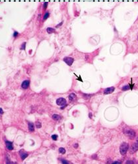
histo: yeast with abundant clear CAPSULE soap bubbles
cryptococcosis
common dz of pigs associated with atrophic rhinitis (pasteurella multocida, bord) with predisposing factors of genetics, environment, nutrional def
porcine progressive atrophic rhinitis

gross lesion: inflamm and atrophy of nasal conchae, DEVIATED SEPTUM, asymmetric conchae, in severe cases complete loss of conchae causing facial deformity
porcine progressive atrophic rhinitis
condition in YOUNG pigs from penetrating wound with swollen snout deformed snout
necrotic rhinitis in pigs

gross lesion: necrosis and suppuration of the nasal cavity and turbinates
necrotic rhinitis in pigs

what is Oestrus ovis in sheep
brown fly that deposits first stage larvae in nostrils of sheep. they mature into maggots and spend their time in nasal passages resulting in inflamm of airway
common dz of FEEDLOT cattle which gains entry thru lesions of viral infections or traumatic injury (from fusobacterium necrophorum aka oppurtunistic pathogen)
necrotic laryngitis

gross lesion: inflammation of mouth or larynx, well demarcated dry yellow gray thick crusted FIBRONECROTIC exudate can can progress ot ulceration
necrotic laryngitis aka calf diptheria

gross lesion: hyperemia and necrosis of nasal, pharyngeal, laryngeal, tracheal mucosa
secondary infection in necrotic regions = formation of thick layer of FIBRINONECROTIC MATERIAL and can turn into pneumonia
infectious bovine rhinotracheitis