BCHM Exam 2
 Call Kai
 Learn
Learn Practice Test
 Spaced Repetition
 Match
Match Flashcards
 Knowt Play
1/60

There's no tags or description
Looks like no tags are added yet.
Name | Mastery | Learn | Test | Matching | Spaced | Call with Kai | Chat |
|---|
No analytics yet
Send a link to your students to track their progress
61 Terms
What is the difference between channel proteins and carrier proteins?
Channel proteins have a fixed structure and a core
Carrier proteins change their structure and do not have a core
Carrier proteins grab and transport molecules in and out of the cell
There is a higher concentration of sodium __________ the cell
outside
Explain how voltage-gated sodium channels work:
Voltage-gated sodium channels do not require activation energy, they are passive as they are activated by changes in membrane voltage (depolarization response)
Na+ ions travel through the plasma membrane via protein channels
these protein channels are a pore with an inactivation gate and an activation gate towards the inside of the cell
the inside of the cell has a resting potential with negative charge
Na+ ions have an electrical attraction to this negative charge
Depolarization will occur
the positive charge of the Na+ traveling through the pore of the membrane opens the activation gate (slow process)
transitions from resting potential to peak potential (positive charge)
Re-polarization occurs next
the sodium travels back out of the cell through the channel
the ball-and-chain structure that’s the inactivation gate swings closed and plugs the pore, closing the gate and inactivating it
transitions from peak potential back to resting potential
Describe the structure of pores:
pores have 4 subunits (labeled P) that surround a core
each P subunit consists of 6 helices (labeled S)
when a charge moves from the outside to the inside of the membrane through a pore, the 3D structure of helices S5 and S6 change (helices closest to the pore channel)

True or False:
Since Na+ and K+ both have a +1 charge, they travel through the same pore channels
False
the geometries of the selectivity filters of Na+ channels are different than the selectivity filters of K+ channels
K+ cannot pass through Na+ channels and vice versa
What distinguishes R groups from one another?
the R groups
__________ R groups are acidic
carboxylate
__________ R groups can covalently bond with each other
Sulfur
True or False:
Our cells contain little vesicles that store amino acids for when they are urgently needed
False
Amino acids can not be stored
we must consume outside protein from our nutrition
True or False:
Proteins can be broken down, and their amino acids recycled
True
List the essential amino acids
histidine
isoleucine
leucine
lysine
methionine
threonine
phenylalanine
valine
tryptophan
What is PDCAAS? What is it’s significance?
PDCAAS = Protein Digestibility Corrected Amino Acid Score
gives us information on which type of foods supply us with which type of amino acids
rates foods on how much amino acid variety they provide
Ex. Egg whites have a PDCAAS of 1
this value is perfect
egg whites supply our bodies with all essential amino acids
What are the 4 categories that amino acids are listed under?
1) Non-polar, Aliphatic
2) Aromatic
3) Polar, Uncharged
4) Charged
There are ____ amino acids that serve as the building blocks of proteins in the human body
20
What is an example of a nutrition disorder involving protein?
Kwashiorkor
Kwashiorkor is a nutrition disorder in which there is a disproportionate consumption of protein to other dietary needs
there’s a lack of consumption of dietary protein
negative Nitrogen balance
a symptom of Kwashiorkor is ascites (accumulation of water that causes bloating of the abdomen)
also negatively impacts the liver
What are zymogens? Give examples
zymogens are the inactive form of an enzyme, protein, or hormone
zymogens have to undergo a biochemical change to activate
Ex. pancreatic hormones are formed as zymogens because they are proteolytic (protein-cleaving)
they are formed as zymogens to prevent them from destroying the cells of the pancreas that produces them
the pancreatic hormone zymogens are secreted by the pancreas and into the small intestine to be activated
if these enzymes are activated too soon in the pancreas, an inflammatory condition called pancreatitis occurs
Examples of zymogens: pancreatic hormones, pepsinogen, insulin
How does the consumption of amino acids impact the liver
the liver needs an adequate amount of amino acids to function property
a negative nitrogen balance and a positive nitrogen balance should both be avoided
negative Nitrogen balance
not consuming enough protein
albumin is the most abundant protein produced by the liver
the amino acids that make up albumin must be consumed in our diet
a low concentration of albumin in the blood causes fluid to leak from the blood vessels which leads to bodily edema
positive Nitrogen balance
consuming too much protein
the break down of amino acids occurs in the liver
too high of a concentration of amino acids causes the liver to convert some to ammonia (NH3) which is converted to urea
someone with liver damage/dysfunction can not always convert this excess ammonia into urea, causing a buildup of ammonia in the bloodstream which becomes toxic
Describe the Sodium dependent cotransport of an amino acid into the cell in the intestines. Why does this occur?
Sodium dependent cotransport can occur to move amino acids into the cell against their concentration gradient by coupling amino acid transport with sodium transport
Sodium binds to the amino acid transporter (a passive process)
this increases the carrier’s affinity for the amino acid, making the amino acid available to bind to the carrier → forms a sodium-amino acid cotransporter
a 3D conformational change occurs in the complex, resulting in the delivery of the sodium and amino acid into the cytosol of the intestinal cell
that sodium then gets pumped back out of the cell

Draw and describe the peptide bonds between two amino acids:
two amino acids (in the trans configuration with steric rotation) are chained together by a peptide bond
Peptide bond
the Nitrogen of one amino acid binds to the carbonyl Carbon of the next amino acid
this is a rigid bond that can NOT rotate
Psi bond
bond that connects the carbonyl Carbon to the alpha Carbon (that is bonded to the R group)
can rotate
Phi bond
bond that connects the Nitrogen atom to the alpha carbonyl Carbon that contains an R group
can rotate

Which bonds can rotate within a peptide bond?
the psi and phi bonds rotate
they rotate around around the peptide bond (they are the bonds that are the next atom over from the peptide bond)
the stearic rotation impacts how proteins fold
How is the shape/rotation of polypeptides determined?
the Levinthal Paradox applies to polypeptides
the folding of a protein happens very quickly and spontaneously, but it is not random, even though there are so many different conformations that are possible
proteins fold so that they are in the most stable, lowest energy conformation
the Ramachandran plot shows how certain bond angles are more likely to be produced when polypeptides are folded because those angles are the most viable stearic rotations
most molecules default to these conformations
certain conformations are eliminated as possibilities because they are physically impossible, have too much stearic hinderance, or they are just not thermodynamically-favorable
When do proteins have their highest energy? When do they have their lowest energy?
highest energy = unfolded states
lowest energy = folded states
Define: amorphous aggregates
proteins that have been incorrectly folded
Define: native state
state of properly-folded proteins
Define: chaperones
proteins that help fold other proteins into their lowest energy state
What are the enantiomers of amino acids? Which one(s) can be metabolize?
there are L amino acid enantiomers and D amino acid enantiomers
we can only metabolize the L amino acids
What is needed to insert an amino acid into a protein?
ATP is needed to insert an amino acid to a protein chain because the ATP molecule will “charge”/activate the amino acid beforehand
What are the different functions of proteins?
function as enzymes
they are transporters (move ions or other proteins)
they are receptors
cell junctions
provide structure (collagen, muscles, etc.)
antibodies
antigens (trigger immune response)
a __________ is released in peptide bond formation
water molecule
the formation of a peptide bond creates __________ entropy
lower entropy when a peptide bond is created because it’s a rigid bond that provides structure/organization
What are the levels of protein structure?
1) Primary Structure
what amino acids are present and in what order they are arranged in
the sequence of amino acids
2) Secondary Structure
the polypeptide backbones begin to fold into one of two structures: alpha helix or a beta-pleated sheet
specifically, Hydrogen bonding occurs between the H and N of one peptide bind, and another Hydrogen bond occurs on the O of the Carbonyl group of the next peptide bond
these hydrogen bonds hold the alpha helix structure or the beta-pleated sheet together
Alpha helix
right-handed spiral structure
Beta-pleated sheet
crimped like a sheet
can be parallel or antiparallel
antiparallel is more stable: there’s less bond angle strain because the bonds are lines up and not angled
two strands of beta sheets are connected by turns
Glycine and Proline are involved in turns
there is no R-group involvement in secondary structures
3) Tertiary Structure
tertiary structure is stabilized by R-groups
the secondary structures fold based on the interactions between the R-groups that become involved
types of R-group interactions:
electrostatic interactions
hydrogen bonds
hydrophobic interactions
disulfide bonds
4) Quaternary Structure
two or more separate polypeptide chains (subunits) assemble and interact non-covalently to form a functional protein complex
this is an allosteric change
not all proteins reach quaternary structure
What causes proteins to denature? What causes proteins to renature?
denaturation occurs under heated conditions
renaturation can occur spontaneously in some proteins, where they refold back into their correct, original shape
Define Supersecondary structures:
supersecondary structures are a combination of a few secondary structures: alpha helices and beta-pleated sheets (so not quite secondary but not quite tertiary structures)
supersecondary structures can get added to proteins to fix it if it misfolds (and the cell won’t have to trash the entire protein)
motifs and domains are supersecondary structures
Motifs
play a structural role only
will fall apart if isolated from protein
types of motifs: beta-alpha-beta (a), meander (b), helix-turn-helix (c), barrel (d), greek key (e), EF hand, zinc finger, leucine zipper
Domains
independent and won’t fall apart if isolated from the protein (higher level of organization than motifs)

Describe the Leucine Zipper:
Leucine Zipper is a 7 amino acid sequence that repeats itself
these 7 amino acids are labeled abcdefg
the Leucine is located at the “d” position
Leucine is hydrophobic, which makes it easier to unzip the helices of DNA
the leucine zipper controls transcription (allows transcription-factor subunits to come together and bind to specified DNA sequences)
Define Microstate vs. Macrostate. Is the Macrostate or the Microstate of a protein consistent?
Macrostate is the arrangement of all the atoms in a protein (how the protein is folded as a whole)
macrostate is consistent
Ex. the entire folded protein
Microstate is the exact arrangement of atoms at a particular instant
microstate is not consistent because proteins can vibrate
Ex. the primary structure
Explain hemes and pyrrole rings
heme groups are a part of myoglobin and hemoglobin proteins, that contain iron to help bond oxygen
hydrophobic
hemes are a linkage of four pyrrole rings (linked by methene bridges)
has Iron in the center
macrophages break down the heme, which gets converted to biliverdin, and then that gets converted to bilirubin
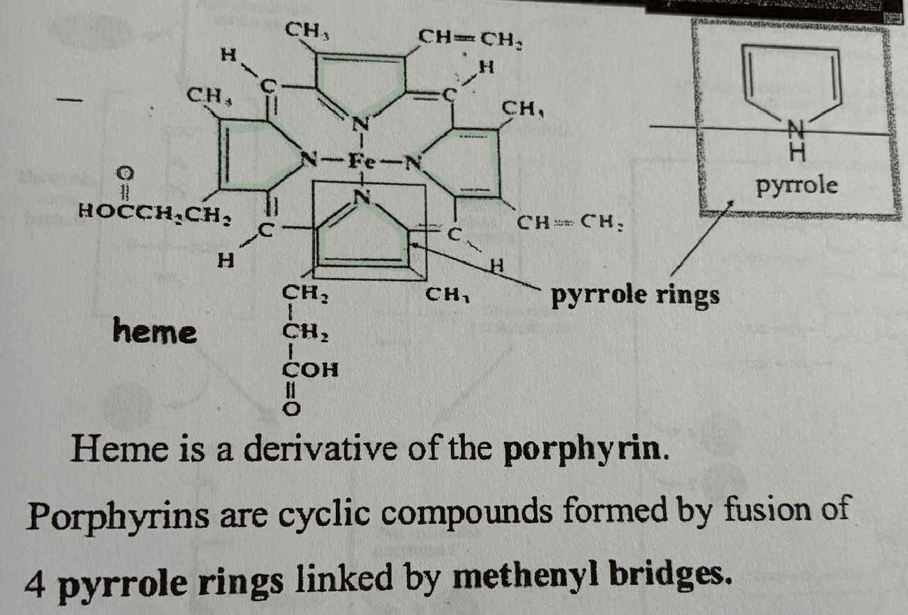
What structures contain hemes and pyrrole rings?
myoglobin and hemoglobin proteins are both contain a linkage of four pyrrole rings as part of their heme group
Describe the structure and function of myoglobin
myoglobin is a storage protein that stores oxygen in the muscle cells
myoglobin has a very high oxygen affinity
myoglobin’s oxygen affinity is NOT affected by pH or CO2 fluctuations
has a hyperbolic oxygen-binding curve
has a heme
has 1 subunit made up of 8 alpha-helix structures
contains two histidine (an amino acid) on opposite sides of the heme plane (one distal histidine and one proximal histidine)
proximal histidine is directly bonded to (and anchors) the Iron at the center of the pyrrole ring heme structure
the proximal histidine pulls the iron into the plane of the heme when oxygen binds to the iron
distal histidine forms a hydrogen bond with the bound O2 molecule to stabilize it
distal histidine helps control access to the heme by other gases (it acts as a “gate” to the heme)
Ex. carbon monoxide should not be let in because it is toxic, even though it wants to bind to the heme’s iron
153 amino acids long
myoglobin will release oxygen when the partial pressure of oxygen drops in the muscles (during muscle contraction, heavy exercise, or when blood flow is restricted)
Describe the structure and function of hemoglobin
hemoglobin carries oxygen from the lungs and to the various tissues of the body
has 4 subunits: 2 alpha helices and 2 beta sheets
so hemoglobin has 2 dimers: α₁β₁ dimer and α₂β₂ dimer
these dimers can rotate (they rotate 15 degreed after oxygen binds)
the dimers surround a central solvent channel, which contains water and 2,3-bisphosphoglycerate
exhibits the Bohr Effect
hemoglobin will be in one of two states: deoxyhemoglobin or oxyhemoglobin
deoxyhemoglobin is in a Taut state (T-state)
will release oxygen to tissues
stabilized by 2,3-bisphosphoglycerate (2,3-BPG), which prevents oxygen from rebinding (Ex. of negative allosterism: prevention)
central channel is wide
bound to little/no oxygen
low oxygen affinity
oxyhemoglobin is in a Relaxed state (R-state)
central channel is narrowed
high oxygen affinity
oxygen binds
positive cooperation occurs in hemoglobin as it transitions from Hb-deoxy state to HbO2 state - this is when a 3D structural change occurs (allosteric change) and this process releases O2
The iron (Fe) in the center of the heme must be in Fe2+ state to bind to oxygen
Oxygen binds to Fe2+ and makes it Fe3+ which pulls the Iron and the proximal histidine up into the same plane as the heme - structural 3D change - 15 degree rotation
Fe3+ gives its electron to oxygen, creating a superoxide ion which is a free radical (which are dangerous), so an extra electron binds to Fe3+ to make it Fe2+ as the oxygen is being released
Describe the gas exchange that occurs in red blood cells:
CO2 from tissues (about 20%) bind to the N-terminus of the hemoglobin molecule, creating HbCO2
creates carbamino hemoglobin
CO2 + Hb → HbCO2
the rest of the CO2 from tissues gets added to water, then carbonic anhydrase reacts those molecules to form carbonic acid
CO2 + H2O —CA→ H2CO3
the carbonic acid dissociates into a bicarbonate ion and a proton
that bicarbonate ion leaves and at that same time Cl- ion enters to balance the charge
the proton binds to hemoglobin, causing an allosteric change
Bohr Effect
oxygen is taken out to be delivered to the tissues

Describe the Bohr Effect
Bohr Effect:
an increase in carbon dioxide concentration or an increase in acidity (decrease in pH) reduces hemoglobin’s affinity for oxygen, causing it to release more oxygen to body tissues by transitioning from deoxy state to oxy state which is an allosteric change
Describe the gas exchange that occurs in the alveoli in the lungs:
CO2 removed from carbamino hemoglobin, then that CO2 gets released
HbCO2 → Hb + CO2
oxygen that was delivered from the lungs displaces the proton on hemoglobin
O2 + HbH+ → HbO2 + H+
bicarbonate ions and protons (released from binding with hemoglobin) are combined to form carbonic acid, which then is turned into water and CO2 (with the help of carbonic anhydrase)
HCO3- + H+ → H2CO3 —CA→ H2O + CO2
that CO2 leaves the lungs
What are cross-bridges?
protons coming out of tissues forms a cross-bridge
cross bridges are formed in hemoglobin between the two parts of the beta subunit and between the beta subunit and the alpha subunit
An increase in partial pressure of oxygen in the tissues will cause a(n) __________ in the amount of oxygen to bind to hemoglobin
increase
What must occur when Oxygen binds to Fe3+?
Fe3+ gives its electron to oxygen, creating a superoxide ion
superoxide ions are free radicals (which are dangerous to our cells)
oxygen is prevented being released in free radical form until an extra electron binds to Fe3+ to make it Fe2+ again as the oxygen is being released
What is the difference between the graphical representation of how readily hemoglobin and myoglobin binds to oxygen (at different partial pressures, which means at different amounts of oxygen availability)?
hemoglobin
hemoglobin has a sigmoidal curve
this is due to hemoglobin’s positive cooperativity
hemoglobin is a quaternary protein with 4 subunits
oxygen binds to the first subunit
a relatively slow process
first subunit’s oxygen affinity is low
when the oxygen binds and the Fe and proximal histidine of the F-helix are moved into the plane of the heme, that structural change is recognized by the second subunit
the second subunit binds with oxygen after that signal, which signals the third subunit to bind, then that signals the fourth subunit to bind
this binding sequentially gets faster each time
this is called positive cooperativity
positive allosteric changes are occuring
the positive allosteric effector is Oxygen (that is the molecule that stimulates a response)
myoglobin
myoglobin has a hyperbolic curve
myoglobin is only made up one one subunit that has a tertiary structure
myoglobin binds oxygen independently, without cooperative binding (only has one binding site, and one heme)

Which binds to oxygen more tightly: hemoglobin or myoglobin
myoglobin because it has a high affinity for oxygen
Which can carry more oxygen: hemoglobin or myoglobin?
hemoglobin can carry more oxygen because unlike myoglobin which only has one subunit (and therefore can only carry 1 oxygen molecule), hemoglobin can carry four oxygens as it has four subunits
makes hemoglobin ideal for transport and myoglobin ideal for storage
What protein structure level is myoglobin?
tertiary
What protein structure level is hemoglobin?
quaternary
Describe sickle-cell anemia:
sickle-cell anemia is a recessive genetic defect within the Beta subunit of hemoglobin
the 6th position amino acid in a healthy individual’s hemoglobin is glutamic acid, but the 6th position is mutated to valine in an individual with sickle cell anemia
in the deoxyhemoglobin state, a hydrophobic protrusion in one Beta subunit finds a hydrophobic pocket a B subunit of another hemoglobin molecule
this causes hemoglobins to clump together in the capillaries
the shape of the red blood cells is impacted when the membrane collapses and loses its smooth disc shape
electrophoresis is used to detect sickle-cell traits and diagnose sickle-cell disease
__________ proteins are more structural
Fibrous
__________ proteins are functional
globular
Which of these proteins are globular? Which are fibrous?
Hemoglobin
Insulin
Collagen
Antibodies
Hemoglobin = globular
Insulin = globular
Collagen = fibrous
Antibodies = globular
Describe the structure of antibodies:
antibodies are in a Y-shape
on the ends of the Y are variable regions (one heavy and one light)
Variable regions are what differentiate antibodies
these are the regions that antigens bind to
towards the base of the Y are constant regions (heavy and light)
constant regions are the same in all antibodies
shows structure/function coordination as this benefits the cell:
when a new antibody needs to be created, only the variable regions have to change - the constant regions always stay the same - so a whole entire new antibody doesn’t have to be made

Describe insulin
Insulin has a globular structure
Insulin is a hormone that’s created as a zymogen
Insulin gets activated when the C-peptide is cut off the insulin
Describe collagen
collagen is a fibrous protein
each collagen molecule is made up of three polypeptide chains wound into a triple helix
collagen molecules line up side-by-side and are cross-linked to form fibrils (that are strong)
the cross-linkages are created out of modifications from the enzyme lysyl oxidase
oxidizes lysine into allysine
the amino acids that are prominent in collagen are:
Glycine: in alpha helices - allows tight packing
Proline: cyclical amino acid that helps maintain the helical structure
Hydroxyproline: helps stabilize the triple helix
Lysine
Describe the process of protein degradation?
built into the amino acid sequences are pest sequences, which signal for degradation
Ubiquitin is the protein that “tags” a misfolded protein to be degraded
Ubiquitin recognizes the pest sequence, binds to it with the help of ATP
That tagged protein gets sent to the proteasome, a large protein chamber where degradation occurs
the cap of the proteasome lets in the protein and unfolds it (this uses ATP)
the unfolded protein moves through the proteasome to the core, where it is chopped up and separated into amino acids that can get reused by the cell
Why must misfolded proteins be degraded or refolded?
our bodies want to break down misfolded proteins to recycle those amino acids
a large amount of misfolded proteins present can be pathological
misfolded proteins can clump together - this can clog neurons and cause Alzheimer’s disease
Explain the process of protein folding:
chaperone proteins help fold proteins properly
Hsp 70 (heat-shock protein) is a chaperone
Hsp 70 binds to the hydrophobic regions of unfolded proteins
Hsp 70 then uses ATP to partially fold proteins (into a partially folded intermediate)
Hsp 70 transfers the intermediate to the chaperonin (a large, barrel-like protein) to finish the folding of the protein using ATP
What are the 3 ways a protein can fold?
1) proteins can spontaneously fold by themselves without a chaperone
2) proteins can fold with the help of a chaperone only
3) proteins can fold with the help of a chaperone and a chaperonin