Anatomy & Radiology of Forelimb (IMSK, VM1 F)
 Call Kai
 Learn
Learn Practice Test
 Spaced Repetition
 Match
Match Flashcards
 Knowt Play
1/90

There's no tags or description
Looks like no tags are added yet.
Name | Mastery | Learn | Test | Matching | Spaced | Call with Kai | Chat |
|---|
No analytics yet
Send a link to your students to track their progress
91 Terms
plane
flat surface of 2 dimensions (medial, sagittal, transverse, dorsal)
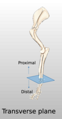
Transverse Plane for forelimb
Is the same transverse plane we learned, but imagine it like the dog has it's arms stretched out lying down splitting the forelimb in proximal and distal portions
Sagittal plane for forelimb
splits forelimb into medial and lateral

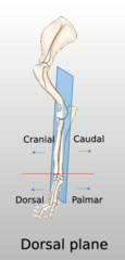
Dorsal Plane for forelimb
splits forelimb into cranial/caudal (and dorsal/palmar once past carpus). again, imagine dog is laying down with legs extended behind itself for it to line up with our planes already learned
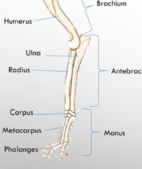
Brachium
osseous and soft tissue structures centered around the humerus

Antebrachium
osseous and soft tissue structures centered around the radius/ulna

Manus
Carpus, Metacarpus, and Phalanges
Caudocranial scapula radiograph of scapula

How to position for caudocranial scapula radiograph

Mediolateral shoulder radiograph
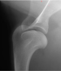
How to position for mediolateral shoulder radiograph

Caudocranial (right) humerus radiograph
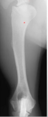
How to position for caudocranial humerus radiograph

Dorsopalmar manus radiograph

How to position for dorsopalmar manus radiograph

Comparative anatomy features to know for forelimb
- horses don't have acromion process (dogs, cats, cows do)
- horses have an intermediate tubercle associated with the proximal aspect of the humerus (most other species only have a greater and lesser tubercle)
- cats have a supracondylar foramen (the brachial artery vein and the median nerve course through this foramen)
- cats and rabbits have a hamate and suprahamate associated with the acromion
Cat hamate and suprahamate processes of scapula

Cat supracondylar foramen of humerus

Horse intermediate tubercle of humerus

Forelimb bones (bones of the thoracic limb)
scapula, humerus, radius, ulna, carpus, metacarpus, phalanges, digits

articular
relating to a joint
the scapula - lateral view
all things in common except acromion process, which is present in bovine and canine, but absent in equine. (note: spine of scapula more or less prominent/palpable depending on nutritional status of animal, especially equine)
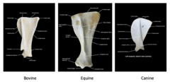
scapula differences across species
acromion process is present in bovine and canine, but not present in equine. equine and bovine have dorsal scapular ligament, but canines do not

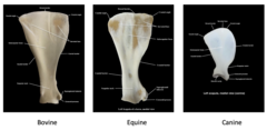
Scapula - medial view
equine and bovine have dorsal scapular ligament, but dogs do not.
clavicle
present in feline, but not in canine, equine, or bovine
Humerus - lateral view
greater tubercle on this side

Humerus - medial view
lesser tubercle & head on this side

Humerus differences between species
in all species, there is an intertubercular groove, but equine have another tubercle - the intermediate tubercle (there tubercles are separated into cranial and caudal parts)
humerus difference (felines): felines have the ________ ________, whereas canines, equine, bovine do not
supracondylar foramen

Radius and ulna - lateral view

Radius and ulna - medial view

because of the _______ _____ there is some supination/pronation possible in dogs
interoccious space
Radius and ulna differences across species
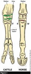
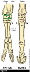
canines have separate radius and ulna. equine have partially fused radius and ulna (interosseous space very small), the ulna only comes to the middle of the radius. bovine have a fused radius and ulna (interosseous space very small), but the ulna goes all the way to the distal end. equine lateral styloid process of radius is actually just the distal epiphysis of ulna (part of the ulna). equine also have a groove for common digital extensor
Carpus - lateral view

Carpus - medial view

Carpus differences across species
organized in 2 rows in all species. canines and equine have 7 carpal bones, bovine have 6.
Canine carpal bones
Proximal row: intermedioradial, ulnar, accessory (accessory is non-weight bearing)
Distal row: C1-C4 (C4 is largest and articulates with digits 4 and 5).

Equine carpal bones
Proximal row: radial, intermediate, ulnar, accessory (accessory is non-weight bearing)
Distal row: C2, C3, C4 (C1 is present in almost 50% of animals, C5 is very rare and only seen in x-rays)
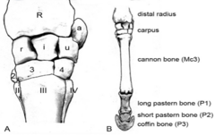
Bovine carpal bones
Proximal row: radial, intermediate, ulnar, accessory (accessory is non-weight bearing)
Distal row: C2 +C3 are fused, C4 (C1 and C5 never present)

Metacarpal bone differences across species
Canine metacarpal bones
MC 1-5

Equine metacarpal bones
MC2 (splint bone). MC3 is the cannon bone. MC4 (splint bone). Splint bones have buttons 3/4 of the way down the cannon bone. The proximodorsal surface of MC3 has a bump called the metacarpal tuberosity
Metacarpal tuberosity is present in which species, on what bone, and for what purpose?
Equine; on the proximodorsal surface of MC3; extensor carpi radialis muscle inserts here
Bovine metacarpal bones
MC3 and MC4 are combined to form one cannon bone (rudimentary MC5 may be present)
Phalanges
P1 (proximal phalanx - base of finger), P2 (middle phalanx), P3 (distal phalanx - tip of finger). All species have extensor process and flexor tubercle/surface (called solar foramina and solar canal in equine)
Canine phalanges
Digits 2-5 have P1, P2, P3. Digit 1 (dew claw) has only proximal and distal phalanx). P3 is the ungual crust

How many digits does a dog have?
all 5
How many digits does a horse have?
just the 3rd digit alone (one)
How many digits does a cow have?
3rd and 4th form the claws. 2 and 5 are present as dew claws. No 1st digit. (4)

Sesamoids of canine
Dorsal: 5 dorsal sesamoids (sesamoid 1 is in abductor digit 1 longus)
Palmar: 2 proximal sesamoids each in digits 2-5 (fetlock joint) and 1 proximal in digit 1
Sesamoids of equine
Palmar: 2 proximal seasmoids (fetlock joint); distal sesamoid (coffin joint)
Sesamoids of the bovine
Palmar: 2 proximal sesamoids for each digit's fetlock joint, 1 distal for each digit's coffin (remember they have 2 digits)
When counting carpus bones and tarsus bones, we go ______ to _______, counting up
medial; lateral
process (processus)
general term for prominence
tuberosity (tuber, tuberositas)
a large, rounded non-articulated projection; a tubercle is a smaller one
trochanter
applied to a few non-articular prominences; e.g. the trochanters of the femur
spine (spina) or spinous process (processus spinosus)
a pointed projection
crest (crista)
a sharp ridge
head (caput)
a rounded articular enlargement at the end of a bone; it may be joined to the shaft by a constricted part, the neck (collum)
condyle (condylus)
an articular eminence which is somewhat cylindrical; a non-articular projection in connection with a condyle may be termed an epicondyle (epicondylus)
trochlea
a pulley-like articular structure
glenoid cavity (cavitas glenoidalis)
a shallow articular depression and a cotyloid cavity or acetabulum is a deeper one
facet
commonly applied to articular surfaces of small extent, especially when they are not strongly concave or convex
fossa, fovea, groove, sulcus, impression
applied to various forms of depressions
foramen
perforation for the transmission of vessels, nerves, etc
sinus
air-cavity within a bone; it is lined with mucous membrane and communicated with the exterior
Flexion
reduction in angle of a joint
Extension
making the angle of a joint bigger
Coffin joint
between P2 and P3

Fetlock joint
between metacarpals and cannon bone (MC3)

Basic anatomy of nails
Basic anatomy of hooves
Basic anatomy of horns
Nails across species comparison
Hooves across species comparison
Horns across species comparison
What are general modifications of the equine forelimb?
● Reduction in the number of digits
● Angulation of joints
● Light but strong bones with reduced medullary cavity
● Check ligaments
● Suspensory apparatus
● Stay apparatus
● Development of the hoof
● Dorsal scapular ligament
● Fusion of the radius and ulna
Bones in the skeleton of the thoracic limb?
● Scapula
● Humerus
● Radioulna (fused radius and ulna, horse), radius/ulna (dog and cow)
● Carpal bones
● Metacarpal bones
● Phalanges
● Sesamoid bones
What purpose does scapular cartilage have in horses and cattle?
It enlarges the area for muscular attachment and distributes concussion. The cartilage becomes increasingly calcified and rigid with age
What is the dorsal scapular ligament in horses and cows?
a cranial extension of the thoracolumbar (lumbodorsal) fascia; it attaches to the scapula and provides attachment sites for various extrinsic muscles of the shoulder
The joints of horses and cattle
● Shoulder
● Elbow
● Carpus ("knee")
● Metacarpophalangeal (''fetlock")
● Proximal interphalangeal ("pastern")
● Distal interphalangeal ("coffin")
Shoulder joint of horse/cow
- Contain no major ligaments (NO collateral ligaments) - tendons of insertions of muscles function as ligaments of this joint.
● The intertubercular (bicipital) bursa can get inflamed and distended withexcess synovial fluid (bursitis). In the horse and ox, there is no communicationbetween this bursa and the joint cavity, however, there is in the dog and sheep.
● Bursitis may occur in infraspinatus bursa where infraspinatus muscle inserts onthe greater tubercle. Affected horses may stand with the limb abducted at theshoulder to relieve the pain.
Elbow joint of the horse/cow
- The lateral and medial collateral ligaments of the elbow lie caudal to the axis of rotation of the joint. This tends to prevent initial flexion of the elbow (lock the joint in extension), thereby assisting the animal to stand with a minimum of muscle action. The ligaments thus comprise part of the stay apparatus of the forelimb (discussed later). The elbow acts as a "snap joint."
● The olecranon bursa (constant) lies between the long head of triceps and the olecranon. A subcutaneous "inconstant (false)" bursa often develops over the olecranon in response to pressure. This painless swelling, which can also occur in large breeds of dog, is called capped elbow, or shoe boil.
The carpus joint (knee) of the horse/cow
- Actually three joints in one (radiocarpal(antebrachiocarpal), middle carpal, and carpometacarpal joints)
● The three joints share a common fibrous joint capsule, but each joint has its own separate synovial joint pouch. The two distal joint pouches (middle carpal and carpometacarpal) communicate between C3 and C4. This is of significance when considering the spread of infection or injected drugs between joints. The radiocarpal joint pouch does not communicate with its distal neighbors.
● In addition to the expected dorsal outpouching of the radiocarpal joint capsule between the extensor tendons, there is an additional out pouching. This lies proximal to Ca and caudal to the lateral digital extensor tendon and is called the palmarolateral pouch.
The fetlock joint of the horse/cow
- between cannon bone and long pastern bone (P1)
- At a slight state of overextension (angullation) at rest. when dealing with heavy loads, this joint tends to be driven into extreme overextension and this leads to injury, despite it having a complex system of ligaments.
- the sesamoid bones, are strongly anchored to it by four sets of distal sesamoidean ligaments. Mc3 and P1 are connected by collateral ligaments.
- the synovial joint cavity extends spacious dorsal and palmar pouches
What are the palmar and dorsal pouches of a horse/cow?
They are extensions of the synovial joint cavity of the fetlock to allow for the joint's extreme mobility (these pouches extend proximally and lie against Mc3 and are easily punctured from the side)
the palmar pouch of the fetlock
extends ¼ of the way (about three finger widths)up the cannon bone immediately deep/cranial to the interosseus. It can bepunctured between the splint button (either Mc2 or Mc4) and the proximalends of the sesamoids. Distensions of the palmar pouch (windgalls) arevisible as puffs here.
dorsal pouch of the fetlock
extends proximally (two finger widths) along the distal end of Mc3, deep to the two extensor tendons. In addition to the dorsal pouch, two bursae are also present here. The bursae lie between each digital extensor tendon and the dorsal fetlock joint capsule.
Pastern joint
at rest it is straight. Overextension at rest is prevented by ligaments connecting Pl andP2, and by the straight sesamoidean ligament
- The joint capsule of the pastern is like that of the fetlock, but the pouches are smaller and only the dorsal pouch is accessible.
- Any inflammatory process near or at the pastern and coffin joints often leads to an excess growth of bone (exostosis) and conditions of high and low ring bones respectively.
Coffin joint
● The coffin joint has a dorsal and a palmar pouch. The palmar pouch extends proximally between P2 and the navicular bone to a level where it abuts against the distal end of the digital sheath. It is separated from this sheath by fibrous connective tissue.
● The dorsal pouch lies between the common digital extensor (CDE) tendon andthe distal part of P2.
Navicular bone
an integral part of the coffin joint. It serves to enlarge the distal articular surface of this joint, and to provide a bearing surface for the deep digital flexor (DDF) tendon. It lies dorsal to the middle third of the frog
- articulates mostly with P2, but there is also some articulation with P3 distally.
- suspended by ligaments attaching to Pl, P2, and P3.
- The collateral (suspensory) navicular ligaments originate from the distal extremity of PI, run along (and insert on) the medial and lateral surfaces of P2, and insert on the ends and proximal border of the navicular bone.
- The distal navicular ligament (or distal sesamoidean impar ligament), is short and wide and connects the distal border of the bone with P3.
- The navicular (podotrochlear) bursa lies between the palmar surface of the navicular bone and the DDF tendon, preventing excessive friction