Pathology of Thyroid & Parathyroid Disease
 Call Kai
 Learn
 Practice Test
 Spaced Repetition
 Match
 Flashcards
 Knowt Play
1/110

There's no tags or description
Looks like no tags are added yet.
111 Terms
Main cause of Thyrotoxicosis
Graves’ disease (85%)
Name some other causes of Thyrotoxicosis
• Toxic (hyperfunctional) multinodular goitre
• Toxic adenoma
• Thyroiditis
• Thyroid carcinoma
• TSH-oma
• Choriocarcinoma / Hydatidiform mole
• Struma ovarii
• Iatrogenic (eg, Amiodarone, L-thyroxine)

Put an up/down arrow next to each one for Thyrotoxicosis

Clinical Features of Thyrotoxicosis
• Anxiety
• Irritability
• Fatigue
• Weight loss
• Good appetite
• Palpitations
• Heat intolerance
• Tremor
• Sweating
• Diarrhoea
• Proximal myopathy
• +/- Eye signs / Dermopathy
Complications of Thyrotoxicosis
• Atrial fibrillation
• Dilated cardiomyopathy
• Osteoporosis
• Amenorrhoea
• Optic nerve compression
• Thyroid storm
Morphology of Graves’ Disease
Diffuse hyperplasia (symmetrical enlargement - not nodular)
2 problems that can go along with Graves’ Disease
• Infiltrative ophthalmopathy
• Dermopathy (15%)
Is there a gender preference in Graves’ Disease
Female:Male = 10:1
Age of risk for Graves’ Disease
Peaks between 20 and 40 years
Other risk factors of Graves’ Disease
• HLA types - HLA-B8 & HLA-DR3
• Associated with other autoimmune disorders
Test to diagnose Graves’
Test radioactive iodine uptake - increased in Graves’
What is gone wrong in Graves’ Disease
Thyroid-specific defect in suppressor T-cell function
What causes the Thyroid-specific defect in suppressor T-cell function in Graves’
• TSH receptor IgG autoantibodies
• Proliferation of CD4+ helper T cells targeting TSH-receptor epitopes
• Thyroid-stimulating antibodies
• Thyrotropin-binding inhibitor immunoglobulin
Histology of Graves’ disease
• Tall columnar follicular epithelial cells
• Increase in number of epithelial cells (hyperplasia)
• Pseudopapillary buds encroach on colloid
• Diminished colloid
• Increased lymphoid tissue in interfollicular stroma
• Increased vascularisation of thyroid
• Pretibial myxoedema – lymphocyte infiltration

What would an Isotope Thyroid scan show in Graves’ disease
Isotope Thyroid scan showing diffusely increased uptake of radiolabelled 99mTechnetium
Name 4 conditions that would cause increased uptake in a Thyroid Radioisotope Scan
Graves’ disease
Toxic multinodular goitre
Solitary toxic adenoma
Hot nodule
How do you differentiate Graves’, Toxic multinodular goitre & Solitary toxic adenoma on a Thyroid Radioisotope Scan
Graves’ disease = Diffuse
Toxic multinodular goitre = Multiple discrete areas
Solitary toxic adenoma = Single area
Name 2 conditions that would cause decreased uptake in a Thyroid Radioisotope Scan
Thyroiditis
Cold nodule
Name 2 types of thyroiditis
Subacute
Postpartum
Do hot/cold nodules have a higher risk of carcinoma
Cold - 20% risk of carcinoma
Malignancy in hot is rare
Causes of Hypothyroidism
• Congenital (cretinism)
• Iodine deficiency (endemic goitre)
• Radioiodine ablation
• Thyroidectomy
• Hashimoto’s thyroiditis
• Subacute thyroiditis
• Drugs (eg, lithium)
• Pituitary lesions
• Hypothalamic lesions
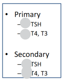
Are these increased or decreased in hypothyroidism

Hypothyroidism - Clinical Features
• Fatigue
• Depression
• Mental slowing
• Weight gain
• Cold intolerance
• Constipation
• Periorbital oedema
• Macroglossia
• Bradycardia
• Menorrhagia
Possible complications of Hypothyroidism
• Congestive cardiac failure
• Pericardial effusion
• Dyslipidaemia
• Psychosis (‘myxoedema madness’)
• Carpal tunnel syndrome
• Infertility
• Hypothermia
• Myxoedema coma
Cause of Hashimoto’s Thyroiditis
Autoimmune
Histology of Hashimoto’s Thyroiditis
Infiltrate of lymphocytes + plasma cells

2 types of Hashimoto’s Thyroiditis
Goitrous or atrophic
Hashimoto’s Thyroiditis initial presentation
Hashitoxicosis initially
Complication of Hashimoto’s Thyroiditis
Most develop hypothyroidism
Pathogenesis of Hashimoto’s Thyroiditis
• Defective thyroid-specific suppressor T cells
• Emergence of CD4+ helper T cells targeting thyroid antigens
• Antibodies to thyroid peroxidase, thyroglobulin, TSH receptors
Male:Female ratio with Hashimoto’s Thyroiditis
Up to 20:1 female predominance
Risk associated with Hashimoto’s Thyroiditis
Small increased risk of B-cell Non-Hodgkin’s lymphoma
Risk factors for Granulomatous (De Quervain’s) Thyroiditis
4th-5th decades of life
Female:Male = 4:1
HLA-B35
Sx of Granulomatous (De Quervain’s) Thyroiditis
Fever | Tender neck | Malaise
Which of these are increased or decreased in the initial phase of Granulomatous (De Quervain’s) Thyroiditis
ESR
T4
T3
Radioactive iodine uptake
Increased: ESR, T4, T3
Decreased: Radioactive iodine uptake
(Inflammation damages the follicles, and the preformed hormone stored in the colloid leaks into the blood → Raises T4 &T3 (The gland isn’t actually making more hormone.) The damaged follicles can’t trap iodine, so uptake is low.)
True/False Granulomatous (De Quervain’s) Thyroiditis is self limiting
True
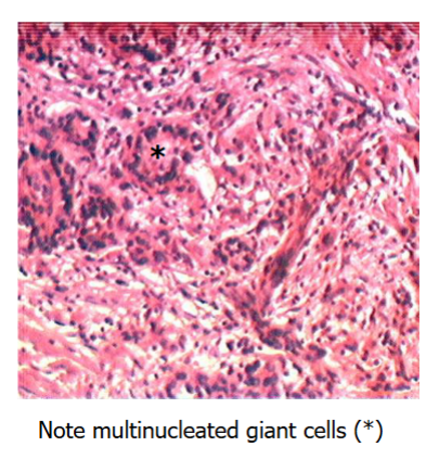
Histology of Granulomatous (De Quervain’s) Thyroiditis
Diffuse Non-toxic (Simple Colloid) Goitre is usually Hypothyroid / Euthyroid / Hyperthyroid
Usually euthyroid
Diffuse Non-toxic (Simple Colloid) Goitre is seen a endemically in what populations
Endemic goitre in Andeans and Tibetans
What’s up with TSH in Diffuse Non-toxic (Simple Colloid) Goitre
Compensatory high TSH
In Diffuse Non-toxic (Simple Colloid) Goitre do you see hypertrophy or hyperplasia
Follicular cell hypertrophy and hyperplasia
Name 2 Goitrogens
cabbage
cassava
Most to least common cause of Diffuse Non-toxic (Simple Colloid) Goitre: endemic, Nonendemic/Sporadic, hereditary
Endemic > Nonendemic / Sporadic > Rare hereditary defects in thyroid hormone synthesis
Histology of Diffuse Non-toxic (Simple Colloid) Goitre
Follicular cell hypertrophy & hyperplasia

Is it common/rare that simple goitres become transformed into multinodular goitres
Common - Most simple goitres become transformed into multinodular goitres
Multinodular Goitre is nontoxic or toxic
May be nontoxic or toxic (induce thyrotoxicosis)
Which of these are present in Multinodular Goitre
ophthalmopathy
dermopathy
cosmetic disfigurement
tracheal compression
retrosternal extension (“plunging goitre”)
cosmetic disfigurement
tracheal compression
retrosternal extension (“plunging goitre”)
Possible complication of Multinodular Goitre
May induce the superior vena cava syndrome
Histology of Multinodular Goitre

Possible causes of Thyroid Nodules - are they generally benign/malignant
• Adenoma
• Carcinoma
• Thyroid cyst
• Parathyroid cyst
• Thyroiditis
• Metastases
• Lymphoma
• Sarcoma
(Less than 1% of thyroid nodules are malignant)
What would Fine-needle aspiration cytology be used for
• All palpable solitary thyroid nodules
• Dominant nodules in a multinodular goitre
Limitation of Fine-needle aspiration cytology diagnostics
Cannot differentiate follicular adenoma & carcinoma
True/False most Tumours of Thyroid Gland are neoplastic
False - Most are not neoplastic
What is the most common type of Tumour of Thyroid Gland
90% are adenomas
Which is more likely to be malignant - a solitary nodule or multiple nodules
Solitary nodule more likely malignant than multiple
Are functioning “hot” or non-functioning “cold” thyroid nodules more likely to be malignant
Non-functioning (cold) nodules more likely to be malignant (but still low risk)
With Tumours of Thyroid Gland, the younger/older the patient the greater the chance of neoplasia
The younger the patient the greater the chance of neoplasia
Which is more suspicious - nodule in a male/female patient
A nodule in a male patient is more suspicious than in a female
Most common type of Thyroid Adenoma
Most are follicular adenomas
What’s the risk like for malignant transformation of Thyroid Adenoma
Negligible risk
Thyroid Adenoma structure
• Encapsulated
• Homogeneous, fleshy cut surface
• Foci of haemorrhage
• Cystic degeneration

Most Thyroid Adenomas are hot/cold on isotope scane
Most are “cold” on isotope scan
Name 6 types of Malignant Thyroid Tumours (& state which is most common)
1. Papillary carcinoma (85%)
2. Follicular carcinoma
3. Medullary carcinoma
4. Anaplastic carcinoma
5. Lymphoma (Rare)
6. Tumours metastatic to the thyroid
Age & gender of risk for Papillary Carcinoma
3rd to 5th decades of life
Female: Male = 3:1
How does Papillary Carcinoma spread
Lymphogenous spread
Is Papillary Carcinoma generally solitary / multifocal
Often multifocal tumours
What genetic mutations are associated with Papillary Carcinoma
Activation of RET/PTC oncogene
Gain-of-function BRAF mutations
Prognosis for Papillary Carcinoma
95% 10-yr survival
Gross appearance of Papillary Carcinoma
Grey-white, firm cut face with foci of calcification or cystic areas
Histology of of Papillary Carcinoma
• Branching papillae
• Concentric Psammoma bodies within papillary cores
• “Orphan Annie eyes” nuclei (no nucleoli)
• Nuclear grooves
Follicular Carcinoma represents what % of thyroid cancers
5-15%
Age of risk for Follicular Carcinoma
4th to 6th decades of life
Name another risk factor for Follicular Carcinoma
Iodine deficiency
Is Follicular Carcinoma usually encapsulated
Yes
How & where does Follicular Carcinoma spread
Vascular invasion
Metastasise to bone, lungs, liver
Anaplastic Carcinoma is what % of thyroid cancers
<5%
Anaplastic Carcinoma age of risk
Elderly patients
Anaplastic Carcinoma presents as
Rapidly enlarging mass
Prognosis for Anaplastic Carcinoma - explain
Metastasise widely → Almost 100% mortality
What cells are affected by Medullary Carcinoma
Neuroendocrine tumours of parafollicular C cells
What do Neuroendocrine tumours of parafollicular C cells secrete
Most secrete calcitonin
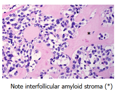
Medullary Carcinoma histology
30% of Medullary Carcinoma cases are associated with what conditions
30% associated with Multiple Endocrine Neoplasia IIa and IIb
Medullary Carcinoma cases are associated with what mutation
Mutation in RET proto-oncogene on chromosome 10q (MEN IIa)
Age of risk for Medullary Carcinoma
5th to 6th decades of life
How does Medullary Carcinoma present
paraneoplastic syndrome (e.g., diarrhoea)
How does Medullary Carcinoma spread
Lymphatic/haematogenous spread
What is different with how tumours present with Multiple Endocrine Neoplasia (4)
• Tumours occur at younger age than sporadic cases
• Tumours arise in multiple organs
• Usually preceded by asymptomatic hyperplasia
• More aggressive and likely to recur
Classic triad of what MEN-1 causes
Primary hyperparathyroidism; Pancreatic tumours; Pituitary adenomas
What does MEN-2 cause - differentiate into 2a/b
Medullary Ca
Phaeochromocytoma
Parathyroid hyperplasia (MEN-2a)
Ganglioneuromas (2b)
Marfanoid habitus (2b)
What mutation is associated with MEN-2
RET mutations
What is the most common cause of symptomatic hypercalcaemia
Malignancy
How does Malignancy → Hypercalcaemia
Paraneoplastic production of PTH-related protein (PTHrP) or osteolysis
Most common cause of asymptomatic hypercalcaemia
Primary hyperparathyroidism
Hypercalcaemia - Clinical Features
• General – lethargy, depression
• Renal – recurrent nephrolithiasis (renal stones), nephrocalcinosis
• Skeletal – osteitis fibrosa cystica
• Gastrointestinal – nausea, vomiting, constipation, peptic ulcer, acute pancreatitis
• Cardiovascular - Hypertension
“Stones, bones, groans, and psychic moans”
Primary Hyperparathyroidism → excess what hormone
Excess Parathyroid hormone
Primary Hyperparathyroidism effects/results
1. ↑ bone resorption
2. ↑ renal tubular reabsorption of Ca2+
3. ↑ renal synthesis of 1,25-(OH)2D3
4. Causes hypercalcaemia
Prevalence of Primary Hyperparathyroidism
25 per 100,000
Age / Gender risk factors for Primary Hyperparathyroidism
Female: Male = 4:1
50+ yrs
Top 4 causes of Primary Hyperparathyroidism
1. Adenoma (85%+)
2. Hyperplasia (10%)
3. Carcinoma (1%)
4. MEN I or IIa (Hyperplasia)