Exam 2: Muscle Structure and Function
 Call Kai
 Learn
 Practice Test
 Spaced Repetition
 Match
 Flashcards
 Knowt Play
1/36

There's no tags or description
Looks like no tags are added yet.
37 Terms
endomysium
surrounds individual fibers in skeletal muscles
fasicles
multiple fibers bundled together in skeletal muscles
perimysium
a collagen sheath that fascicles are bound by in skeletal muscle
epimysium
surrounds all fascicles of an entire muscle in skeletal muscle
deep fascia
binds muscles into functional groups in skeletal muscle
sarcomere
basic functional and contractile unit of striated muscle tissue; repeating makes up a myofibril
g actin
monomeric form (makes up f actin); active site where actin interacts with myosin; tropomyosin wraps around
f actin
polymerized form, double helix; string of g actin
tropomyosin
covers and uncovers attachment sites for myosin on actin myofilaments; regulatory protein
troponin
provides calcium binding sites on actin filaments; causes tropomyosin to make way for myosin when calcium binds to it
rod region of myosin
backbone of thick filaments; parallel to myosin filament
globular heads of myosin
attach to actin; bind to ATP and break it down into ADP + P; when ADP is released from head it attaches to ATP and releases that from actin; involved in crossbridging; is what performs the power stroke
calcium during relaxed state of muscle sliding
calcium concentration in sarcoplasm is low and is stored in sarcoplasmic tubules
tropomyosin during relaxed state of muscle sliding
troponin-tropomyosin complex attached to actin filament, tropomyosin positioned to block myosin binding sites on actin filament
energy during relaxed state of muscle sliding
ATP and inactive ADPase bound to myosin head; low energy configuration; binding to actin is not possible
step one of events during contraction
nerve impulse (afferent signal) from motor neuron generates action potential in nerve cell; action potential propagated along sarcolemma and down t tubules
step two of events during contraction
myosin ATPase activated; ATP splits; high energy myosin-ADP complex; myosin ATPase is active at low levels even when muscle contraction is not occurring
step three of events during contraction
action potential causes release of calcium from sarcoplasmic reticulum
step four of events during contraction
calcium binds to troponin; molecular shape of troponin changes; tropomyosin is removed from binding site of myosin on the actin filament; myosin attaches to actin
step five of events during contraction
contraction (power stroke): potential energy stored in high-energy configuration is used to pivot myosin head; myosin head bends as it pulls on actin; inorganic phosphate are released from myosin
step six of events during contraction
new ATP attaches to myosin head replacing the ADP from prior power stroke; cross bridge simultaneously detaches; following death there is not ATP and muscle fibers cannot relax; rigor mortis
step seven of events during contraction
if no new impulse, calcium is pumped back into sarcoplasmic reticulum; relaxation occurs
step eight of events during contraction
if calcium is present from additional impulse, cycle repeats; myosin head “steps” to next binding site on actin
a-bands
dark bands; thick filament region where actin and myosin overlap; anisotropic; polarizes light; center of each sarcomere

what is highlighted
a-band
i-band
light bands; two regions of thin filament (actin) bisected by z-disc protein anchoring thin filaments

what is highlighted
i-band
H band / H zone
within A band; no thin filaments; myosin only, contains M line

what is highlighted
H band / H zone
M line
structural protein that runs down the middle of the sarcomere; where myosin attaches
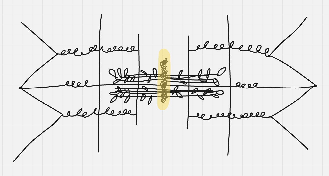
what is highlighted
M line
Z disc
membrane; midline in i band; where sarcomere starts/ends; where actin attaches

what is highlighted
Z disc
titan filament
spring-shaped elastic protein that attaches to the thick filament and anchors it to the z disc; contributes to elasticity

what is highlighted
titan filament
actin
thin protein; made out of 2 woven strands; have active sites; has tropomyosin on it
active sites
locations where myosin and actin interact with each other and do contractions