Genetics Chapter 3
 Call Kai
 Learn
Learn Practice Test
 Spaced Repetition
 Match
Match Flashcards
 Knowt Play
1/65

There's no tags or description
Looks like no tags are added yet.
Name | Mastery | Learn | Test | Matching | Spaced | Call with Kai | Chat |
|---|
No analytics yet
Send a link to your students to track their progress
66 Terms
Zygote
First cell that leads to development of a new individual is a fertilized ovum
Forms when sperm cells from maleand an oocyte from a female joins
Gametes
Sperm and oocytes
Sex cells
Each reproductive system has:
Gonads: paired structured (where sperm and oocytes are made)
Tubes that transport these cells
Hormones and secretions that control reproduction
Testes (male gonad)
Packed with sperm-producing seminiferous tubules and cells that secrete testosterone
Formed in abdominal cavity during embryonic development and then descends into scrotum before birth
Accessory Glands
Prostate gland, seminal vesicles, and bulbourethral gland have secretions that all combine into seminal fluid
Fluid is alkaline and carries sperm
Maintains sperm viability and motility
Semen = Sperm + Seminal fluid
Where doessperm mature?
Epididymis
Ovaries (female gonad)
Fetal development: Oocyte development begins before birth, but pauses during prophase of meiosis 1 at birth
Puberty: Meiosis resumes once puberty begins
Ovulation: One secondary oocyte is released from an ovary during each menstrual cycle
Oviduct
Site of fertilization
Uterus
Growth of fetus
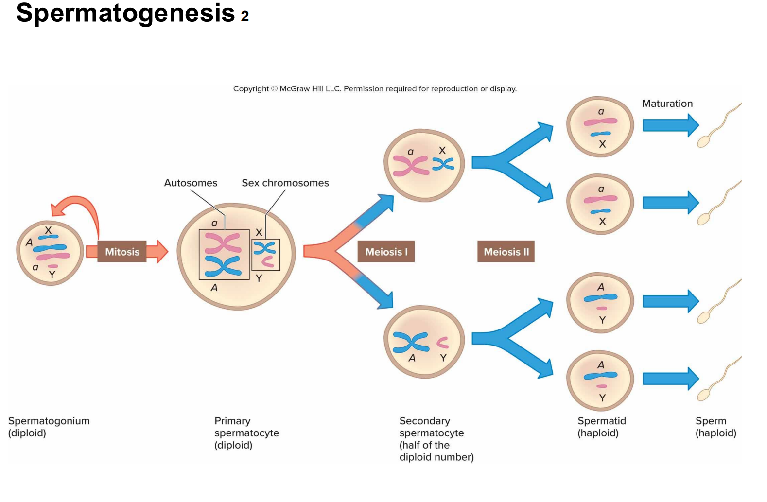
Meiosis
Meiosis 1 (reduction division): reduces the number of replicated chromosomes from 46 to 23
Meiosis 2 (equational division): produces 4 haploid from the two cells from the first division by splitting the replicated chromosome

Homologous Pairs
Same genes in the same order, BUT different alleles, or variants, of the same gene

Gametes
Haploid (1n)
Somatic cell
Diploid (2n)
Meiosis only occurs in ____ cells.
Gametes
Creates 4 genetically different cells
Two divisions
Two major sources of genetic variation in meiosis:
Crossing Over
Independent assortment of chromosomes
When does crossing over occur?
Prophase I of meiosis.
What happens during Meiosis I?
Homologous chromosomes separate, reducing the chromosome number from diploid to haploid.
What happens during Meiosis II?
Sister chromatids separate, producing four haploid cells.
What happens during prophase?
Chromosome condenses
Nuclear envelope breaks down
Spindle forms
What happens during metaphase?
Chromosomes line up at the middle of the cell.
What happens during anaphase?
Chromosomes/chromatids separate and move toward opposite poles.
What happens during Telophase?
Nuclear envelopes assemble, spindles disappears.
Cytokinesis
Divides cells.
What does Meiosis II lack?
DNA replication.
How many mature sperm come from one primary spermatocyte?
4 mature sperm
Sperm midpiece
Has many mitochondria
Splits ATP molecules, which releases energy that powers sperm cell to swim inside female reproductive tract
Oogenesis
Begins with a diploid oogonium
Mitosis: Primary Oocyte (diploid)
Meiosis 1: primary oocyte divides unequally forming a small polar body (dies) and a large secondary oocyte
Meiosis 2 (if fertilization occurs): secondary oocyte divides to form a second polar body and a mature ovum (egg)

When is the secondary oocyte completing meiosis II?
Fertilization triggers completion of meiosis II.
How many mature ova result from one primary oocyte?
One functional ovum (egg).
When are primary oocytes arrested?
In prophase I of meiosis.
Oocytes start meiosis 1 (replicate their DNA, pair up their homologous chromosomes, and do crossing over (recombination)) before a female is even born, but they pause partway through and stay paused for years until PUBERTY.
AFTER Puberty: Meiosis 1 continues in one or several secondary oocytes each month, but stops again at Metaphase 2 (ovulation = when oocyte is released and unfertilized)
Meiosis is only completed if the ovum is fertilized
Where does fertilization happen?
Uterine (fallopian) tube
After fertilization, meiosis 2 continues and a zygote with 46 chromosomes is formed
What is the largest Human cell?
Oocyte
can be seen with naked eye
Fertilization Steps
Fusion of Oocyte and sperm
Formation of zygote
Formation of the blastocyst (fluid filled ball of cells) by cell division (mitosis: blastomere → morula (ball of cells)
Inner cell mass will form the cells of the fetus
As implantation begins: Trophoblast will form the structures that support the fetus
during pregnancy
Implantation
Blastocyst embeds into the endometrial lining of the uterus
Trophoblast: secrete human chorionic gonadotropin (hCG), a hormone that prevents menstruation.
Hormone detected in woman’s urine/blood = pregnant
First Trimester
Organ Formation
Second Trimester
Organ Maturation
Third Trimester
Rapid Growth
How long does ovulation last?
12-48 hours
Acrosomal Enzymes
Aid sperm penetrate the protective layers around the oocyte
Bore through protective layer (zona pellucida)
Capacitation
Capacitation: activates sperm and the oocyte secretes a chemical that attracts sperm
Chemical and electrical changes in the oocyte surface
block entry of more sperm

Gastrula
Three-layered embryo primary germ layer form)
End of second week
Dizygotic Twins (DZ)
Fraternal twins
2 Sperm fertilize 2 oocytes
Same genetic relationship as any two siblings
Monozygotic Twins (MZ)
Identical twins
Single fertilized ovum
Types of Identical Twins
Identical twins with separate amnions AND and chorions (1/3)
Identical twins that share an amnion and chorion (1 percent)
Identical twins that share a chorion BUT have separate amnion (2/3)

Conjoined Twins
RARE
Embryo divides into twins after the point at which the two groups of cells can develop as two individuals
Between at 13-15
Organogenesis
Transformation of simple three germ layers into distinct organs
Primitive Streak
Third week of prenatal development, a band appears along back of embryo
First sign of nervous system
Day 14: implantation completes
Rise to connective tissue progenitor cells and the notochord
Notochord
Induces sheet of overlying ectoderm to fold into the hollow neural tube
Neural tube = brain and spinal cord (Central Nervous system)
Week 8 of Gestation
All organs will be present in the newborn have begun to develop
It is now a fetus
Fetal Period
Body proportions approach those of a newborn
Bone replaces softer cartilage
Nerve and muscle functions become coordinated, fetus moves
SRY Gene
Sex-determining region of the Y
Anatomical difference between the sexes appear at week 6, after SRY gene is expressed in males
Week 12 (3 months)
Sucks thumb, kicks, makes fists and faces, and has the beginnings of teeth
Week 16 (4 months)
Has hair, eyebrows, lashes, nipples, and nails
Week 18
Vocal cords formed
Fetus makes no sound because it doesn’t breath air
End of Second Trimester (27 weeks)
Woman feels kicks and jabs
May detect fetal hiccup
Final Trimester
Fetal brain cells link into networks as organs elaborate and grow
Fat fills out the skin
Digestive and respiratory system mature last
Birth
Hormonally induced
Oxytocin: uterine contraction
Cervical dilation
Uterine contractions: expel the fetus and the placenta
Critical Period
Time during prenatal development when genetic variants, toxic substances, or viruses can alter a specific structure
2/3 of birth defects develop during the embryonic period (3-8 weeks)
MORE SEVERE than those that arise during the fetal period
Phocomelia
Congenital disorder caused by a mutation that acts at a specific point in prenatal development
Mutation halts limb development from 3rd to 5th week of the embryonic period (no arms & legs)
Teratogens
Chemicals or other agents that cause birth defects (congenital disorders)
EX:
Thalidomide (no limbs)
opioids
alcohol: Fetal alcohol syndrome (FAS) → small head & flat faces (behavior problems)
Nutrient (vitamins): Accutane (vit A) causes spontaneous abortion and hear, nervous and face problems
Viral Infections: zika virus (microcephaly: small head), HIV (prematurity, low weight, stillbirth), rubella (congenital disorder & stillbirths)
Aging
Age 30 = turning point for decline
Human body becomes less efficient by 0.8 percent each year
Diseases that begin in adulthood have genetic components (COMPLEX)
Many years for environmental exposures to alter gene expression
Malnutrition
Powerful environmental influence: create risks that appear later
Increase of cardiovascular and diabetes later in life
Single-Gene disease
Strike in childhood, tend to be recessive
Delayed effects of fetal malnutrition
Dominantly Inherited Conditions
Affects health in early to middle adulthood
Progeroid syndromes
single-gene disorders that speed aging-associated changes
Progerias: shorten life expectancy (SEVERE)
Inability to adequately repair DNA
Hutchinson-Gilford Progeria Syndrome
Most SEVERE rapid aging condition
Normal cells divide 50 times before dying
BUT cells from syndrome die after 10-30 divisions
Caused by mutation in LMNA (lamin) gene
Lamin A: called progerin triggers rapid programmed cell death (apoptosis)
Telomeres shrink → cell ages quick → nuclear membrane collapse