Lecture 7: Nervous System Organization and Function of the Cerebrum
 Call Kai
 Learn
Learn Practice Test
 Spaced Repetition
 Match
Match Flashcards
 Knowt Play
1/80

There's no tags or description
Looks like no tags are added yet.
Name | Mastery | Learn | Test | Matching | Spaced | Call with Kai | Chat |
|---|
No analytics yet
Send a link to your students to track their progress
81 Terms
Cerebrum
Largest part of brain (top/front part) which is split into left/right hemispheres
Cerebral cortex
Outermost layer of grey matter that covers the cerebral hemispheres of the brain.

What is the cerebral cortex responsible for?
Thinking, memory, voluntary movement, conscious sensor perception
Ex: consciously grasping a pencil; perceiving sesnsations from the skin/joints

Diencephalon
Sensory and motor relay centre involved in autonomic functions.

Brainstem
Connects cerebrum/cerebellum to spinal cord; autonomic functions and cranial nerve pathway

Cerebellum
Coordination of movement and balance

Spinal cord
Motor output, sensory input, reflexes, communication with the PNS

Grey matter
Neuronal cell bodies and supporting glial cells located on the outside of the cerebral cortex.

Deep grey matter
Basal nuclei and limbic system

Nucleus
Distinct cluster/mass of grey matter (nerve cell bodies) inside the CNS.
White matter
Myelinated neuronal axons located inside the cerebrum that transmit info based on the type of fiber.
Fiber types: commissural, association, projection

Commissural fibers
Connect the two cerebral hemispheres
Ex: corpus callosum, anterior commissure

Association fibers
Connect regions within the same hemisphere.
Ex: connections between Wernicke’s and Broca’s area, allowing them to communication with each other.

Projection fibers
Connect the cerebral cortex with distant CNS regions like the brainstem and spinal cord.
Ex: internal capsule

How is the arrangement of grey/white matter different in the spinal cord?
It is reversed → grey matter inside, white matter outside.
What does the contralateral cerebrum refer to?
Each hemisphere controls/receives sensory info from the opposite side of the body.
What is the left cerebral hemisphere responsible for?
Processing motor/sensory + visual info from the right side of body.
Logical processing
Language areas
Wernicke’s area: language comprehension
Broca’s area: language production
Aphasia
Damage to language areas, causing difficulty understanding language (Wernicke’s) or producing speech (Broca’s).
Global aphasia → both areas damaged.
What is the right hemisphere responsible for?
Processing motor/sensory + visual info from the right side of body.
Spatial orientation and object recognition
Artistic/visual processing → like interpreting 2D drawings as 3D
Ability to recognize emotional context in speech
Lateralization
The specialization of certain functions in one hemisphere (ex: hand/eye dominance).
How is lateralization different in males and females?
Less pronounced in females due to more developed commissural pathways (corpus callosum and anterior commissure)
Corpus callosum
Large commissural fibers connecting the cerebral hemispheres
Gyri
Raised raidges/folds of the cerebral cortex

Sulci
Shallow grooves between gyri

Fissures
Deep grooves that separate the hemispheres or certain lobes

Longitudinal fissure
Separates the left and right cerebral hemispheres

Central sulcus
Separates the frontal and parietal lobes

Lateral (Sylvian) sulcus
Separates the temporal lobe from the frontal and parietal lobes

Precentral gyrus
Contains the primary motor cortex

Postcentral gyrus
Contains the primary somatosensory cortex

Features of the corticol homunculus
Large hands, lips, tongue → require more motor control/sensory procesing.
Larger head/upper limbs than trunk/lower limbs.

What would a stroke affecting the left ACA cause?
Motor/sensory deficits in the right lower limb.
What would a stroke affectng the right MCA cause?
Motor/sensory deficits in left face and left upper limb.
Premotor cortex
Plans movements before execution
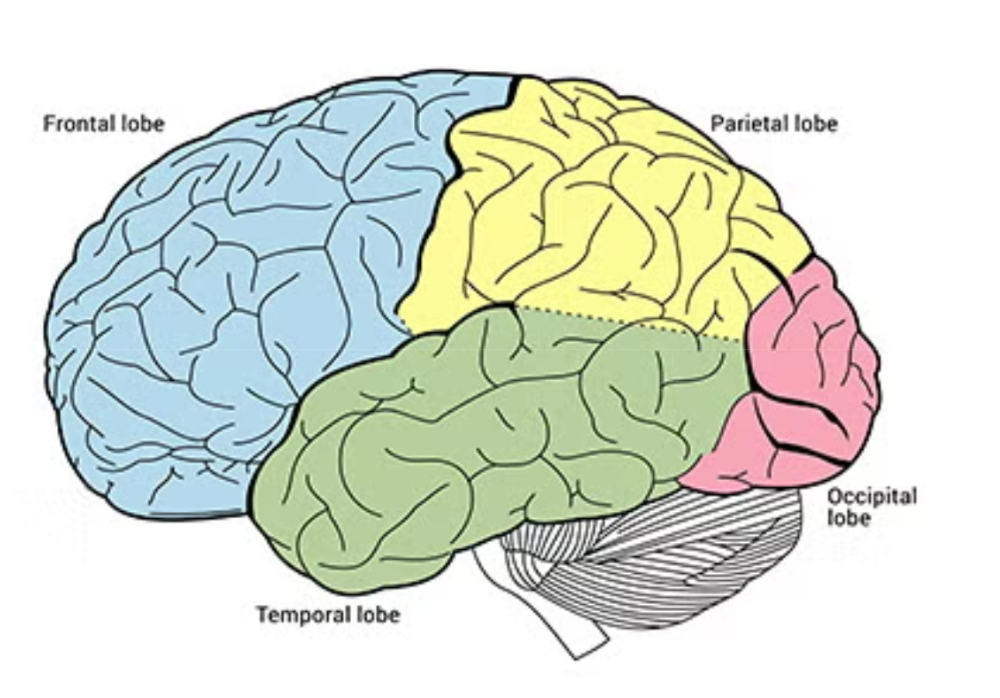
Located in the frontal lobe

Primary motor cortex
Executes voluntary movement
Located in the frontal lobe
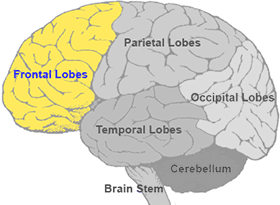
Where are the premotor cortex and primary motor cortex located?
Frontal lobe

What is the frontal lobe responsible for?
Movement planning and execution → contains premotor cortex and primary motor cortex
Intellect → thinking, reasoning, morals, personality, ethics
Frontal eye field → conscious, coordinated movement of the eye
Language production → contains Broca’s area

Primary somatosensory cortex
Consciously processes general body sensations (touch, pressure, temp, pain, vibration) but not special senses!
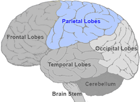
Located in the postcentral gyrus of the parietal lobe.
Association cortex
Stores/interprets somatosensory information
Located in all lobes

Common integrative area
Combines general body sensations, special senses, and other sensory info to create an integrated understanding of the environment.
Located in parietal lobe

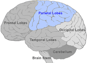
What is the parietal lobe responsible for?
Spatial awareness, body orientation, object recognition → contains associate cortex and common integrative area.
Processes general sensory info → contains primary somatosensory cortex
Auditory cortex
Processes and interprets info from ears (conscious perception of sound)
Located in temporal lobe
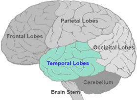
What is the temporal lobe resposible for?
Auditory processing → contains auditory cortex and association cortex
Memory storage → contains hippocampus
Language comprehension (written/oral) → contains Wernicke’s area

Visual cortex
Processes visual information → conscious perception of the field
Located in occipital lobe

What is the occipital lobe responsible for?
Visual processing → vontains auditory cortex and association cortex
Functions of basal nuclei
Deep collections of grey matter that:
Initiates/terminates motor movements and eliminates unnecessary movements.
Involved in procedural memory (learned subconscious habits/skills)
Involved in attention, memory, planning, initiation/termination of cognitive processes, regulation of some emotional behaviours.
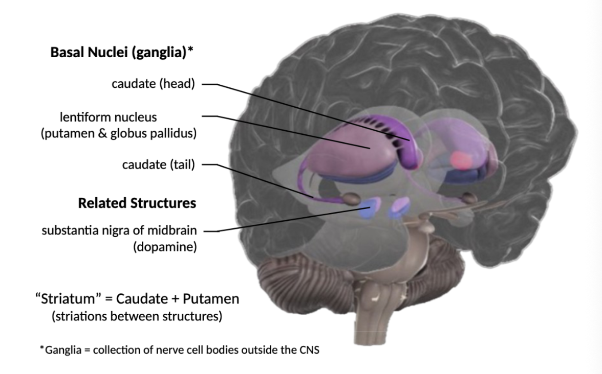
Striatum
Largest structure and main input station of the basal nuclei
Contain caudate nucleus and putamen

Steps in motor planning
Prefrontal cortex plans an intended movement.
Prefrontal cortex communicates with basal nuclei to access learned motor routines.
The info passes through the thalamus before returning to the cortex.
Allows the cortex to use rapidly use established movement patterns.

Substantia nigra
Dark-pigmented nucleus located in the midbrain, containing dopamine-producing neurons.
Role of dopamine in the nigrostriatal pathway
Dopamine is released in the substantia nigra and sent to the basal uclei to regulate intiation/termination of movement.
Cause of Parkinson’s disease and its symptoms
Loss of dopamine-producing neurons in the substantia nigra → inhibits basal nuclei function → difficulty initiating/terminating movements.
Cause of Huntington’s chorea and its symptoms
Neurodegeneration in the striatum → excessive involuntary movements
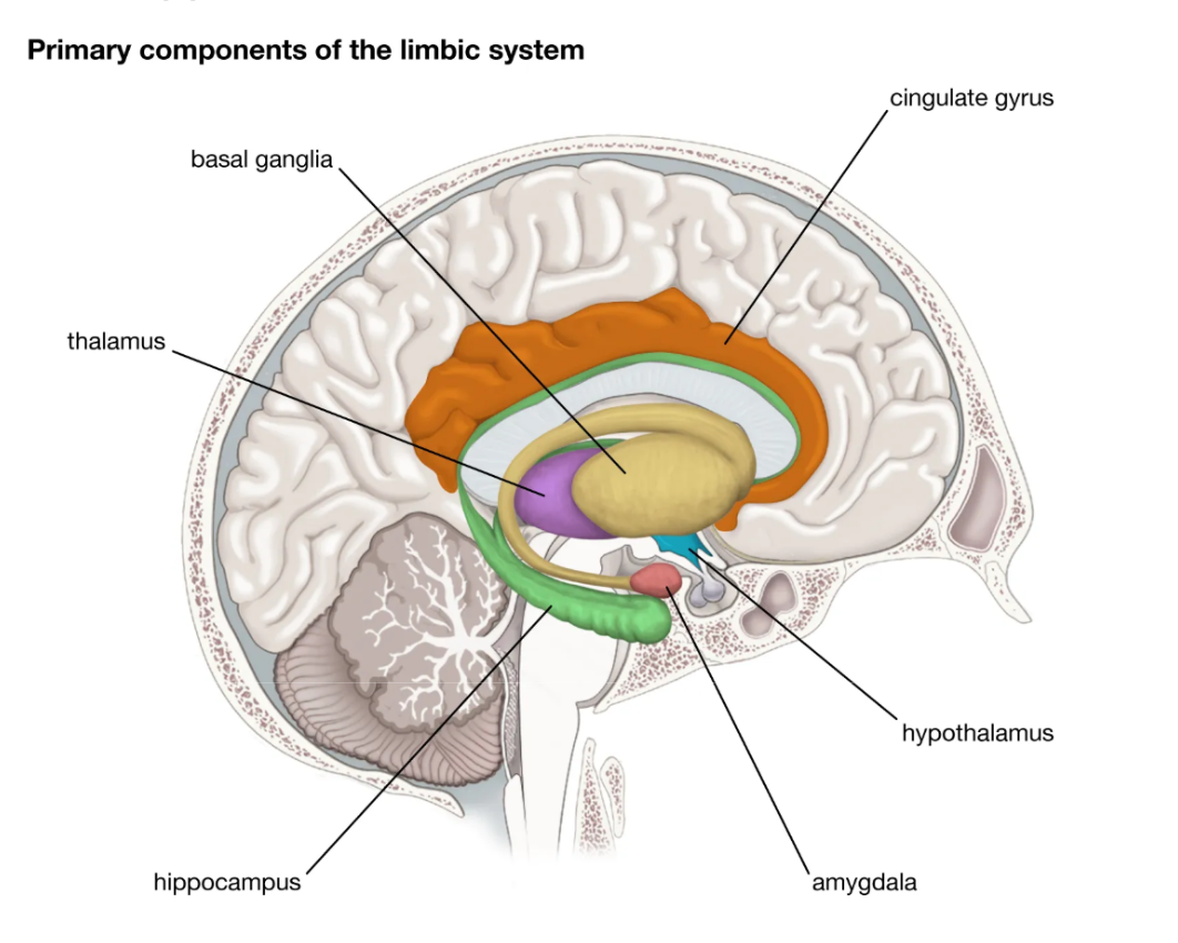
Function of the limbic system
Emotional and memory center of the brain, involved in:
Memory formation
Emotional processing
Responses to significant stimuli

Structures in the limbic system
Hippocampus, entorhinal cortex, amygdala, mammillary bodies, fornix, cingulate cortex.

Function of the hippocampus
Encording and consolidating memories (organize info about who, what, where, when)
Encoding
First step in memory processing → transforms incoming info and neural activity into a form that can be stored/retrieved later
Consolidation
Integrating different components of an experience (sight, smell, etc)
All somatic sessations and special senses must pass through the hippocampus.
Stabilization of memories of long-term storage

Entorhinal cortex
Brain’s “GPS” → spatial memory and navigation; helps associate memories with spatial/contextual info
Located in the temporal lobe
Function of the amygdala
Subconsciously assesses potential threats
Produces fear responses by communicating with the hypothalamus to influence autonomic function (activation of SNS → dilated pupils, muscle trembling, sweating, faster heart rate)
Interpret fear/anger in other people
Add emotional signifiance to memories

How does alcohol affect the amydala?
Impairs its ability for threat assessment/analysis, leading to risky behaviours.
Disordered amygdala function may lead to
Anxiety-related disorders
Function of mammillary bodies
Participate in memory processing by linking sensory experiences (especially smell) and emotions to memories.

Which pathway are mammillary bodies connected to?
Olfactory
Fornix
Major fiber pathway → mammillary bodies receive input from hippcampus via fornix.

Cingulate cortex
C-shaped region that wraps around the corpus callosum

Declarative memory
Conscious, intentional recollection of facts, events, and specific information.
Two types of declarative memory
Semantic memory
Episodic memory
Semantic memory
Memory for facts and general knowledge
Episodic memory
Memory for personal experiences/events
Non-declarative memory
Long-term memories that you use and store without conscious awareness
What brain regions regulate declarative memories?
Limbic system
What brain region regulates non-declarative memories?
Cerebellum, basal nuclei, thalamus
Role of cerebellum in non-declarative memories
Learning new motor proecdures
Pathway for learning new motor procedures
Sensory info → cerebellum → thalamus → premotor cortex
Role of basal nuclei in non-declarative memories
Develop and execute familiar motor routines → help movements become automatic through repeated experience.
Pathway for familiar motor movements
Early experience → basal nuclei → thalamus → premotor cortex
Role of thalamus in non-declarative memories
Regulates ability to switch between the:
Cerebellum, when adjusting movement in response to changing sensory info.
Basal nuclei, when executing well-learned movement routines.
Amnesia
Memory loss of facts or experience
Either anterograde or retrograde
Anterograde amnesia
Inability to form new memories
Retrograde amnesia
Loss of memories formed before brain injury
Why can anterograde vs. retrograde amnesia and declarative vs. procedural memory formation occur independently?
Different brain regions are responsible for different memory systems.