Leg and Foot Anatomy Exam 2
 Call Kai
 Learn
Learn Practice Test
 Spaced Repetition
 Match
Match Flashcards
 Knowt Play
1/180

There's no tags or description
Looks like no tags are added yet.
Name | Mastery | Learn | Test | Matching | Spaced | Call with Kai | Chat |
|---|
No analytics yet
Send a link to your students to track their progress
181 Terms

#6?
Cuboid

#7?
Lateral Cuneiform

#3?
Navicular

#4?
Talus

K?
Calcaneus

J?
Talus

I?
Navicular

H?
Cuboid
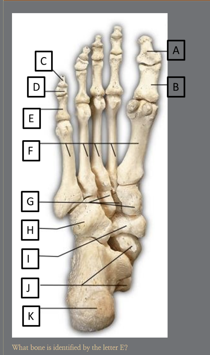
E?
Proximal Phalanx of the 5th Phalange

D?
Middle Phalanx of the 5th Phalange

F?
Dorsal cuboideonavilcuar ligament

I?
Bifurcate ligament

H?
Interosseous talocalcaneal ligament

D?
Anterior talofibular ligament

B?
Anterior tibiofibular ligament

D?
Tibiocalcaneal ligament
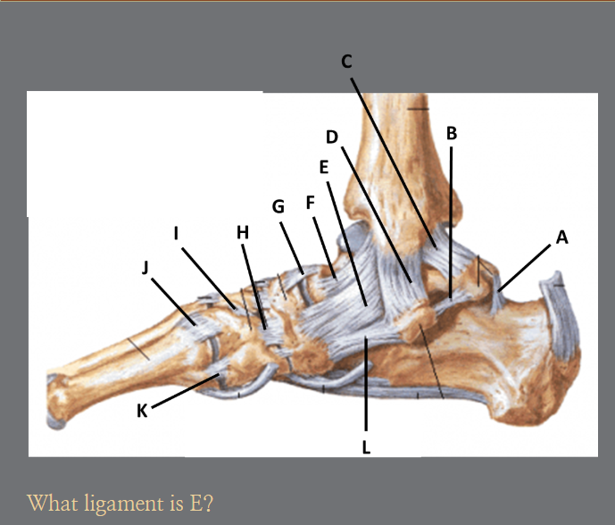
E?
Tibionavicular ligament

F?
Anterior tibiotalar ligament
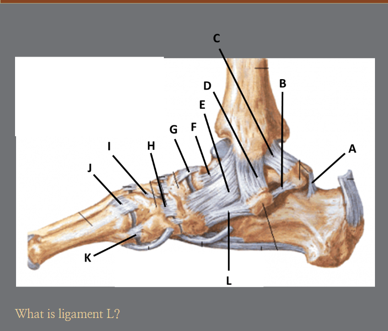
L?
Plantar calcaneonavicular ligament

J?
Dorsal tarsometatarsal ligament
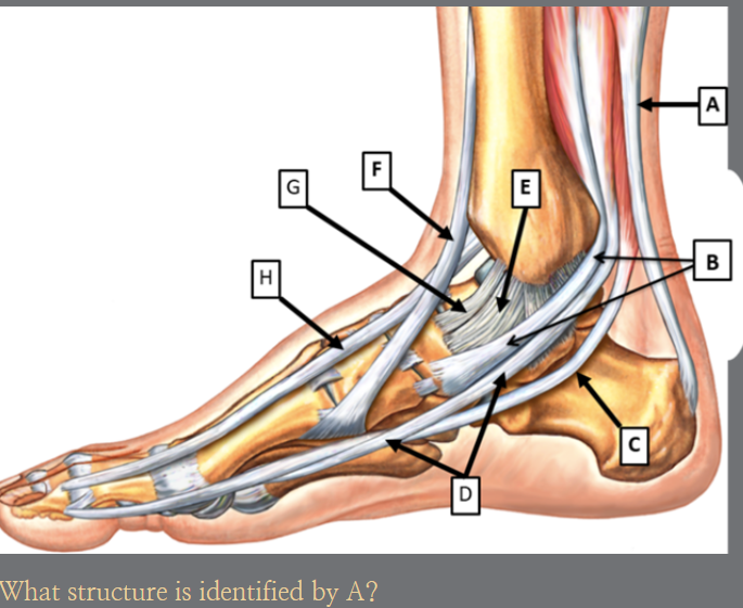
A?
Calcaneal Tendon
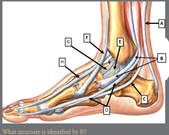
B?
Tibialis posterior tendon

C?
Flexor hallucis longus tendon

F?
Tibialis anterior ligament

G?
Anterior tibotalar ligament
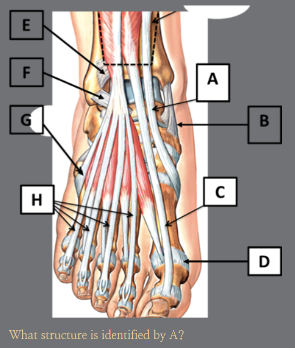
A?
Tibialis anterior tendon

B?
Tibonavicular ligament

C?
Extensor hallucis longus tendon
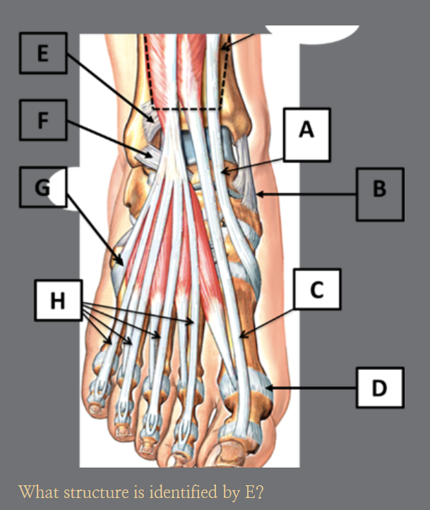
E?
Anterior tibiofibular ligament
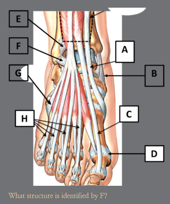
F?
Anterior talofibular ligament

G?
Fibularis tertius tendon

1?
Posterior tibial tendon
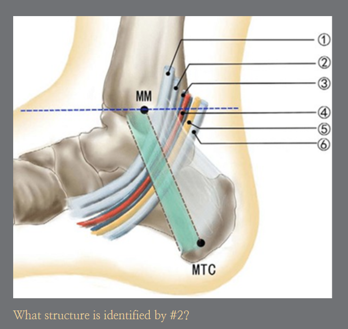
2?
Flexor digitorum longus tendon
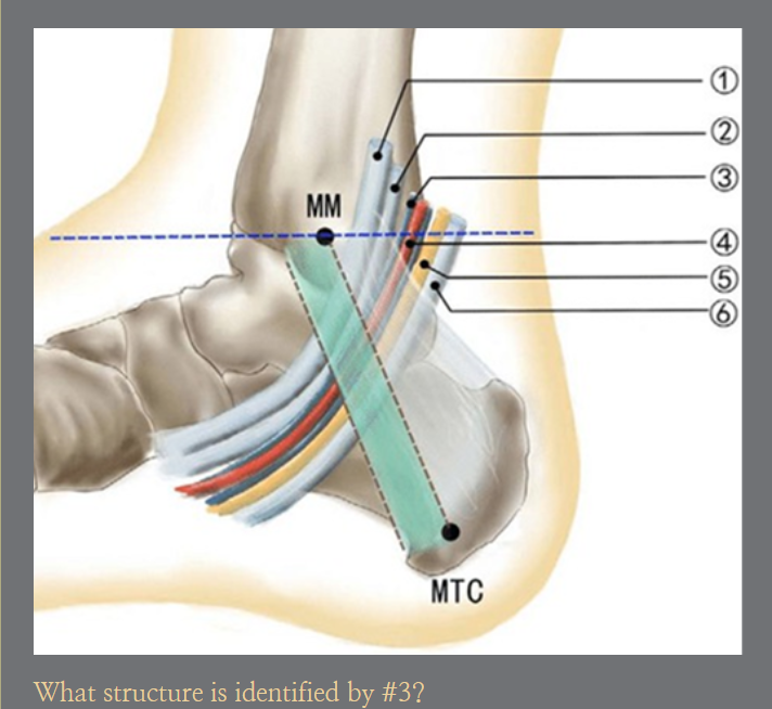
3?
Posterior tibial artery

4?
Posterior tibial vein

5?
Posterior tibial nerve

6?
Flexor hallucis longus tendon

4?
Quadratus Plantae

5?
Lumbricals

6?
Flexor digiti minimi brevis
Every toe in the foot is made up of 3 bones, distal, middle and proximal phalanx EXCEPT the,
1st Phalange
The base of the first metatarsal articulates with what tarsal bone?
Medial Cuneiform
The sustentaculum tali is a landmark located on the:
Calcaneus
Which structure is also known as the styloid process of the 5th metatarsal?
Tuberosity of the 5th metatarsal
Which tarsal bone articulates directly with the tibia at the ankle joint?
Talus
The articulation between the phalanges and metatarsal bones is known as the,
Metatarsal Phalangeal Joint (MTP)
The articulation between the metatarsal bones and the cuboid and all the cuneiform bones is known as the,
Tarsometatarsal (TMT) joint

What structure is identified by the letter B?
Proximal phalanx of the first phalange

What structure is identified by the letter K?
Navicular Bone
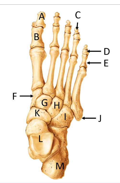
What structure is identified by the letter I?
Cuboid
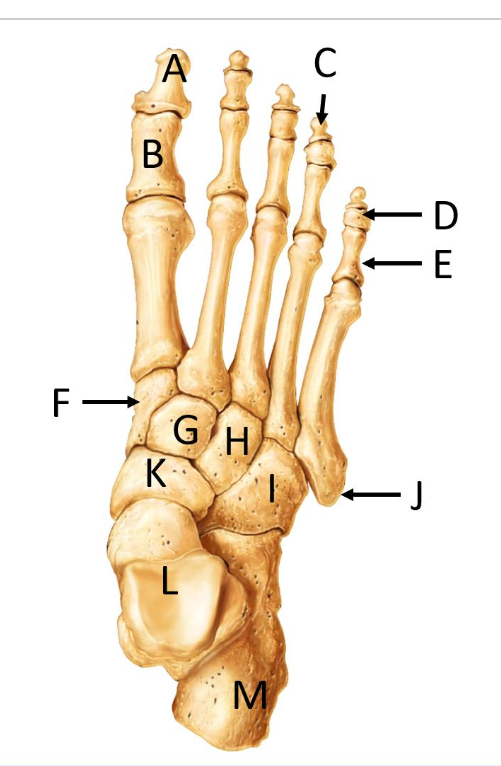
What structure is identified by the letter H?
Lateral Cuneiform

What structure is identified by the letter L?
Talus

Where are the sesamoid bones?
D

What anatomical structure is identified by the number 1?
First metatarsal phalangeal joint

What anatomical structure is identified by the number 5?
Navicular tuberosity

What anatomical structure is identified by the number 8?
Calcaneonavicular ligament
A lateral ankle sprain would be an injury to what structure?
Calcaneofibular Ligament (CFL)

What anatomical structure is identified by the number 6?
Transverse head of the abductor Hallucis
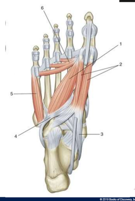
What anatomical structure is identified by the number 5?
Flexor digiti minimi brevis

What anatomical structure is identified by the number 2?
Flexor Hallucis Brevis

What anatomical structure is identified by the number 1?
Abductor Hallucis

What anatomical structure is identified by the number 2?
Flexor digitorum brevis

What anatomical structure is identified by the number 3?
Abductor digiti minimi

Which muscle is identified by the number 6?
Flexor Hallucis Longus
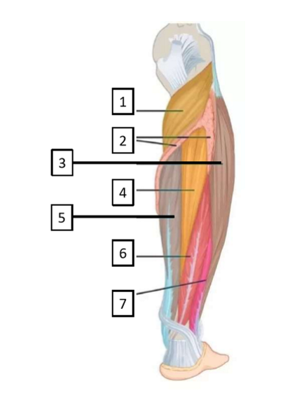
Which muscle is identified by the number 4?
Tibialis Posterior

Which muscle is identified by the number 7?
Extensor Hallucis Longus

Which muscle is identified by the number 4?
Tibialis Anterior
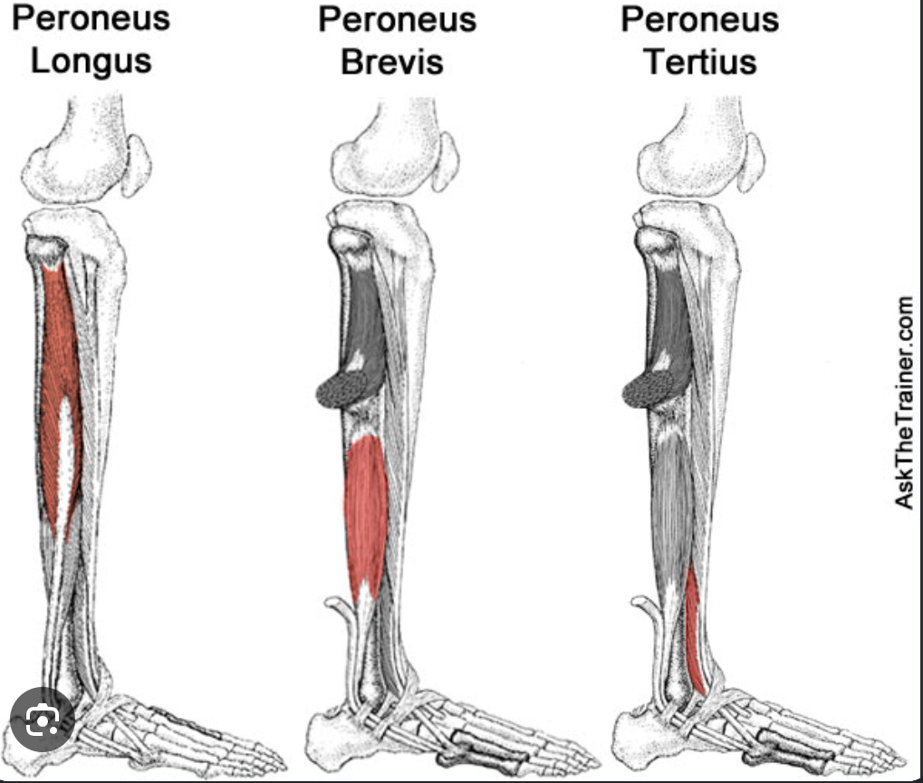
What muscle does NOT do ankle eversion?
Tibialis Anterior

Which muscles DO ankle eversion?
Peroneus Brevis
Peroneus Longus
Peroneus Tertius
Extensor Digitorum Longus
Anterior group includes:
Tibialis Anterior
Extensor digitorum longus
Extensor Hallucis longus
Fibularis (peroneus) Tertius

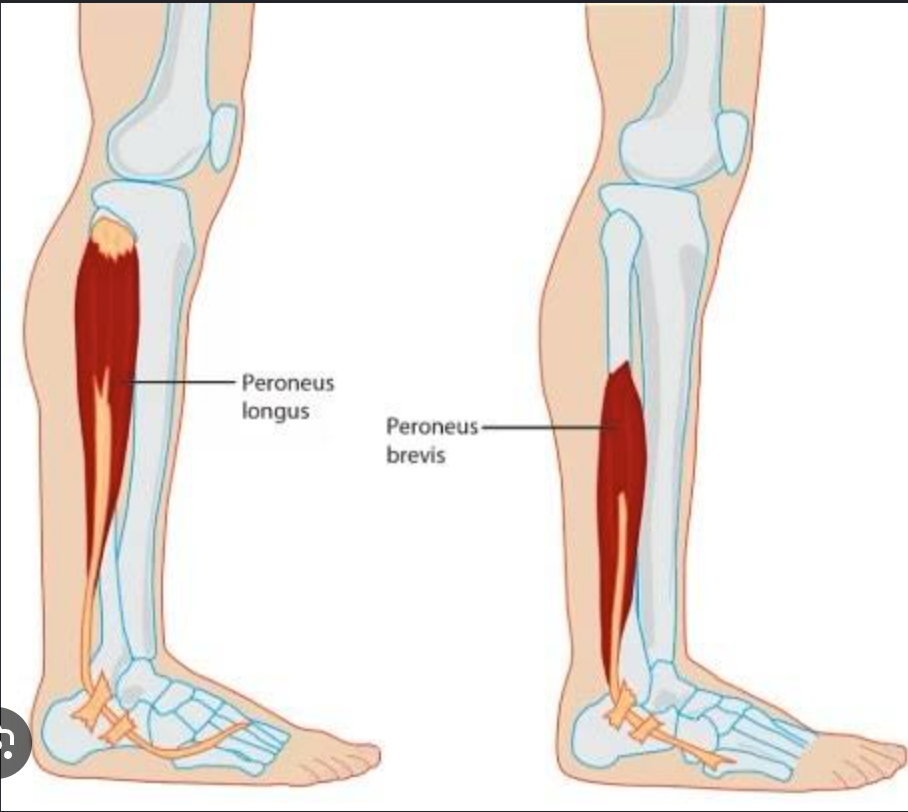
Lateral Group includes:
Fibularis (peroneus) longus
Fibularis (peroneus) Brevis
Posterior group (superficial) includes:
Gastrocnemius
Soleus
Plantaris

Posterior group (Deep Layer) includes:
Popliteus
Tibialis Posterior
Flexor digitorum Longus
Flexor hallucis longus

Tibialis Anterior
Origin: Lateral condyle of tibia; proximal, lateral surface of tibia; and interosseous membrane
Insertion: Medial cuneiform and base of the first metatarsal
Action: Invert the foot; dorsiflex the ankle (talocrural joint). (Also provides medial support to the transverse arch
Extensor Digitorum Longus
Origin: Lateral condyle of tibia; proximal, anterior shaft of fibula; and interosseous membrane
Insertion: Middle and distal phalanges of second through fifth toes
Action: Extend the second through fifth toes (metatarsophalangeal and interphalangeal joints); dorsiflex the ankle (talocrural joint); evert the foot
Extensor Hallucis Longus
Origin: Middle, anterior surface of fibula and interosseous membrane
Insertion: Distal phalanx of first toe
Action: Extend the first toe (metatarsophalangeal and interphalangeal joints); dorsiflex the ankle (talocrural joint); invert the foot
Fibularis (peroneus) Tertius
Origin: Anterior surface of the distal fibula
Insertion: Dorsal shaft of the 5th metatarsal
Action: Evert the foot; assist to dorsiflex the ankle (talocrural joint)
Fibularis (peroneus) Longus
Origin: Head of fibula and proximal two-thirds of lateral fibula
Insertion: Base of the first metatarsal and medial cuneiform
Action: Evert the foot; assist to plantar flex the ankle (talocrural joint) (Also provides lateral support to the transverse arch)
Fibularis (peroneus) Brevis
Origin: Lateral surface of the fibula
Insertion: Base / tuberosity of the fifth metatarsal
Action: Evert the foot; assist to plantar flex the ankle (talocrural joint)
Gastrocnemius
Origin: Posterior surfaces of the condyles of the femur
Insertion: Calcaneus via the calcaneal (Achilles) tendon
Action: Flex the knee (tibiofemoral joint); plantar flex the ankle (talocrural joint)
Soleus
Origin: Soleal line; proximal, posterior surface of tibia; and posterior aspect of head of fibula
Insertion: Calcaneus via the calcaneal (Achilles) tendon
Action: Plantar flex the ankle (talocrural joint)
Plantaris
Insertion & Action: Inserts into the calcaneus via the calcaneal tendon to perform plantar flexion of the ankle
Popliteus
Origin: Lateral condyle of the femur
Insertion: Proximal, posterior aspect of tibia
Action: Medially rotate the flexed knee (tibiofemoral joint); flex the knee (tibiofemoral joint)
Tibialis Posterior
Origin: Proximal, posterior shafts of tibia and fibula; and interosseous membrane
Insertion: Navicular, all cuneiform bones, cuboid, calcaneus, and bases of second through fourth metatarsals
Action: Invert the foot; plantar flex the ankle (talocrural joint)
Flexor digitorum Longus
Origin: middle, posterior surface of tibia
Insertion: distal phalanges of second through fifth toes
Action: Flex the second through fifth toes (metatarsophalangeal and in interphalangeal joints) weak plantar flexion of ankle (talocrural joint); invert the foot
Flexor Hallucis Longus
Origin: Middle half of posterior fibula
Insertion: Distal phalanx of first toe
Action: Flex the first toe (metatarsophalangeal and interphalangeal joints); weak plantar flexion of ankle (talocrural joint); invert the foot
Intrinsic Muscles of the Foot (Dorsal group)
Extensor Digitorum Brevis
Extensor Hallucis Brevis

Extensor digitorum Brevis
Origin: Dorsal surface of calcaneus
insertion: Second through fourth toes via the extensor digitorum longus tendons
Action: Extend the second through fourth toes (metatarsophalangeal and interphalangeal joints)
Extensor Hallucis brevis
Origin: Dorsal surface of calcaneus
Insertion: Proximal phalanx of first toe
Action: Extend the first toe (metatarsophalangeal joint)
Intrinsic Muscles of the Foot (Plantar Group-1st layer)
Abductor Hallucis
Flexor digitorum Brevis
Abductor digiti Minimi

Abductor Hallucis
Origin: Medial process of calcaneus and plantar aponeurosis
Insertion: Proximal phalanx of first toe (medial surface) and medial sesamoid bone
Action: Abduct the first toe (metatarsophalangeal joint); assist to flex the first toe (metatarsophalangeal joint)
Flexor digitorum Brevis
Origin: Medial process of calcaneus and plantar aponeurosis
Insertion: Middle phalanges of second through fifth toes
Action: Flex middle phalanges of the second through fifth toes (proximal interphalangeal joints)
Abductor Digiti Minimi
Origin: Lateral process of calcaneus and plantar aponeurosis
Insertion: Proximal phalanx of fifth toe (lateral surface)
Action: Flex the fifth toe; assist to abduct the fifth toe (metatarsophalangeal joint)
Intrinsic Muscles of the Foot (Plantar Group-2nd layer)
Quadratus Plantae
Lumbricals
Quadratus Plantae
Origin: Medial and lateral sides of plantar surface of calcaneus
Insertion: Posterior, lateral aspect of flexor digitorum longus tendon
Action: Assist flexor digitorum longus to flex 2nd–5th toes
Lumbricals
Origin: Tendons of flexor digitorum longus
Insertion: Bases of proximal phalanges of 2nd–5th toes and extensor digitorum longus tendons (on dorsal surface of the toes)
Action: Flex the proximal phalanges of the 2nd–5th toes at the metatarsophalangeal joints; extend the middle and distal phalanges of the 2nd–5th toes at the interphalangeal joints
Intrinsic Muscles of the Foot (Plantar Group-3rd layer)
Adductor Hallucis
Flexor Hallucis Brevis
Flexor Digiti mini Brevis
Opponens Digiti minimi
Adductor Hallucis
Origin: Oblique head: Bases of second through fourth metatarsals.
Transverse head: Plantar ligament of third through fifth metatarsophalangeal joints
Insertion: Lateral surface of base of proximal phalanx of first toe
Action: Adduct the first toe; assist to maintain transverse arch of foot; assist to flex the first toe
Flexor Hallucis Brevis
Origin: Plantar surfaces of cuboid and lateral cuneiform
Insertion: Medial and lateral surfaces of base of proximal phalanx of first toe
Action: Flex the first toe (metatarsophalangeal joint)
Flexor digiti minimi brevis
Origin: Base of fifth metatarsal
Insertion: Base of proximal phalanx of fifth toe
Action: Flex the fifth toe (MTP joint)