Learn: Skeletal System Anatomy
 Call Kai
 Learn
Learn Practice Test
 Spaced Repetition
 Match
Match Flashcards
 Knowt Play
1/374

There's no tags or description
Looks like no tags are added yet.
Name | Mastery | Learn | Test | Matching | Spaced | Call with Kai | Chat |
|---|
No analytics yet
Send a link to your students to track their progress
375 Terms
cervical vertibrae
neck

Mandible
lower jaw bone, only bone of the skull not joined by sutures

Sternum
breastbone, forms the middle of the front of the rib cage and is divided into three parts

Radius
the thicker and shorter of the two bones in the forearm, "on the thumb side"

Phalanges
bones of the fingers and toes

Ribs
form the thoracic cage, 12 pairs

Tibia
the thicker and innermost of the two bones in the lower leg, "shin bone"

Fibula
the thinner and outermost of the two bones in the lower leg, "calf bone"

Scapula
shoulder blade

Carpals
wrist bones

Metacarpals
bones of the palm

Coxal
hip bone

Femur
largest bone in the body, "thigh bone"

Tarsals
ankle bones
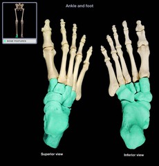
Metatarsals
bones of the foot

Patella
knee cap

Clavicle
collar bone

Humerus
upper arm bone

Ulna
the thinner and longer of the two bones in the forearm, "on the pinky side"

Maxilla
upper jaw bone

Zygomatic
a projection of the temporal bone, found on each side of the maxilla

Frontal
forehead bone

Temporal
two bones on each side of the head that form the lower portion of the skull

Occipital
bone that forms the back of the skull

Parietal
two bones on each side of the head that form the upper portion of the skull

Ilium
the upper, wing-shaped portion of the hip bone

Ischium
the lowest portion of the hip bone, connects to the pubic symphysis
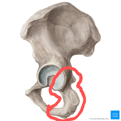
Pubis
the middle, thinnest portion of the hip bone

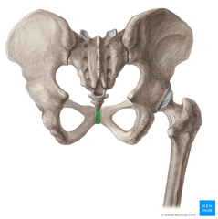
Pubis Symphysis
point where the right and left coxal bones join together
Cervical Vertebrae
Behind the neck, C1-C7

Thoracic Vertebrae
Behind the thoracic cage, T1-T12

Lumbar Vertebrae
Behind the lower back, L1-L5

Atlas
1st cervical vertebra

Axis
2nd cervical vertebra

Sacrum
Formed by 5 fused sacral vertebrae

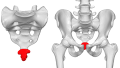
Coccyx
Formed by 4 fused coccygeal vertebrae, "tail bone"
Ethmoid
located between the two eye sockets

Sphenoid
two bones of each side of the head, anterior to the temporal bones
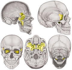
Inferior Nasal Concha
bones on the inferior lateral sides of the nasal cavity

Lacrimal
teardrop shape bone on either side of the nasal cavity

Nasal
bones that form the bridge of the nose
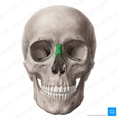
Palatine
bones on the superior medial portion of the nasal cavity

Vomer
the small, thin bone separating the right and left nasal cavities

206
number of bones in the skeletal system
Axial and Appendicular
two large groups of the skeletal system
Protect and support the brain, spinal cord, and vital organs housed in the thorax
functions of the axial skeleton
Sutures
immovable joints of the skull
True Ribs
ribs attached directly to the sternum, pairs 1-7
False Ribs
ribs not attached directly to the sternum, pairs 8-12
Floating Ribs
ribs that are only connected to the vertebrae, pairs 11-12
Involved in locomotion and the manipulation of the environment
functions of the appendicular skeleton
Ilium, Ischium, and Pubis
the 3 bones that fuse to form the coxal bones
Articulation
a location where two bone surfaces come together
Projection
an area of a bone that projects above the surface of the bone
Tubercle
small, rounded process or bump
Tuberosity
rough process, larger than a tubercle
Spine
sharp process
Crest
ridge
Fibrous, Cartilaginous, and Synovial
3 major types of joints
Flexion
decreases the angle of a joint

Extension
increases the angle of a joint

Hyperextension
increases the angle of a joint beyond normal range
Abduction
movement away from the median plane

Adduction
movement toward the median plane

Rotation
the turning of a structure around its axis
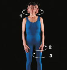
Pronation
movement that positions body parts to the back/down

Supination
movement that positions body parts to the front/up

Plane joints
produce multiaxial and gliding movements, restricts rotation, bones are flat or slightly curved

Examples of plane joints
intercarpal and intertarsal joints, articular processes between vertebrae
Hinge joints
produce uniaxial movements, one bone moves while the other remains stationary

Examples of hinge joints
elbow and knee joints
Pivot joints
produce uniaxial movements, a projection or process on one bone fits into and rotates within an opening on another bone

Examples of pivot joints
atlantoaxial and radioulnar joints
Saddle joints
produce biaxial movements, concave and convex portions of bones fit together

Examples of saddle joints
carpometacarpal joints
Ball-and-socket joints
produce multiaxial movements, provides the greatest range of motion

Examples of ball-and-socket joints
shoulder and hip joints
Condyloid joints
produce biaxial movements, modified ball-and-socket joints (oval shape versus spherical)

Examples of condyloid joints
metacarpophalangeal and atlantooccipital joints
Head
prominent rounded surface
Facet
flat surface
Condyle
rounded surface
Holes
opening or groove in a bone that allows blood vessels and nerves to enter or pass through the bone
Sulcus
groove
Canal
passage in bone
Foramen
hole
Sinus
air-filled space
Dorsiflexion
decreases joint angle between the front of the leg and the top of the foot, "flexing the foot"
Plantar Flexion
decreases joint angle between the back of the leg and the sole of the foot, "pointing the foot"
Coronal Suture
the suture between the parietal and frontal bones of the skull

Lambdoid Suture
where the parietal bones meet the occipital bone posteriorly

Sagittal Suture
suture between the two parietal bones

Anterior Cranial Fossa
formed by the frontal bone and part of the sphenoid bone

Middle Cranial Fossa
sphenoid, temporal and occipital bones

Posterior Cranial Fossa
contains the cerebellum and brainstem

Foramen Rotundum
Houses the maxillary nerve
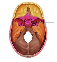
Foramen Ovale
Houses the mandibular nerve

Carotid Canal
passageway for internal carotid artery

Cribiform Plate
superior surface of the ethmoid; perforated by a foramina which allows passage of the olfactory nerves

Optic Canal
allows the optic nerve and opthalmic artery to pass to the eye
