Neuroscience Exam 2
 Call Kai
 Learn
 Practice Test
 Spaced Repetition
 Match
 Flashcards
 Knowt Play
1/103

There's no tags or description
Looks like no tags are added yet.
Name | Mastery | Learn | Test | Matching | Spaced | Call with Kai | Chat |
|---|
No analytics yet
Send a link to your students to track their progress
104 Terms
what does the somatosensory system detect
touch, pressure, vibration, limb position (proprioception), heat, cold, itch, and pain
somatosensory afferent neurons are …
psuedounipolar; cell body is midway along the path of the axon, not at the beginning

in somatosensory afferent neurons where do the action potentials begin and travel to
begin at receptor endings, turn into acons that travel to CNS; AP bypasses cell body
where do afferent somatosensory neurons reside
dorsal root ganglion (spinal cord) and trigeminal ganglia (brainstem)
sensory transduction
converting energy in a stimulus into an electrical signal
steps to trigger an action potential in somatosensory afferent neurons
stimulus causes cation channels in afferent fiber endings to open, channel openings create receptor potential, if large engough receptor potential triggers AP
what are mechanorecpetive afferent fiber endings encapsulated by
mechanoreceptor cells - specialized cells that help tune responses to a particular somatic feature
what do mechanoreceptor cells allow for
greater sensitivity to stimuli
what are nerves that lack mechanoreceptor cells called
free nerve endings
afferent properties: touch and proprioception vs pain and temperature
larger diameter and myelinated vs smaller diameter and little to no myelination
receptive field
area of skin over which stimulation causes a significant change in firing rate of action potentials; largely realted to branching area of fiber ending
two point discrimination
minimum distance between two tactile points on skin that is percieved as being from two distinct points
what determines the two point discrimination threshold
receptor density and field size
T/F: tactile spatial acuity vaires sustantially across body
true
rapidly adapting afferent somatosensory neurons
fire rapidly at beginning of stimulus, then cease with continued stimulation
what do rapidly adapting afferent somatosensory neurons convey
information about changes in ongoing stimulation (ex. putting on clothes and not feeling them as day goes on)
slowly adapting afferent somatosensory neurons
fire rapidly at beginning of stimulus, then steadily at a lesser rate with continued stimulation
what do slowly adapting afferent somatosensory neurons convey
information about static spatial attributes like size and shape (ex. feeling continuous weight of a heavy object)
what are the differences in sensitivity to somatosensory submodalities (temperature, light touch, vibration, etc.) determined by
types of ion channels and mechanoreceptor cells associated with afferent
glabrous skin
hairless skin of mammals predominantly found on palms, feet, and lips
what is glabrous skin specialized for
disciminative touch → use with object recognition and to provide feedback when gripping and reaching (sensations from touching an object alone are often enough to form a “neural image” and identify it)
4 types od mechanoreceptive afferents in glabrous skin
merkel cells, meissner corpuscles, pacinian corpuscles, ruffini corpuscles
merkel cells characteristics
slowly adapting, located in epidermis at border of dermis, mechanosensitive ion channels in the membrane of the fiber surrounded by merkel cell
which cell has the highest spatial acuity
merkel cells - highly sensitive to points, edges, and curvature
meissner corpuscles characteristics
rapidly adapting, located in dermis at border with epidermis, made of several layers (center contains 2-6 afferent fibers)
T/F: merkel cells are 4 times more sensitive than Meissner corpuscles
False; Messierners corpuscles are more sensitive with less spatial acuity
what are meissners corpuscles highly sensitive to
low frequency (3-40 Hz) vibrations - ex. vibrations of textured object slipping from hand
pacinian corpuscles characteristics
rapidly adapting, located deep in dermis or subcutaneous layer, made of several concentric layers of membranous around 1 afferent fiber
what are pacinian corpuscles most sensitive to
mechanoreceptor afferents, can detect deflections as small as 10 nanometers (very large receptive fields) - suited for detecting high frequency vibrations transmitted through grasped objects
ruffini corpuscles characteristics
slowly adapting, located deep in skin and in tendons and ligaments, parallel to strech lines in skin
what are pacinian corpuscles sensitive to
cutaneous stretch produced by finger movement - thought to provide information about finger and hand position
hairy skin
the 90% of skin of mammals outside of the palms, lips, and feet (includes regions with very little hair)
what does hairy skin transmit
information on discriminative touch and non-discrimminative affective touch (touch that evokes emotional response)
what tactile recpetors are in hairy skin
ruffini and pacinian corpuscles
which mechanoreceptive afferents innervate hair follicles
merkel cells innervate touch domes surrounding follicles
what wraps around the base of the follicle
circumferential endings and longitudinal lanceolate endings
what are the types of proprioceptive mechanoreceptors
muscle spindles, golgi tendon organs, joint receptors
intrafusal vs extrafusal muscle fiber function
sense muscle stretch vs produce force of muscle contraction
muscle spindles activation
muscle stretch → stretch of intrafusal fibers → activation of mechanically gated ion channels in afferent endings
what afferents innervate muscle spindles
group 1a: rapidly adapting (signal limb dynamics) and group II: slowly adapting (signal static position)
would big muscles with coarse movements like the quads have more or less density of muscle spindles than small muscles with fine movements like the eyes
lesser density of muscle spindles
how are golgi tendon organs arranged and what innervated them
in series with a tendon and group of 10-20 extrafusal muscle fibers; group Ib afferents (fast)
what do golgi tendon organs signal changes in
muscle tension
joint receptors location
mechanorecpetors in and around joints - activated at extremes of joint position (protective role) when near the limits of normal range of motion
primary somatosensory cortex (S1)
somatosensory neurons in thalamus mainly project to postcentral gyrus in parietal lobe
brodmann’s area
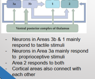
neighboring cortical regions also process somatosensory input
what was the result when scientests recorded with electrode from one point in cortex and would touch different areas of the body until a neural response was evoked
each brodmann’s area contains a complete somatotopic map
T/F: cortical representation of somatosensory input shows partial segregation of tactile and proprioceptive processing
true
plasticity of the cerebral cortex due to lack of stimulation
immediately after peripheral lesion (ex. removing a finger), corresponding cortical area is unresponsive - after weeks the same brain area is responsive to stimulation of body areas neighboring the lesion
plasticity of the cerebral cortex due to overstimulation
monkey brain trained on a task that requires it to use digits 2,3, and 4 - after training the cortical area devoted to those digites expand at the expense of other corical areas
nociceptors
somatosensory afferents with free nerve endings that transduce painful stimuli into electrical signals; slow conduction
what do nociceptors innervate
skin, deep tissue, and internal organs
when are nociceptors vs thermoreceptors activated
extreme stimulus levels vs lower stimulus levels
what are the two responses to pain
sharp, localized 1st pain and a delayed, diffuse, longer-lasting 2nd pain
what fibers eliminate 1st pain vs 2nd pain
A gamma vs C
A gamma fiber types
I: respond to dangerously intense chemical or mechanical stimulation; II: respond to dangerously intense thermal stimulation
C fibers
most respond to all types of nociceptive stimuli, some respond to noxious heat, chemical irritant, acidic substances, or cold
human threshold for painful heat applied to skin vs cold applied to skin
43 C (110 F) vs 15 C (59 F)
what type of channels are sensitive to temperature
transient receptor potential
T/F: channels are typically only sensitive to one modalitiy of nociceptive stimuli
False; example - acids elicit a “burning” sensation, perhaps by activating TRPV1, which mainly transduces noxious heat
what happens when TRPs (transient receptor potential) activated
allow influx of Na+ and Ca2+ → depolarization
T/F: several channels are known to be activated by exogenous chemicals
true - TRPM8 activated by menthol and TRPV1 activated by capsaicin
central pain pathway steps (4)
cell bodies of nociceptors and thermoreceptive afferents in dorsal root ganglia alongside spinal cord
afferents enter spinal cord through dorsal root, split into ascending and descending collaterals, then synapse on neurons in dorsal horn of nearby spinal segments
dorsal horn neurons decussate, then ascend spinal cord through lateral column
dorsal horn neurons synapse on various targets in brainstem and thalamus
anterolateral system
bundle of axons of dorsal horn neurons traveling up the lateral column of the spinal cord; contains informationon pain, temperature, non-discriminative affective touch, itch
2 parallel central pain pathways
sensory-discriminative aspects of pain and affective-motivational aspects of pain
sensory-discriminative pathway
location, intensity, and quality of painful stimuli; nociceptive dorsal horn projects to thalamus, then project to somatosensory cortex
affective-motivational pathway
fear and anxiety, unpleasent feeling, “fight or flight” reaction; nociceptive dorsal horn neurons project to various brainstem areas, plus a thalamic relay to cingulate and insular cortices
hyperalgesia
the phenomenon where stimuli in the area of an injury that would normally be considered slightly painful are percieved as very painful; cut on finger and areas by it are sensitive to pain
causes of hyperalgesia
peripheral and central sensitization
peripheral sensitization
“inflammatory soup” of substances released by nociceptors or immune cells in areas of damaged tissue, they interact with receptors or ion channels on nociceptors and augment their response
central sensitization
increase in the excitability of dorsal horn neurons following high level of activation of nociceptive afferents; several different mechanisms including enhancement of synaptic responses due to activation of NMDA receptors in the dorsal horn
modulating the transission of pain to the brain
local modulation at the level of the spinal cord and modulation via descending projections
local modulation at the level of the spinal cord
tactile afferents with receptive fields that overlap with nociceptive afferents inhibit transmission of the pain signal by dorsal horn neurons via an inhibitory interneuron
modulation via descending projections
in dorsal horn, an interneuron synapses on the presynaptic terminals of nociceptive C fibers, inhibiting excitatory transmission to dorsal horn neurons
the placebo effect
the beneficial effect following administration of a sham pharmeceutical substance
how does the placebo effect work
mediated by the activation of descending projections (opiod-based) based on the anterolateral system
retina
innermost layer at posterior eye containing photoreceptors that transduce light stimuli into electrical signals
pathway of light through the eye
cornea → aqueous humor → pupil → lens → vitreous humor → retina
cornea
highly transparent tissue at the front of the eye; highly refractive → serves to bend the path of light
lens
highly transparent tissue; refractive, but less so than cornea; refractive power can be dynamically altered by changing the thickness of the lens
pupil
opening in the middle of iris; can dilate or constrict to alter amount of light iluminating the retina
aqueous humor
clear fluid in front of lens; supplies nutrients and oxygen to lens and cornea
vitreous humor
clear, gelatinous substance behind lens; maintains shape of eye; contains phagocytes that remove cellular debris that could obstruct light passage
what do the cornea and lens serve to do
focus light from one point in the visual field onto one point in the retina (one-to-one mapping)
pupillary light reflex
contracts pupillary constrictor muscle (sphinctor), decreases pupil size → decreases amount of light entering the eye
accommodation
reflexive changing of the shape of lens to increase or decrease refraction; bringing near or far objects into focus on the retina
what is accommodation controlled by
edinger-westphal nucleus; triggers reflexive contraction or relaxation of the ciliary muscles, which is attached to lens by zonule fibers
in the retina how many basic classes of neurons are arranged in a well-defined, layered circuit
5
photoreceptors
neurons that transduce light energy into electrical signal
photoreceptor components
inner segment contains nucleus, outer segment houses membranous discs containing photopigment (where phototransduction occurs)
photopigment types
rods and cones
main, feed-forward information flow
photoreceptors → bipolar cells → ganglion cells; axons of ganglion cells comprise the optic nerve
lateral information flow
horizontal cells ←> photoreceptor/bipolar cell synapse; role in adapting response to different background light intensities
what is the first strange thing about phototransduction
light stimulation does not cause action potentials in photoreceptors, but causes a graded change in membrane potential with a corresponding change in the release of synaptic vesicles; retinal cells are very close → less need for axons, which are useful for transmitting signals over long distances
what is the second strange thing about phototransduction
light stimulation causes membrane hyperpolarization, not depolarization; at rest, membrane is depolarized → voltage gated Ca2+ channels near synapse activated → high rate of release of neurotransmitter; in response to light, membrane is transliently hyperpolarized → transient closing of Ca2+ channels → transient decrease in NT release
photopigment
retinal molecule enclosed by opsin; light causes a double bond in retinal to break, changing it from cis to trans isomer which is its active state
different types of opsin
rhodopsin (rods) and cone ospins (cone)
phototransduction cascade (7)
light activates rhodospin (G-protein-coupled receptor)
rhodospin activates transducin (G-protein)
transducin activates a phosphodiesterase (effector protein)
phosphodiesterase hydrolyzes the cGMP (2nd messenger)
cGMP levels decrease in cytosol of outer segment
less cGMP causes closing of cGMP-gated ion channels-these channels are permeable to Na+ and Ca2+
decrease of inward current → hyperpolarization
signal amplification in the phototransduction cascade
1 activated rhodopsin → closure of 200 channels → 1 mV of hyperpolarization