Myofilaments, Sarcomere, Sliding Filament Theory
 Call Kai
 Learn
 Practice Test
 Spaced Repetition
 Match
 Flashcards
 Knowt Play
1/18

There's no tags or description
Looks like no tags are added yet.
19 Terms
Myofilaments
Protein bundles that make up myofibrils
There are three types:
thick filaments, thin filaments, and elastic filaments
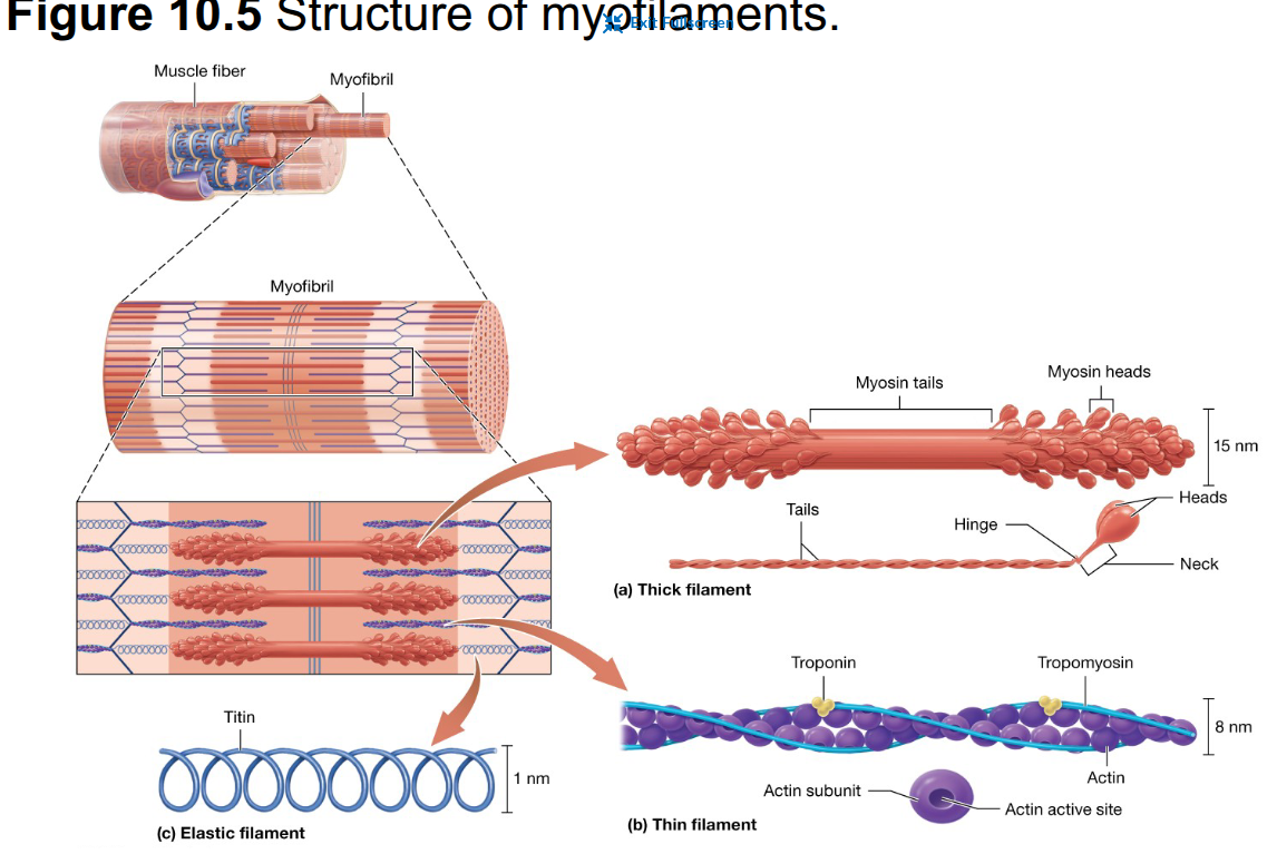
Three functional types of muscle proteins in myofilaments
Contractile proteins produce tension
regulatory proteins control when contraction can occur
structural proteins hold myofilaments in place and stabilize the muscle fiber
Thick filaments
Largest-diameter myofilaments
composed primarily of the contractile protein myosin
Contractile protein of thick filaments
each molecule has two globular heads and two intertwining chains forming a tail
The myosin heads interact with actin during contraction
Contain contractile, regulatory, and structural proteins including actin, tropomyosin, troponin, and nebulin
Actin
Bead-shaped contractile protein of thin filaments
Each actin subunit has an active site that can bind to a myosin head
actin “string” appears as two intertwining strands in the functional thin filament
Tropomyosin
Long, ropelike regulatory protein that spirals around two actin strands
at rest, it covers the active sites on actin and prevents myosin from binding
Troponin
small globular regulatory protein that helps hold tropomyosin in place
works with tropomyosin to switch muscle contraction on and off
Nebulin
Structural protein believed to help determine the length of the thin filament by limiting how many actin subunits can make up the filament
similar to the string that holds the beads of a necklace
Elastic filaments & titin
Elastic filaments are the thinnest myofilaments and are composed of the springlike structural protein titin
Titin stretches and recoils, resists excessive stretching, and helps the muscle fiber return to its original length
titin runs through the core of the thick filament

Sarcomere
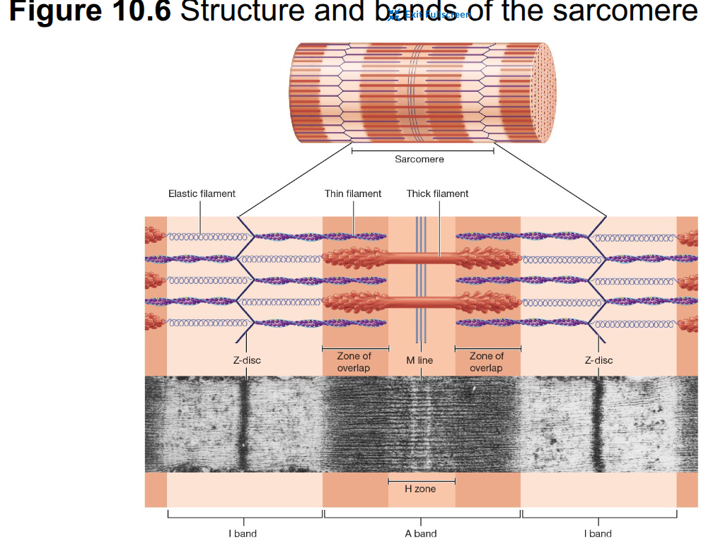
Repeating functional unit of a myofibril
The dark and light areas noticeable through the microscope are I, A, H, M, Z
extends from one Z-disc to the next Z-disc and shortens during muscle contraction
I band
Light region of a striation of the sarcomere that contains only thin filaments, so more light passes through

A band
Dark region of a striation
contains thick filaments which block more light
portions of the A band also contain overlapping thin filaments
Middle portion of the A band that contains only thick filaments
M line
Dark line in the middle of the A bands
consists of structural proteins that hold thick filaments in place
Z-discs
composed of structural proteins
Form the boundaries of each sarcomere
anchor thin filaments
provide attachment for elastic filaments
attach neighboring myofibrils to one another
Zone of overlap
Region where thick and thin filaments overlap, allowing myosin heads to interact with actin

Sarcomere from Z-disc to center
Z-disc → I band → A band → H zone → M line
the pattern is mirrored on the other half of the sarcomere

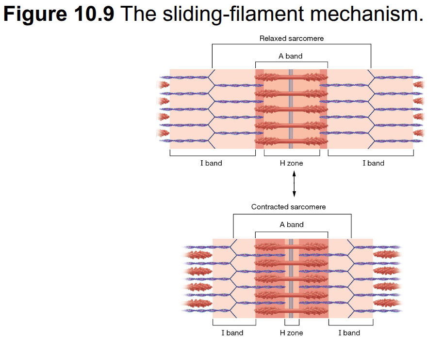
Sliding-filament mechanism
Thin filaments slide past thick filaments toward the M line, generating tension and shortening the sarcomere
the filaments themselves do NOT shorten
The Z-discs move closer together as the sarcomere shortens
The I band narrows as the thin filaments slide toward the M line
The H zone narrows as the thin filaments move farther into the center of the sarcomere
The A band remains the same width because the thick filaments do not change length
The zone of overlap between the thick and thin filaments increases
Neither thick or thin filament shortens
The thin filaments slide past the thick filaments toward the M line
How does shortening of sarcomeres shorten the whole muscle fiber?
Sarcomeres are arranged end to end within each myofibril
When many sarcomeres shorten simultaneously, the myofibrils and ultimately the entire muscle fiber shorten