Chp 6
 Call Kai
 Learn
Learn Practice Test
 Spaced Repetition
 Match
Match Flashcards
 Knowt Play
1/81

There's no tags or description
Looks like no tags are added yet.
Name | Mastery | Learn | Test | Matching | Spaced | Call with Kai | Chat |
|---|
No analytics yet
Send a link to your students to track their progress
82 Terms
articular cartilage
thing layer of cartilage covering an epiphysis, reduces friction and acts as a shock absorber
Appendicular Skeleton
126 bones that form the upper and lower limbs, as well as the shoulder and pelvic girdles that attach them to the central axis of the body

Appositional cartilage growth
new cartilage growth at the outer edge of cartilage, expanding outward, adding new layers to surface
articulation
where two bone surface meet
Axial skeleton
consists fo 80 bones that form the central core and vertical axis of the body

bone
hard, dense connective tissue that forms the structural elements of the skeleton
bone lining cells
flat cells on bone surface that help osteocytes maintain matrix
External surface: Periosteal cells
Internal: Endosteal cells
Bone markings
sites of muscle, ligaments and tendon attachment on external surfaces
Three types (projection, depression, opening)
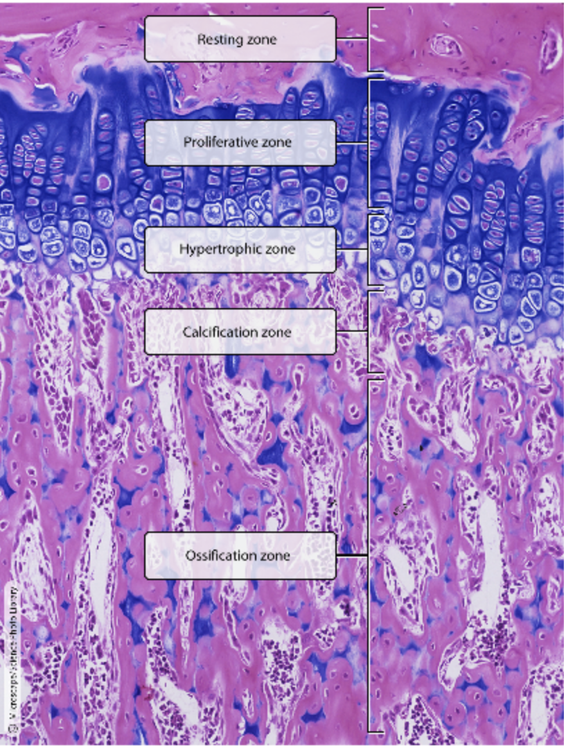
Calcification Zone
(Epiphyseal plate 5 zones)
at the epiphysis-diaphysis junction, the cartilage matrix calcifies and the chondrocytes die and deteriorate

canaliculi (singular = canaliculus)
hair line canals that connect lacunae to each other and to central canal. Allows communication and nutrients moving.
channels within the bone matrix that house one of any osteocytes many cytoplasmic extensions that it uses to communicate and receive nutrients

cartilage
semi-rigid connected tissue found in the skeleton in areas where flexibility and smooth surface support movement
Blood supply comes from pericondrium surrounding cartilage
Made up of chondrocytes
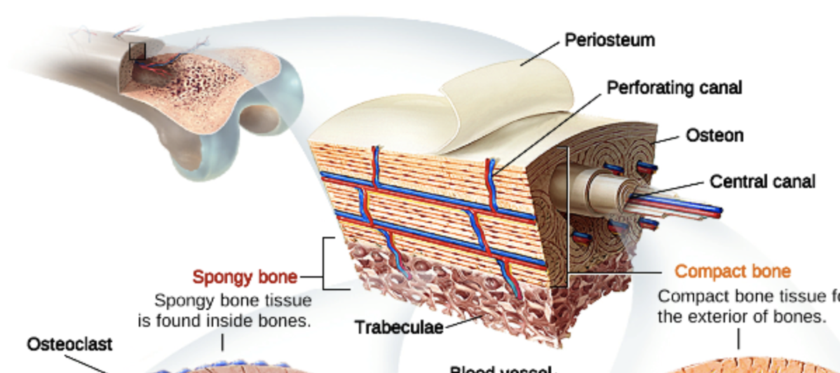
central canal
runs through core of osteon
longitudinal channel in the center of each osteon, contains blood vessels, nerves, and lymphatic vessels also known as the haversian cancal

closed reduction
manual manipulation of a broken bone to set it into its natural position without surgery
Comminuted fracture
when a bone is crushed into three or more pieces

compact bone
dense osseous tissue that can withstand compressive forces
Structural unit - osteon (haversion system) each osteon consists of concentric lamellae enclosing a central canal through which blood vessels passed

diaphysis
tubular shaft that runs between the proximal and distal ends of a long bone
diploë
layer of spongy bone, that is sandwiched between the two layers of compact bone found in flat bones
endochondral ossifciation
process in which bone forms by replacing hyaline cartilage
bones are called cartilage bones (endochondral) bones at this point, form most of skeleton
endosteum
delicate memnranous lining o a bones medullary cavity
epiphyseal plate
(growth plate) sheet of hyalin cartilage in the metaphysis of a immature bone; replaced by bone tissue as the organ grows in length

epiphysis
wide section at each end of a long bone; filled with spongy bone and red marrow
external callus
collar of hyaline cartilage and bone that forms around the outside of a fracture
flat bone
thin and curved bone; serves as a point of attachment for muscles and protects internal organs
Consists of thin plates of spongy bone inside and covered by compact bone on the outside. Bone marrow scattered throughout, no bone marrow cavity.
Cranial bones, scapulae, sternum, ribs
fracture hematoma
blood clot that forms at the site of a broken bone
Fracture: nondisplaced vs displaced
Nondisplaced: ends retain normal position
Displaced: ends of of normal alignment
Fracture: Complete vs Incomplete
Complete: broken all the way through. Incomplete: not borken all the way through
Fracture: Open vs Closed
Open/Compound: skin penetrated
Closed/Simple: skin not penetrated
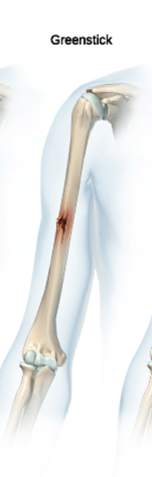
Greenstick fracture
partial breaks in a bone that occur as a result of bending
hematopoiesis
production of blood cells, which occurs in the red marrow of the bones
hole
opening or depression in a bone
hypercalcemia
condition characterized by abnormally high levels of calcium
hypertophic zone
(Epiphyseal plate 5 zones)
chondrocytes enlarge and the lacunae erode
Impacted fracture
one end of a broken bone is driven into the other end

Inorganic components
hydroxyapatites (mineral salts)
65% of bone by mass, responsible for hardness and resistance to compression
Half as strong as steel by resisting compression
Strong as steel by resisting tension
Lasts long after death because of mineral composition
interna callus
fibrocartilaginous matrix, in the endosteal region, between the two ends of a broken bone
intramembranous ossification
proccess by which bone forms directly from fibrous membrane, bones are called membrane bones at this point
interstitial cartilage growth
growth of cartilage from inside, existing chondrocytes indie the tissue divide and push matrix apart to expand internally
irregular bone
bone of a complex shape; protects internal organs from compressive forces
Consists of thin plates of spongy bone inside and covered by compact bone on the outside. Bone marrow scattered throughout, no bone marrie cavity.
Vertebrae & pelvic bones
lacunae (singular = lacuna)
spaces in a bone that house osteocyte

Lamellae
Interstitial - between osteons (fill gaps between them)
Circumferential - extend around entire circumference of bone

long bone
cylinder shaped bone that is longer that it is wide; functions as a lever
Upper limbs: Humerus (upper arm), radius and ulna (forearm), metacarpals (palm), and phalanges (fingers).
Lower limbs: Femur (thigh), tibia and fibula (leg), metatarsals (foot), and phalanges (toes).
Other areas: Clavicle (collarbone).
Shaft = diaphysis
Bone end = Epiphyses
Outerlayer of compact bone surrounding the spongy bone

medullary cavity
hollow region of the diaphysis; filled with yellow marrow
modeling
process during bone growth, by which bone is resorbed on one surface of a bone and deposited on another
Resorbed - break down old bone and Deposited - lay down new bone matrix
Nutrient foramen
small opening in the middle of the external surface of the diaphysis, through which an artery enters the bone to provide nourishment
Organic Components
Osteoid (made mostly of collagen fibers, glycoproteins, and proteoglycans)
open reduction
surgical exposure of a bone to resent a fracture (surgical wires or pins placed)
orthopedist
doctor of musculoskeletal includes bones, joints, ligaments, tendons, muscles and nerves
osseous tissue
bone tissue; a hard dense connective tissue that forms the structural elements of the skeleton
ossification (also osteogensis)
bone formation
ossification center
cluster of osteoblasts found in the early stages of intramembranous ossification
Ossification Zone
(Epiphyseal plate 5 zones)
osteoclasts dissolve the calcified cartilage and osteoblasts lay down bone matrix in place of the cartilage

osteoblasts
cell responsible for forming new bone (actively mitotic)
secretes osteoids

osteoclast
cell responsible for resorbing bone
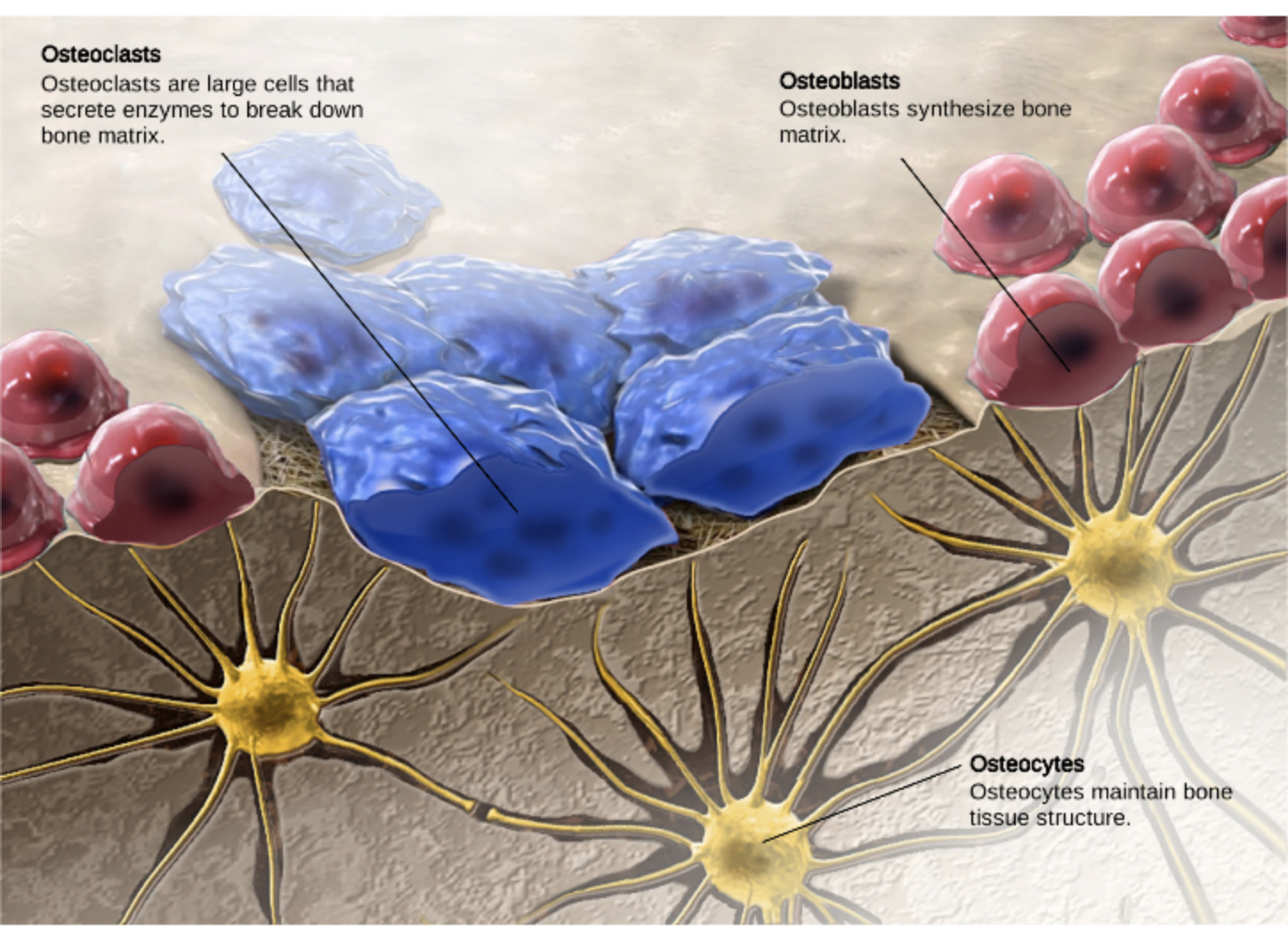
osteocyte
primary cell in mature bone; responsible for maintaining the matrix
No longer divide, respond to stimuli

osteogenic cell (osteoprogenitor cells)
Mitotically active stem cell; the only bone cells that divide; they differentiate and develope into osteoblasts
osteoid
uncalcified bone matrix secreteted by osteoblasts
Osteomalacia & Rickets
bone is inadequately mineralized
results in soft week bones, pain upon bearing weight
Osteomalacia of children is rickets: Vit D deficiency insufficient dietary calcium. bone ends are enlarged and abnormally long
osteon (also, haversian system)
basic structural unit of a compact bone; made of concentric layers of calcified matrix
consist of and elongated cylinder that runs parallel to long axis of bone. Osteon cylinder consists of several rings of bone matrix called lamellae
osteoporosis
disease characterized by a decrease in bone mass; occurs when the rate of bone resorption exceeds the rate of bone formation, common occurrence as the body ages
osteoclasts constantly remove old bone whole osteoblasts generate new bone
Osteoporotic bone (bone pain, tenderness, weakend and brittle bones)

Pagets Disease
excessive and haphazard bone deposit and resorption cause bone to grow fast and develop poorly
very high ratio of spongy to compact bone and reduced mineralization
Cause unknown, Moth-Eaten appearance
perforating canal (Volkmann’s canal)
channel that branches off from the central canal and houses vessels and nerves that extend to the periosteum and endosteum

perichondrium
memnbrane that covers cartilage
periosteum
fibrous membrane covering the outer surface of bone and continuous with ligaments

primary ossification center
region, deep in the periosteal collar, where bone development starts during endochondral ossification
projection
bone markings where part of the surface sticks out above the rest of the surface, where tendons and ligaments attach
proliferative zone
(Epiphyseal plate 5 zones)
chondrocytes divide via mitosis, lengthening the bone
Area of cartilage on diaphysis side of epiphyseal plate that is rapidly dividing
New cells moving upward pushing ephysis away from diaphysis causing lengthening of bone
region of the epiphyseal plate what makes new chondrocytes to replace those that die at the diaphyseal end of the plate and contributes to a longitudinal growth of the epiphyseal plate

red marrow
connective tissue on the interior cavity of a bone where hematopoiesis (produce new blood cells) takes place
remodeling
process by which osteoclasts resorb (bone resorption) old or damaged bone at the same time as and on the same surface where osteoblasts form new bone to replace that which is resorbed
reserve/resting/quiescent zone
(Epiphyseal plate 5 zones)
resting zone
area of cartilage on epiphyseal side of epiphyseal plate that is relatively inactive
region of the epiphyseal plate that anchors the plate to the osseous tissue of the epiphysis

secondary ossificatoin center
region of the bone development in the epiphyses
sesamoid bone
small, round bone embedded in a tendon; protects the tendon from compressive forces
patella
short bone
cube-shaped bone that is approximately equal in length, width, and thickness; provides limited motion
Areas that need strength, support and stability
Wrist (Carpals): Bones like the scaphoid, lunate, triquetral, hamate, pisiform, capitate, trapezoid, and trapezium.
Ankle (Tarsals): Bones like the calcaneus (heel), talus, navicular, cuboid, and cuneiforms
Consists of thin plates of spongy bone inside and covered by compact bone on the outside. Bone marrow scattered throughout, no bone marrie cavity.
skeletal system
organ system composed of bones and cartilage that provides for movement, support and protection
spongy bone (cancellous bone)
trabeculated osseous tissue that supports shifts in weight distribution
made up of honeycomb of small needle like pieces
trabecular confer strength

trabeculae (singular = trabecula)
spikes or secretions of the lattice-like matrix in spongy bone
strength to bone (like cables to bridge)

yellow marrow
connective tissue in the interior cavity of a bone where fat is stored
zone of calcified matrix
region of the epiphyseal plate closest to the diaphyseal end; functions to connect the epiphyseal plate to the diaphysis
zone of maturation and hypertophy
region of the epiphyseal plate where chondrocytes from the proliferative zone grow and mature and contribute to the longitudinal growth of the epiphyseal plate
5 types of bones
long, short, flat, irregular, sesamoid

4 Major Stages of Bone Repair
Hematoma formation - torn blood vessels hemorrhage causing hematoma
Fibrocartilaginous callus formation - capillaries grow into hematoma, phagocytic cells clear the debris of the fracture hematoma and dead bone cells. Next the fibroblasts secrete collagen fibers that connect broken bone ends. Then osteogenic cells differentiate into chondroblasts and osteoblasts to rebuild tissue. Chondroblasts produce the cartilage matrix that combines with the collagen fibers, this forms the fibrocartilaginous callus that fills the fracture gap
Bony callus formation - within one week, new trabeculae appear in fibrocartilaginous callus. Callus is converted to bony callus of spongy bone
Bone remodeling - begins during bony callus formation and continues for several months. Excess material on diaphysis exterior and within medullary cavity is removed, compact bone is laid down to reconstruct shaft walls, final structure resembles original structure
Flat bone formation

Long bone formation & growth
