(MMI) Lectures 1 - 7
 Call Kai
 Learn
Learn Practice Test
 Spaced Repetition
 Match
Match Flashcards
 Knowt Play
1/196

There's no tags or description
Looks like no tags are added yet.
197 Terms
Classes of Microorganisms
Bacteria, fungi, protozoa, algae, virus
Study of bacteria
Bacteriology
Study of fungi
Mycology
Study of viruses
Virology
Study of infectious protozoa and parasitic worms
Parasitology
Beneficial microbes compete against harmful microbes
Probiotics
Largest photosynthesizers (microbes) in the ecosystem
Phytoplankton
Decomposers of organic waste
Fungi
He looked at the structure of cork and reported that living things were composed of little boxes or cells (in 1665). He proposed that the cell was the smallest component or building block of living things
Robert Hooke
Aseptic techniques prevent
- Infections as a result of surgery
- Food spoilage
- Contamination in microbiology laboratories
He developed a very simple microscope and used it to describe the nature and behavior of live microorganisms that he observed in teeth scrapings, rain water, etc... (1673-1723)
Antonie van Leeuwenhoek
Life is created from non-living matter by some "vital force"
Spontaneous generation theory
Life can only arise from pre-existing living matter
Biogenesis theory
In 1745, John Needham put boiled nutrient broth into covered flasks. It resulted in microbial growth. This suggests
Spontaneous generation
In 1765: Lazzaro Spallanzani boiled nutrient broth solutions in flasks. It resulted in no microbial growth. This suggests
Biogenesis
In 1861, his experiment showed that microorganisms are present in the air.
Louis Pasteur
He used a chemical disinfectant to prevent surgical wound infections after looking at Pasteur's work showing microbes are in the air, can spoil food, and cause animal diseases.
Joseph Lister
He proved that a bacterium causes anthrax and provided the experimental steps, known as "Koch's postulates", to prove that a specific microbe causes a specific disease.
Robert Koch
He made the first vaccine (for smallpox)
Edward Jenner
Vaccination is derived from this latin word which means cow
Vacca
Chemicals produced by bacteria and fungi that inhibit or kill other microbes.
Antibiotics
Malaria was treated using (Collected from tree bark)
Quinine (a natural bitter alkaloid derived from the bark of the cinchona tree)
Chemotherapeutic agents used to treat infectious disease
Synthetic drugs/antibiotics
He discovered the first antibiotic.
Alexander Fleming
What antibiotic did The fungus Penicillium made?
Penicillin
The antibiotic penicillin helps to fights against which microbe?
Staph. aureus
The study of how the host responds to the infecting micro-organism
Immunology
Four types of microscopy
- Light Microscopy (Brightfield, Darkfield, Phase contrast, Differential interference contrast (DIC))
- Electron microscopy (Scanning & Transmission)
- Fluorescence microscopy
- Confocal microscopy
A simple microscope such as that used by van Leeuwenhoek has only one lens.
Light microscopy
In a compound microscope, the image from the objective lens is magnified again by the
Ocular lens
Objective lens x ocular lens
Total magnification
The ability of the lenses to distinguish between two points
Resolution
Shorter wavelengths of light provide
Greater resolution
A microscope with a resolving power of 0.4 nm can distinguish between two points
≥ 0.4 nm
Due to the relatively long wavelength of light in a compound light microscope, the effective limit on resolution is
200nm (= 0.2 μm)
Dark objects are visible against a bright background. Contrast staining is often required to see objects.
Brightfield microscopy
Light objects are visible against a dark background because only light reflected off the specimen enters the objective lens. Staining usually not required.
Darkfield microscopy
Accentuates diffraction of the light that passes through a specimen. Can visualize living specimens without staining.
Phase-contrast microscopy
It is similar to phase-contrast microscopy but has a higher resolution because it uses two beams of light. Prisms split the light beams adding contrasting colours to the specimen.
Differential Interference Contrast (DIC) Microscopy
It uses electrons instead of light. The shorter wavelength of electrons gives greater resolution.
Electron microscopy

Transmission electron microscopy (TEM)
- Magnification 10 - 100 000x
- Resolution ~ 2.5 nm
- Need ultra-thin sections of specimen to allow electron passage.
- Specimens may be stained with heavy metal (electron dense) salts such as uranium or osmium tetroxide.
Scanning electron microscopy (SEM)
- Magnification 1 - 10 000x
- Resolution ~ 20 nm
- Examine whole cells or surface structures
- Electron beam is sprayed over the sample scattering electrons from its surface structures which are collected and amplified to produce a 3d image.
- Specimens may also be stained with heavy metal (electron dense) salts such as uranium or osmium tetroxide to provide contrast.
It uses ultraviolet (U.V.) light and the cells may be stained using fluorescent dyes.
Fluorescence microscopy
It uses fluorochrome dyes to stain the cells and a laser light. The laser illuminates and captures an image for each plane in a specimen.
Confocal microscopy
Two photons of long-wavelength (red) light are used to excite the dyes so it can examine thicker samples/cells (up to 1 mm deep).
Two-photon microscopy
A thin film of a solution of microbes is spread on a slide.
Smear
Use of a single basic dye.
Simple stain
It may be used to hold the stain or coat the specimen to enlarge it.
Mordant
Staining the background instead of the cell
Negative staining
It classifies bacteria as either gram-positive or gram-negative.
Gram staining
A means of discriminating one type or class of cell from another.
Differential stains
Appears purple under microscope
Gram-positive bacteria
Appears red under microscope
Gram-negative bacteria
Gram staining procedure
- Prepare the slide using heat fixing
- Apply Crystal Violet (stain) and let it sit for 1 minute. Wash it off with water.
- Flood the slide with Gram's iodine (mordant) and wash it using water after 1 minute.
- Rinse the slide using Ethanol (decolorizer) and wash it with water immediately after.
- Apply Safranin and wash it off after 1 minute.
Cells that retain a basic stain in the presence of acid-alcohol.
Acid-fast
Very hydrophobic structures that enhance the virulence of certain microbes.
Capsules
What staining is useful for observing capsules?
Negative staining
It requires a mordant to make the flagella wide enough to see.
Flagella staining
It is used to detect the presence of endospores in bacteria.
Endospore staining
This term comes from the Greek words for prenucleus. It includes bacteria and cyanobacteria.
Prokaryote
This term comes from the Greek words for true nucleus. It includes fungi, algae, protozoa, plants & animals.
Eukaryote
Spherical cells
Cocci (coccus)
Rod shaped cells
Bacilli (bacillus)

Curved (spiral) cells
Vibrio
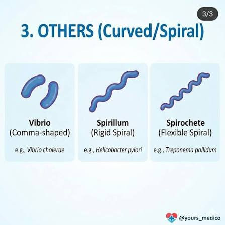
Helical, flexible cells
Spirochetes
Helical, rigid cells
Spirilla (spirillum)
Unusual e.g. star shaped or rectangular
Archaea
Means double
Diplo-
Means twisted chain
Strepto-
Means cluster
Staphylo-
Means group of four
Tetrad
Means bundle
Sarcinae

Bacteria with one shape throughout life.
Monomorphic
Bacteria with several morphologies
Pleiomorphic
Outside components of prokaryotic cells
Glycocalyx, Cell wall, Flagella, Fimbriae, & Pili
Inside components of prokaryotic cells
Cell membrane, Cytoplasm, Nucleoid, Ribosomes, Endospores & Inclusion bodies
Simple polysaccharide chains layered just outside cell wall. It protect cell from drying out; protect against phagocytosis; allow cell to colonize a new niche.
Glycocalyx
Flagella (flagellum) is composed of chains of a protein called
Flagellin
A single flagellum at the end of the cell.
Monotrichous
A single flagellum or a tuft of flagella at both ends of the cell.
Amphitrichous
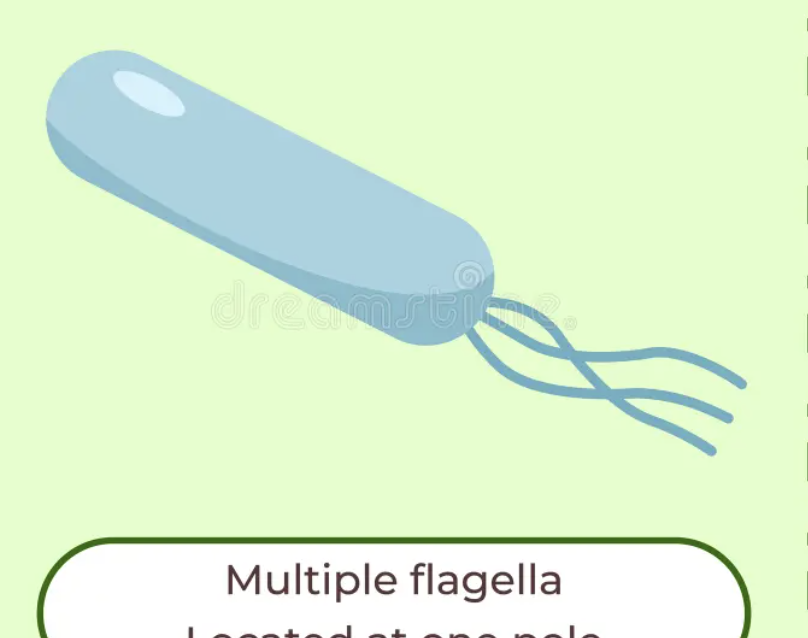
A tuft of flagella at the end of the cell.
Lophotrichous
An all-over, random flagellar arrangement.
Peritrichous
Axial filaments (they're found in spirochetes) are also known as
Endoflagella
They're not used for motility. They're shorter and straighter than flagella
Fimbriae
They're longer than a fimbriae. They're used to transfer DNA from one cell to another.
Pili (pilus)

The process of transferring DNA from one cell to another.
Conjugation
It provides support and protection; determines cell shape. It also prevents bursting of cell (in hypotonic solution).
Bacterial cell wall
Lipid A (lipopolysaccharide) is an example of an
Endotoxin (toxic molecules made of (LPS) that form the outer cell wall of Gram-negative bacteria)
It provides some protection from host defenses (phagocytes, complement) and antibiotics. It is composed of lipopolysaccharides, lipoproteins, phospholipids.
Gram negative outer membrane
They're not present in eukaryotic cells therefore treatments which selectively attack this will not affect human or animal cells.
Peptidoglycan: a tough, mesh-like polymer made of sugars and amino acids that surrounds the plasma membrane
A gel-like solution of water and organic and inorganic substances; contains all internal structures.
Cytoplasm
It functions as a selective barrier to limit entry and exit of materials.
Plasma membrane
Similar structure between prokaryotes and eukaryotes
- Phospholipid bilayer
- Peripheral proteins
- Integral proteins
- Transmembrane proteins
A passive transport process in which a molecule passes directly across the membrane down its concentration gradient with no expenditure of cell energy.
Simple diffusion
A passive transport process in which a molecule crosses the membrane down its concentration the gradient with the aid of premise.
Facilitated diffusion
An energy-requiring process in which a transport protein pumps a molecule across the membrane against its concentration and/or electrical gradient.
Active transport
It contains single, long, almost always circular thread of double stranded DNA.
Nucleoid
Separate smaller strands/circles in the nuclei.
Plasmids
They're present in both eukaryotes and prokaryotes. They're the protein synthesis machinery of the cell.
Ribosomes
Two subunits of the ribosomes are composed of (small subunit 30S + large subunit 50S = Complete 70S ribosome).
Protein and ribosomal RNA