Neuro: Management of Patients with Neurologic Dysfunction
 Call Kai
 Learn
 Practice Test
 Spaced Repetition
 Match
 Flashcards
 Knowt Play
1/140

There's no tags or description
Looks like no tags are added yet.
141 Terms

Blood: Supplies the brain with oxygen and nutrients (glucose) and removes waste.
CSF (cerebrospinal fluid): Cushions and protects the brain and spinal cord and maintains a stable environment.
Blood-brain barrier (BBB): Controls which substances pass from the blood into brain tissue, helping protect the brain from harmful substances.
Clinical Assessment - LOC
Arouse to wakefulness
Call name
Touch
Shake firmly
Assess orientation
Person, place, time, situation
Behavior changes: restless, irritable, combative
Clinical Assessment - Noxious Stimuli
Nail bed pressure: Apply pressure to the nail bed to assess the response to pain.
Supraorbital pressure: Apply pressure above the eye at the supraorbital notch; avoid if facial injury is suspected.
Trapezius squeeze: Squeeze the trapezius muscle to assess the response to pain.
Sternal rub: Rub the sternum to assess the response; may cause bruising or skin injury.
Assess the response:
Purposeful movement (localizes pain)
Withdrawal from pain
Abnormal flexion or extension (posturing)
No response
Normal Pupils

Pupillary Changes
Pupillary size:
Blown pupil: Fully dilated.
Pinpoint pupil: Very small, constricted.
Pupillary reaction:
Sluggish: Reacts slowly to light.
Non-reactive: Does not respond to light.
Unequal pupils: Pupils are different sizes.
Irregular shape: Pupils are not round; may occur after cataract or other eye surgery.
Medications:
Opioids → constrict pupils.
Anticholinergics → dilate pupils.
Pupillary Assessment
Observe both pupils together and compare their size and reaction to light.
Measure and document pupil size in millimeters (mm).
The optic nerve is located at the back of each eye and carries visual information to the brain.
When assessing eye opening in an unresponsive patient, use appropriate technique, such as pressure on the supraorbital ridge when indicated.
Disconjugate gaze:
Eyes point in different directions instead of moving together.
Oculocephalic reflex (Doll’s eyes):
Rapidly turn the patient’s head side to side only if cervical spine injury has been ruled out.
Normal: Eyes move opposite the direction of the head movement.
Abnormal: Eyes move with the head or do not move as expected.
Oculovestibular reflex (cold caloric test):
Instill cold water (e.g., 20 mL ice water, per protocol) into the ear to assess eye movements in an unresponsive patient.
Normal: Eyes move toward the ear receiving cold water.
Abnormal: No eye movement response.
Clinical Assessment - Motor
Strength of muscles groups
Bilateral hand grips, then release
Strength/Movement of extremities
5 – full strength, no deficit or weakness
4 - holds up without wavering
3 – holds up with wavering
2 – slides along surface
1 – flicker or trace of movement
0 – no movement
Reflexes
Primitive reflexes: Present at birth; may reappear with brain dysfunction. They are among the last reflexes to disappear with normal aging or neurological decline.
Examples: sucking, startle, and grasp reflexes.
Corneal reflex (blink): Touch the cornea with a sterile swab or wisp of cotton; normal response is blinking.
Gag reflex: Stimulation of the back of the throat causes gagging.
Babinski reflex:
Normal adult: Toes curl downward.
Abnormal adult: Big toe moves upward and other toes fan out.
Infants: Upward big toe and fanning are normal.
Deep tendon reflexes (DTRs): Assess reflex responses to tendon tapping, such as the knee-jerk reflex.
Decorticate Posturing

less severe
Decerebrate Posturing

Glasgow Coma Scale
≥13 mild injury
9-12 moderate
≤8 severe impairment
Eye Opening (E) | Verbal Response (V) | Motor Response (M) |
4=Spontaneous | 5=Normal conversation | 6=Normal |
Total = E+V+M | ||
Intracranial Pressure (ICP)
Pressure inside the skull created by three components:
Brain tissue
Blood volume
Cerebrospinal fluid (CSF)
Compensation (Monro-Kellie doctrine): If one component increases, the body compensates by reducing the volume of the others to maintain pressure.
Shifting CSF out of the skull.
Increasing CSF absorption.
Decreasing cerebral blood volume.
Intracranial Pressure (ICP) - Normal Range
10-20 mmHg
Cerebral Perfusion Pressure (CPP)
Pressure that drives blood flow to the brain.
CPP = MAP − ICP
Cerebral Perfusion Pressure (CPP) - Normal
70–100 mmHg
MAP (Mean Arterial Pressure):
Average pressure in the arteries during one cardiac cycle.
MAP = (SBP + 2 × DBP) ÷ 3
SBP = systolic blood pressure; DBP = diastolic blood pressure.
CPP < 50 mmHg
can cause inadequate brain perfusion and potentially irreversible neurological damage.
CPP > 100 mm Hg
permanent ischemic injury
Example: Calculating MAP and CPP
Given:
SBP = 120 mmHg
DBP = 80 mmHg
ICP = 15 mmHg
MAP = 93.3 mmHg; CPP = 78.3 mmHg.
Increased ICP: Signs & Symptoms Early
Altered LOC
Headache
Restlessness/confusion
Drowsiness
Pupil changes
Vomiting/projectile/possibly without nausea
Vomiting = ICP is going up
Visual disturbances
Increased respiratory effort
Increased ICP: Signs & Symptoms Late
Stuporous/Unarousable
Loss of reflexes
Projectile vomiting
Comatose
Abnormal motor responses
Decorticate
Decerebrate
Increased ICP: Cushing’s Response
The body's attempt to maintain cerebral perfusion when ICP rises.
Compensation: The body increases BP to try to maintain blood flow to the brain.
Increased ICP: Cushing’s Triad (Late Sign)
Increased systolic BP with widened pulse pressure.
Bradycardia: Decreased heart rate.
Abnormal respirations: Slow, irregular breathing; may include Cheyne–Stokes respirations.
Increased ICP: Complications & Monitoring
Widened pulse pressure: Increased difference between systolic and diastolic BP.
Example: 180/60 = 120 mmHg pulse pressure.
Severe increased ICP: Can compress brain tissue, cause brain herniation, and lead to irreversible damage.
Monitoring: Watch for changes in BP, heart rate, respirations, LOC, and pupils.
ICP Monitoring - Purpose:
Identify increased ICP early.
Directly measure pressure inside the skull.
Drain and collect CSF for testing when appropriate.
ICP Monitoring - Types of ICP monitors:
Ventriculostomy – lateral ventricle
Subarachnoid bolt – subarachnoid space
Epidural or subdural catheter
Fiberoptic monitor – either
ICP Monitoring: ICP Waveforms
A waves: Sustained pressure elevations; may indicate severely reduced intracranial compliance and impaired cerebral perfusion.
B waves: Rhythmic ICP fluctuations that may be associated with increased ICP.
C waves: Small, rapid ICP fluctuations related to changes in blood pressure and respirations.

Nursing Management of Increased ICP - Goals
Decrease cerebral edema (brain swelling).
Maintain cerebral perfusion pressure (CPP).
Reduce CSF and intracranial blood volume.
Early treatment improves outcomes.
Nursing Management of Increased ICP - Decrease cerebral edema:
Mannitol (osmotic diuretic): Pulls fluid out of brain tissue to reduce swelling.
Corticosteroids (dexamethasone): Reduce vasogenic edema, especially with brain tumors.
Fluid restriction: Only as prescribed; avoid dehydration.
Lower body temperature: Treat fever to reduce cerebral metabolic demand.
Nursing Management of Increased ICP - Maintain CPP:
Dobutamine: Increases cardiac contractility and cardiac output when indicated.
Nursing Management of Increased ICP - Mannitol — Nursing considerations:
Administer IV as prescribed.
Monitor BP, vital signs, fluid balance, electrolytes, kidney function, and urine output.
Check for crystals; follow product instructions for warming and filtration before administration.
Excreted by the kidneys.
Nursing Management of Increased ICP - Corticosteroids — Nursing considerations:
Monitor blood glucose, weight, and fluid retention.
Monitor temperature and signs of infection; steroids may mask infection.
Monitor liver and kidney function as indicated
Ex: methyprednisone, hydrocortisone, cortisone, hydrocortisone
Nursing Management of Increased ICP - Dobutamine — Nursing considerations:
Monitor ECG, BP, heart rate, respiratory rate, and fluid balance.
Avoid extravasation because it can cause tissue ischemia.
Nursing Management of Increased ICP - Control fever:
Administer antipyretics (e.g., Tylenol/acetaminophen).
May be administered through an enteral feeding tube if prescribed and appropriate for the formulation.
Rectal suppositories may be used when appropriate.
Never give oral medication to an unconscious patient because of aspiration risk.
Use a cooling blanket as prescribed.
Prevent shivering because it increases metabolic demand and oxygen consumption.
Nursing Management of Increased ICP - Maintain oxygenation:
Monitor ABGs, oxygen saturation, and hemoglobin.
Monitor ventilator settings and respiratory status.
Assess for bleeding at arterial blood gas (ABG) puncture sites.
Nursing Management of Increased ICP - Reduce metabolic demands:
Barbiturates: Pentobarbital (Nembutal) may reduce cerebral metabolic activity in selected patients.
Paralyzing agents: Vecuronium and pancuronium reduce muscle movement.
Important:
paralyzing agent: can’t move/see, but don’t affect consciousness, pain perception, or hearing
Sedating agent: affect consciousness, pain perception, or hearing
Have to use both
Nursing Management of Increased ICP - Maintain head and neck alignment:
Keep the head in a neutral position.
Avoid neck flexion or rotation to promote venous drainage.
When moving patients, use a cervical collar if indicated and log-roll while maintaining spinal alignment.
Nursing Management of Increased ICP - Monitor:
BP, ECG, and vital signs.
Intake and output (I/O).
Blood glucose.
IV site and intracranial catheter/bolt insertion site.
Glasgow Coma Scale (GCS), pupils, and neurological status.
ABGs and ventilator function.
Complications of Increased ICP
Herniation
Brain tissue shifts due to increased pressure; can compress the brainstem and become life-threatening.
Seizures
Respiratory failure (respiratory drive/failure to maintain airway)
Damage to brain centers controlling breathing or inability to maintain the airway.
Diabetes insipidus
lack of ADH
Polyuria
Polydipsia – craving for water, ice chips
dehydration
replace volume – IV fluids
replace hormone - Vasopressin
SIADH syndrome of inappropriate antidiuretic hormone
too much ADH
oliguria
water intoxication
cerebral edema
fluid restriction
Fever
Increases metabolic demand and oxygen consumption.
ICP - Seizure Precautions: Nursing Implications
Prepare the environment:
Keep the bed in a low position.
Pad side rails according to facility policy.
Remove sharp or hazardous objects.
Keep suction and oxygen equipment available.
Identify triggers: Ask about flashing lights, stress, sleep deprivation, and seizure history.
Monitor medications: Check therapeutic drug levels when indicated.
Observe for auras: Recognize warning signs before a seizure when present.
During a Seizure
Stay with the patient.
Protect the head with a soft object.
Loosen tight clothing around the neck.
Turn the patient onto their side when possible to help maintain the airway.
Time the seizure from beginning to end.
Observe and document movements, responsiveness, and breathing.
Do not restrain the patient.
Do not put anything in the patient's mouth.
Do not force the patient into a position during active convulsions.
After a Seizure (Postictal Care)
Assess airway, breathing, and circulation (ABCs).
Provide oxygen as needed and per protocol.
Reorient the patient; confusion and sleepiness may occur.
Allow the patient to rest and recover.
Check neurological status and vital signs.
Notify the healthcare provider according to the patient's condition and facility protocol.
Document the seizure's duration, movements, responsiveness, interventions, and recovery.
Monitor ABGs if clinically indicated.
Increase ICP: Related Nursing Diagnoses….
Ineffective tissue perfusion
Ineffective breathing pattern
Ineffective airway clearance
Risk for injury r/t LOC
Ineffective thermoregulation
Impaired elimination: bowel/urinary
Interrupted family processes
Risk for impaired tissue integrity
Sensory deprivation/stimulation
Craniotomy
Opening the skull to gain access to intracranial structures
Purposes:
Remove a brain tumor.
Relieve elevated ICP.
Evacuate a blood clot (hematoma).
Control intracranial hemorrhage.
Bone flap storage: If the brain is swollen, store the skull bone in the abdomen until it can be replaced.
Preoperative Management of Craniotomy
- Diagnostic tests
CT: Detects fractures, bleeding, and blood clots.
MRI: Provides detailed images of soft tissues; identifies brain tumors and other abnormalities.
Endocrine studies: Assess pituitary gland function.
PET scan: Shows metabolic activity when indicated.
- Prophylaxis
Anticonvulsants: Prevent seizures when indicated.
Corticosteroids: Reduce cerebral edema, especially tumor-related swelling.
Antibiotics: Prevent infection when indicated, especially with contamination risk.
- Medications
Anxiolytics (e.g., Valium): Reduce anxiety.
Diuretics (e.g., IV mannitol, furosemide/Lasix): Reduce cerebral edema or ICP when prescribed.
- Imaging safety
CT: Usually takes about 5–15 minutes.
MRI: Often takes 30–60 minutes or longer.
PET: Often takes 60–90 minutes.
MRI: Screen for metal implants or foreign bodies and inform the MRI technologist.
Craniotomy: Family Education
Pre-Op teaching
Deficits post-op
What will they see post-op?
Shaved head
Large head dressing
Facial swelling
Swollen eyes
Equipment
Ventilator/A-line
Urinary catheter
Ventriculostomy/burr holes
Communication
Craniotomy: Pre-Op Assessment
Baseline Neuro Assessment
LOC
Responsiveness to stimuli
Paralysis
Vision
Speech
Bladder/Bowel Function
Motor Strength
Position is important – lay on non operative side and monitor operative site
Intracranial Surgery:
Craniotomy – surgical opening in head
Transphenoidal – through the nose
Supratentorial – side of head
Infratentorial – nape of skull
Craniectomy – excision of portion of skull
Cranioplasty – repair with plastic or metal
Burr holes – holes made for exploration
Craniotomy: Postoperative Management
Reduce cerebral edema: Administer prescribed medications and monitor neurological status.
Monitor ICP: Use a ventricular catheter connected to a drainage system, if present.
Observe for CSF leakage: Clear CSF drainage may form a halo sign on the pillow; report suspected leakage.
Prevent seizures:
Administer anticonvulsants on time to maintain therapeutic levels.
Phenytoin (Dilantin): Therapeutic blood level is typically 10–20 mcg/mL.
Diazepam (Valium): Used for acute seizure control when indicated.
Relieve pain and discomfort:
For transsphenoidal surgery, nasal packing may cause mouth breathing.
Provide frequent oral care to prevent dry mouth.
Provide emotional support:
Support the patient and family.
Encourage coping and promote a positive self-image.
Craniotomy: Nursing Care and Monitoring
Respiratory, Neurological Status, and Vital Signs
Monitor respiratory status and signs of respiratory infection to prevent cerebral ischemia.
Assess level of consciousness, responsiveness, and Glasgow Coma Scale (GCS).
Compare neurological status with preoperative baseline.
Monitor vital signs and prevent fever using prescribed medications, ice packs, or hypothermia blanket.
Perform frequent assessments.
Shivering increases metabolic rate and oxygen consumption.
Positioning
Turn every 2 hours using a draw sheet.
Avoid extreme head rotation; maintain neutral neck alignment.
Infratentorial: Side-lying, small firm pillow, log-roll, elevate head slowly as tolerated.
Supratentorial: Back or non-operated side, one pillow, elevate HOB according to ICP and surgeon's preference.
Increased ICP and Other Complications
Increased ICP signs: ↑ BP, ↓ pulse, respiratory failure.
Bleeding under bone flap is life-threatening; watch for failure to awaken and new neurological deficits.
Monitor fluids and electrolytes, especially with pituitary involvement.
Watch for SIADH and DI.
Corticosteroids and H2 blockers may be prescribed.
Infection, Seizures, and Edema
Monitor temperature and signs of infection; administer prescribed antibiotics.
Maintain seizure precautions and administer antiseizure medications as ordered.
Periorbital edema: Apply cold compresses if permitted; report worsening swelling to the provider.
Later Complications
DVT and pulmonary embolism (PE).
Infection.
Pressure ulcers.
Self-Care and Family Care
Include family in care and allow expression of feelings and concerns.
Identify strengths, weaknesses, and limitations.
Use a protective helmet when indicated if part of the skull is absent.
Provide PT, OT, and speech therapy as needed.
Keep scalp dry until sutures are removed, following postoperative instructions.
A hat, scarf, or wig may be used.
Head Injuries
Because the brain cannot store oxygen and glucose, even minor injuries to the brain can have grave consequences.
Closed head injury
Brain injury without an open skull wound.
Acceleration/deceleration: Brain moves rapidly inside the skull.
Coup/contrecoup: Injury occurs at the impact site and/or on the opposite side of the brain.
Rotational injury: Twisting movement of the brain.
Whiplash: Rapid back-and-forth head movement can cause brain injury.
Open head injury
Injury involves an opening in the skull or penetration.
Simple/linear fracture: Straight break in the skull.
Comminuted fracture: Skull breaks into multiple pieces.
Depressed fracture: Broken bone pushed inward.
Concussion
Temporary neurological dysfunction, sometimes with brief loss of consciousness.
Symptoms: seeing stars or spots, confusion, and amnesia.
Requires assessment and monitoring; observation depends on symptoms and provider instructions.
Contusion
Bruising of brain tissue, often caused by a direct blow.
May cause abnormal vital signs, abnormal eye movements, and changes in consciousness.
Severe injury can progress to coma or death.
Closed Head Injury (CHI): Subdural Hematoma
Bleeding between the dura mater and the brain, usually due to trauma; may also occur with coagulopathy (impaired blood clotting).
Acute: Develops within 24–48 hours; often associated with major head injury, contusion, or laceration.
Subacute: Develops within 48 hours to 2 weeks; associated with less severe head trauma.
Chronic: Develops over 2–3 weeks or longer after a minor head injury.

Basal Skull Fracture - Linear fracture: Base of cranium
Linear fracture at the base of the skull; may be associated with a dural tear.
Basal Skull Fracture - Signs and symptoms:
Battle’s sign: Bruising behind the ear over the mastoid.
CSF otorrhea: Cerebrospinal fluid (CSF) leaking from the ear.
CSF rhinorrhea: CSF leaking from the nose.
Hemotympanum: Blood in the middle ear.
Raccoon eyes: Bruising around the eyes (periorbital ecchymosis).
Cranial nerve deficits: Loss of smell (anosmia), hearing loss, or double vision.
Basilar Skull Fracture: Nursing care:
Assess the ear canal with an otoscope as appropriate.
Clear drainage from the nose may be tested for glucose per protocol; beta-2 transferrin is more specific for identifying CSF.
Monitor for CSF leakage and neurological changes.
Avoid inserting anything into the nose or ear or packing a suspected CSF leak.
Important: Basilar skull fractures can be serious because of possible CSF leakage, infection, and brain injury.
Brain Injury: Diagnostic Testing
CT
Detects bleeding, skull fractures, and acute brain injuries.
Assess kidney function and contrast reactions if IV contrast is used.
Oral contrast may be needed for certain scans.
Time: 5–15 minutes.
MRI
Provides detailed images of brain and soft tissues.
Remove all metal objects.
Screen for pacemakers, implants, and metal in the body.
Assess kidney function if contrast is used.
Time: 30 minutes–2 hours.
PET
Detects metabolic activity in the brain.
NPO for 4–6 hours before exam, except water, as instructed.
Monitor blood glucose if indicated.
Encourage fluids afterward if permitted.
Time: 60–90 minutes.
X-Ray
Brain Injury: Signs & Symptoms
Seizures
Paralysis
Permanent brain dysfunction
Persistent vegetative state
Coma>1 month
Survive d/t nursing care
Brain death
Support paralyzed limbs-with orthotics, braces, etc
Assessment of Brain Injuries
- Initial assessment
Assess ABC (airway, breathing, circulation).
Assess level of consciousness (LOC): anxiety, irritability, dizziness, confusion, lethargy, obtundation, stupor, or coma.
Watch for difficulty awakening or speaking, severe headache, vomiting, one-sided weakness, visual disturbances, and CSF leakage from the ears or nose.
- Older adults
TBI symptoms may be mistaken for normal aging.
Even ground-level falls can cause serious or fatal injuries.
Older adults may fall without reporting it.
- Neurological checks every 1–4 hours or as ordered
LOC: Alertness, attention, orientation, behavior, and ability to follow commands.
Face and tongue: Check symmetry.
Strength: Compare bilateral hand grips and foot push/pull.
Sensation: Have the patient identify which extremity is touched with eyes closed.
Pupils: Assess size, equality, and reaction to light.
Vital signs: Monitor for changes.
- Priority: Report any new or worsening neurological changes immediately and perform a more complete assessment as indicated.
Nursing Care in Traumatic Brain Injury (TBI)
- Monitor neurological changes
Perform frequent neurochecks, including cranial nerve exams.
Observe for seizures; administer anticonvulsants as ordered (phenytoin/Dilantin, sodium valproate).
- Maintain airway and breathing
Watch for airway obstruction (snoring respirations) and low SpO₂.
Monitor for neurogenic pulmonary edema.
- Cardiovascular support
Monitor heart rate, dysrhythmias, and BP to maintain cerebral perfusion.
Watch for Cushing’s triad: increased BP, decreased HR, and irregular respirations.
Monitor electrolytes (Na⁺, K⁺, Mg²⁺), serum osmolality, and urine specific gravity for fluid imbalances.
- Prevent complications of immobility
Elevate HOB as ordered.
Perform passive range-of-motion (PROM) every 4 hours as ordered.
Use multipodus boots to prevent foot drop, hand rolls, mitts if needed for safety, and positioning supports.
Turn and maintain proper body alignment to prevent pressure injuries.
- Gastrointestinal support
Provide adequate nutrition; use enteral feedings if needed.
Use high-fiber, calorie-adequate feeding as appropriate.
Keep HOB at 30° or higher during tube feedings; pause feedings when the head must be flat, according to protocol.
Promote bowel function with prescribed stool softeners (e.g., docusate) and monitor constipation.
- Urinary support
Monitor Foley catheter output for signs of SIADH or diabetes insipidus (DI).
Secure and handle the catheter properly to reduce infection risk.
- Family support
Explain the patient's condition and provide emotional support.
Offer spiritual counseling and connect family with support systems.
- Long-term care and rehabilitation
Provide PT, OT, and speech therapy as needed.
Recovery may involve inpatient or outpatient rehabilitation, assisted living, independent living, or long-term care.
Some patients recover fully; others may have permanent physical, cognitive, or personality changes.
Brain Death
Irreversible loss of all brain function
EEG and CBF studies
Potential organ donation
Life Link of Georgia
It is possible to transplant 25 different organs and tissues, including corneas, heart, heart valves, liver, kidneys, bone and cartilage, bone marrow, skin, pancreas, lungs, intestine and more.
organ donation
Approximate preservation times for the following organs and tissues are:
Kidney 48 to 72 hours
Liver Up to 24 hours
Heart 4 to 6 hours
Heart/Lung 4 to 6 hours
Pancreas 12 to 24 hours
Lung 4 to 6 hours
Corneas 7 to 10 days
Bone Marrow Up to 3 years
Skin 5years or more
Bone 5 years or more
Spinal Cord Injuries
Major Health Problem
200,000 people in US
11,000 new injuries yearly
Males>Females
16-30 yrs
African American > Caucasians
Goal: Primary Prevention!
ETOH/Drug Abuse
MVA 35%
Violence 30%
Falls 19%
Sports 8%
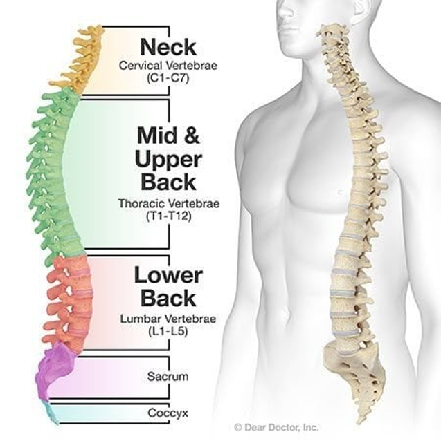
5th, 6th, & 7th Cervical
12th Thoracic
1st Lumbar
Spinal Cord Injury SCI
Injury to spinal cord
Trauma, penetrating wound
Vertebral injuries
Spinal Cord infarction
Primary injury
Occurs at time of event
Usually permanent
Secondary
Follows primary
Etiology unknown
Spread of injury
Can be mitigated with treatment
Emergency Management SCI
Speed and correct handling is paramount to preserving neurological function
Anyone having MVA, diving injury, contact sports injury, fall or direct trauma to head and neck is assumed to have SCI until otherwise proven.
Initial care includes
Extrication
Rapid assessment
Immobilization
Stabilization
Transportation to trauma center
Emergency Management SCI
Immobilize
Backboard
Neck brace
Neutral position
One person controls head
Four persons to move client to backboard
Emergency Management SCI
Respiratory support
O2 to keep PO2 high
High cervical injuries interfere with innervation of diaphragm (T1-T11)
Apnea/hypoventilation
Intubation and ventilation
Cardiovascular support
Maintain perfusion
Pharmacology
High dose corticosteroids within 8 hrs of injury
Steroid early on – tend to do better
C3-C4 – everything below is paralyzed
higher chance to stop breathing
Management SCI
Immobilization and reduction of fractures
Cervical fractures
Crutchfield Tongs – Burr holes
Gardner-Wells Tongs – noninvasive
Halo traction/Halo vest
Surgical management
Cord compression
Unstable vertebrae
Cord penetration
Bony fragments in spinal canal
Deteriorating neurologic status
Spinal Cord Injury: Signs and symptoms
Loss of sensation and/or motor ability below level of injury
Paralysis
Paralysis (plegia): Complete loss of voluntary movement; sensation may or may not be lost.
Paresis: Partial weakness with some movement remaining; sensation may or may not be affected. Recovery determined by extent of secondary injury
Function determined by location and whether complete or partial
C1-C3 fx generally incompatible with life
Spinal Cord Injury SCI: Complete
Usually acute
Usually cervical
Loss of motor, sensory, autonomic and reflexes below level of injury
Spinal Cord Injury SCI: Partial
Some motor or sensory function remains below the level of injury.
May be associated with lumbar injuries.
Paralysis - Flaccid paralysis
No muscle tone (limp muscles).
No voluntary movement.
Absent reflexes.
Urinary and bowel incontinence.
Muscle wasting (atrophy).
Example: Polio.
Paralysis - Spastic paralysis
Increased muscle tone (stiff muscles).
No voluntary movement.
Urinary and bowel retention.
Unregulated reflex activity (hyperreflexia).
Upper extremities (UE): Flexion.
Lower extremities (LE): Extension.
Paraplegia:
Paralysis of both lower extremities (legs).
Hemiplegia:
Paralysis of one side of the body (arm and leg).
Quadriplegia (Tetraplegia):
Paralysis of all four extremities (both arms and legs), usually due to cervical spinal cord injury.
Nursing Care for SCI
Adequate breathing & airway clearance
Ventilator management & oxygenation
Improving mobility
Adaptation to sensory & perceptual alterations
Urinary elimination
Bowel elimination
Skin integrity
Spinal/Neurogenic Shock
Sudden loss of reflex activity below the level of spinal cord injury (SCI).
Duration: Hours to weeks.
Can occur with any type of SCI, even if spastic paralysis develops later.
Loss of autonomic nervous system (ANS) function: Disrupts regulation of heart rate, blood pressure, breathing, digestion, and sweating.
Sympathetic nervous system: Normally helps the body respond to stressful situations; disruption can cause vasodilation, hypotension, and bradycardia.
Spinal/Neurogenic Shock - Signs and symptoms:
Absent reflexes.
Flaccid paralysis.
Loss of bowel and bladder control.
Venous pooling in extremities.
Peripheral vasodilation.
↓ BP → ↓ cardiac output.
↓ HR (bradycardia).
No perspiration below the lesion.
Autonomic Dysreflexia
Occurs after spinal/neurogenic shock resolves.
Exaggerated sympathetic nervous system (SNS) response.
Usually occurs with spinal cord injuries at T6 or above.
Can cause stroke, seizures, or death if untreated.
Why it happens
The SNS triggers the fight-or-flight response.
A stimulus below the injury causes an exaggerated sympathetic response, leading to vasoconstriction and a sudden increase in BP.
The brain attempts to lower BP, which can cause bradycardia, but signals cannot adequately control the response below the injury.
Autonomic Dysreflexia - Causes / Triggers
Distended bladder (common trigger)
Distended bowel or constipation
Skin stimulation or irritation
Other irritating stimuli below the level of injury
In spinal cord injury patients, many different stimuli can trigger this response.
Autonomic Dysreflexia - Signs and Symptoms
Severe hypertension (sudden increase in blood pressure)
Severe headache
Flushed face
Sweating above the injury
Cold, pale skin below the injury
Goosebumps (piloerection) below the injury
Bradycardia (slow heart rate)
Nausea
Nasal congestion
Nursing Care Autonomic Dysreflexia
Rapid assessment: Check BP and assess for signs and symptoms.
Remove the triggering stimulus.
Usually urinary or bowel related.
Check for a distended bladder or blocked catheter.
Empty the bladder or bowel as appropriate.
Place the patient in a sitting position.
Promotes venous pooling in the lower extremities, helping decrease BP.
Administer prescribed antihypertensive medication.
Hydralazine IV (Apresoline) 10–20 mg slowly, per order and facility protocol.
Label MR
Teach the family how to recognize, prevent, and respond to autonomic dysreflexia.
Treat as an emergency.
Long Term Effects of SCI
Loss of bladder and bowel function
Possible ventilator dependency
Respiratory complications
Loss of sexual function
Postural hypotension
Deep vein thrombosis
Sensory deprivation
Peripheral edema
Skin breakdown
Contractures
Rehabilitation
Long Term SCI Rehabilitation:
The Shepherd Center is a private, non-profit hospital in Atlanta, Georgia. Founded in 1975, the hospital focuses on rehabilitation for people with spinal cord injury and disease, acquired brain injury, multiple sclerosis, chronic pain and other neuromuscular problems.
Long Term Nursing Management SCI
Increase mobility
Specialty beds and positioning devices
Encourage mobility and rehabilitation as appropriate
Promote skin integrity
Use gel pads and pressure-relieving devices.
Reposition regularly and inspect skin for pressure injuries.
Bladder and Bowel management
Self-catheterization
manual digital stimulation of anus
high fiber
hydration
exercise
Coping
Depression
alterations in cognition
Anger
Suicidal
Sexual function
penile implants
Prevent Complications
Spasticity: Splints and other prescribed interventions.
Infection and sepsis: Monitor for pulmonary, skin, and urinary infections.
Pulmonary complications: Encourage coughing, deep breathing, and turning/repositioning.
Urinary complications: Maintain appropriate bladder-emptying routines.
Skin complications: Continue pressure relief and skin assessments.
Home care
Home health nurse
specialty care givers
family
Management of Patients with Neuromuscular Disorders
Multiple Sclerosis
Parkinson’s Disease
Myasthenia Gravis
Guillain-Barre
Multiple Sclerosis MS
Chronic demyelinating disease of the central nervous system (CNS).
The immune system damages myelin, disrupting or slowing nerve impulse conduction.
MRI may show lesions (white spots) in the CNS.
Cause: Thought to be autoimmune.
Common age: 20–40 years.
More common in women than men.
Disease pattern: Exacerbations (flare-ups) and remissions (symptoms improve or decrease).
Multiple Sclerosis MS - Signs and Symptoms
Fatigue: Pervasive/common symptom.
Spasticity: Muscle stiffness and spasms.
Ataxia: Poor coordination and balance.
Paresthesias/pain: Tingling, numbness, or pain.
Multiple Sclerosis MS - Symptoms by CNS Area
Area affected | Possible symptoms |
|---|---|
Optic nerve | Blurred vision, diplopia (double vision), and sometimes severe vision loss |
Cerebrum | Emotional lability, euphoria, depression, and possible suicidal thoughts |
Cerebellum | Ataxia, poor coordination, and tremors |
Brainstem | Motor and sensory problems; swallowing or other functions may be affected |
Spinal cord | Spasticity, weakness, and sensory changes |
Multiple Sclerosis MS - Treatment
Corticosteroids: Shorten the duration of acute exacerbations; they do not cure MS.
Disease-modifying therapies (including certain immunosuppressive or immunomodulating medications): Reduce disease activity and may slow progression.
Supportive care: Manage fatigue, spasticity, pain, mobility problems, and other symptoms.
Multiple Sclerosis MS - Prognosis
MS varies greatly from person to person.
Many people live for decades with MS and may die from unrelated causes.
Severe disease can cause complications, including respiratory problems, though respiratory failure is not the usual outcome for most patients.
Multiple Sclerosis MS - Primary Symptoms
Fatigue
Depression
Weakness
Numbness
Impaired coordination/loss of balance
Pain
Multiple Sclerosis MS - Secondary Symptoms
UTI
Constipation
Pressure ulcers
Contractures
Decreased bone mass
Dependent pedal edema
Pneumonia
Reactive depression
Multiple Sclerosis - Diagnosis
MRI – visualizes plaques
CSF – fluid (presence of immune elements)
Neuropsychological testing
in-depth assessment of skills and abilities linked to brain function.
Urodynamic studies
bladder, sphincters, and urethra hold and release urine. These tests can show how well the bladder works and why there could be leaks or blockages.
Sexual history
Management of Multiple Sclerosis (MS) -
No cure
Goals:
Delay disease progression.
Reduce the severity and duration of exacerbations.
Manage symptoms and prevent complications.