Histology Prelab - Digestive Glands
 Call Kai
 Learn
 Practice Test
 Spaced Repetition
 Match
 Flashcards
 Knowt Play
1/191

There's no tags or description
Looks like no tags are added yet.
192 Terms
Endocrine: Islets of Langerhans (pancreatic islets)
Exocrine: Large interlobular ducts
Pancreatic lobules containing:
-numerous pure serous acinar cells
-Intercalated ducts
-Centroacinar cell
-Intralobular duct
What are the key histological features of the pancreas?
AKA pancreatic islets
Pale clusters of cells interspersed within lobules and surrounded by acinar units
No ducts associated
What are islets of Langerhans?
Large diameter with simple columnar to stratified surrounded by DICT
What are pure serous acinar units?
Smallest duct drains the serous acinar unit
Contains simple squamous/cuboidal epithelium and centroacinar cell
Secretes bicarbnate ion
Drains into intralobular duct
What are intercalated ducts?
Found in center of acinar unit
Light blue/gray stained with central nucleus
Represents the start of the intercalated duct (part of it)
Secretes bicarbonate ion
What are centroacinar cells?
Larger than intercalated duct but still within lobule
Typically cuboidal with thin layer of CT
No striated duct in pancreas, only found in salivary glands
Transport function only
What is an intralobular duct?
the serous acinar cells of the lobule
The ducts that transport digestive enzymes to the duodenum originates within ___________________
Islets of Langerhans (Pancreatic islets)
The endocrine function of the pancreas is associated with _______________________ (histological structure)
This is the location of the rER and the nucleus
Why do serous cells have a basophilic stain at the basal region?
Digestive enzymes
Serous pancreatic acinar cells contain vesicles that contain _____________________
Islets of Langerhans (pancreatic islets)
Centroacinar cells
What are two features that distinguish the pancreas from salivary glands?
Intercalated duct
What is the smallest duct within a pancreatic lobule?
Found in DICT between lobules
DICT often contains BVs and nn.
What are interlobular ducts?
Alpha, beta and delta cells
What cells are found in pancreatic islets?
Secrete glucagon in response to low blood glucose
Works with adrenal gland hormones and responds to sympathetic stimulation
What do alpha cells do in the pancreatic islets?
Secrete insulin in response to high blood glucose
Inhibition of insulin when low blood glucose
What do beta cells do in the pancreas?
Elevated blood glucose by the break down of glycogen, lipids, and proteins as well as release of glucose into blood
What is the outcome of alpha cells secreting glucagon?
Stimulated uptake of glucose into liver cells, neurons, skeletal muscle, and adipose
Glucose stored in cells as glycogen, proteins, or lipids if not used
What is the outcome of beta cells releasing insulin?
Secrete somatostatin in response to ACh, ghrelin, and high glucose
Can inhibit beta cells to prevent hypoglycemia
What do delta cells do in the pancreas?
Hormones released by enteroendocrine cells in duodenum:
CCK - stimulation of bile release
Secretin - simulation of bicarbonate release
When chyme enters the duodenum, how are pancreatic cells stimulated?
Intramural or intrapancreatic ganglia
Parasympathetic ganglia in the pancreas are known as ________________________
Outside the walls of the organs in the celiac and mesenteric ganglia
Where are sympathetic fibers found in the pancreas?
Splanchnic nerve fibers
What are the long, postganglionic sympathetic nerve fibers called in the pancreas?
A CT capsule and serosa (visceral peritoneum)
What covers the outside of the liver?
Hexagonal shape
Centrally placed vein
Several peripherally located portal areas between adjacent lobules
What is do liver lobules look like?
Portal triad:
Portal vein (if small - venule)
Hepatic artery (if small - arteriole)
Bile duct (if small - ductule)
What do portal areas contain in the liver?
From portal area to central vein
What is the direction of the flow of blood within liver lobules?
From center of lobule (near central vein) to portal area
What is the direction of the flow of bile in liver lobules?
Sinusoids between hepatocytes
Portal vessels are drained by what type of capillary?
Sinusoids
What vessels are inter-hepatic?
Portal venule and arteriole
What vessels are interlobular in the liver?
4-6
How many portal areas are there per lobule?
AKA terminal hepatic vein
Located at center of lobule
Receives blood from portal area vessels via sinusoids
What is the central vein of liver lobules?
Epithelial cells in the liver
Represent sinusoids
Responsible for bile and cholesterol synthesis and bilirubin production
What are hepatocytes?
Nothing
What accompanies the central vein in liver lobules?
3, distribution of oxygen and nutrients
Functionally, the liver is said to have __ regions that reflect ______________________________
Zone 1 - periportal area
Zone 2 - mid-lobular area
Zone 3 - centrilobular area
What are the three functional regions within the liver?
Periportal area
What zone in the liver has the most nutrients and oxygen?
Centrilobular area
What zone in the liver has the least nutrients and oxygen?
Centrilobular area
What zone is most susceptible to drug toxicity in the liver?
Periportal area
What zone is best suited for oxidative metabolism?
Bile canaliculi (singular = canaliculus)
Where does bile that is synthesized by hepatocytes first enter?
Canal of Hering
After entering bile canaliculi, where does bile travel?
Bile
"What is the principal exocrine product of the liver?
Closest to central vein
If bile blockage occurs, bile will accumulate in the liver cells closest to the point of blockage. If it is a mild block, where would you see this accumulation?
Closest to portal area
If bile blockage occurs, bile will accumulate in the liver cells closest to the point of blockage. If it is a large block (main hepatic duct), where would you see this accumulation?
Where the blockage occurs
Where is the point of accumulation of bile determined by in a blockage?
Gallstones
What is the most common etiology of bile obstruction?
Pain, nausea/vomiting, jaundice
How is bile obstruction manifested?
Stagnation of bile due to impaired secretion or obstruction of bile flow anywhere along flow path
What is cholestasis?
Intrahepatic, extrahepatic
If the blockage of the bile duct occurs inside the liver, if is considered ________________________. If it occurs outside the liver, it is considered to be ____________________
Bile acids and bilirubin accumulate
Bilirubin accumulates as bile pigment in hepatocytes, bile canaliculi, and in severe cases, bile ductules
Eventually, bilirubin pigments and bile acids spill into blood causing jaundice
What happens if the bile duct is blocked?
Store and concentrate bile synthesized by hepatocytes
What is the function of the gallbladder?
The common bile duct
Bile is delivered to the gallbladder from the liver via ________________________
the 2nd part of the duodenum, hepatopancreatic ampulla, sphincter of hepatopancreatic ampulla (AKA sphincter of Oddi)
To be used, bile is released into ________________________ at the _____________________________ controlled by the ______________________________
Simple columnar epithelium with apical microvilli
Mucosa highly folded - but no true villi and plicae
Crypts are absent
Submucosa and muscularis mucosa absent
muscularis externa present, but no distinct layers
Both adventitia and serosa present
What are the key histological features of the gallbladder?
Absorption of water increases concentration of bile
Why does epithelium in the gallbladder have an absorptive function?
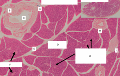
Blood vessel
Identify the structure labeled A

DICT
Identify the type of tissue labeled B

Interlobular duct
Identify the structure labeled C

Pancreatic lobule
Identify the structure labeled D

CT (not specified)
Identify the type of tissue labeled E
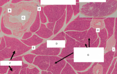
Pancreatic islets (islets of Langerhans)
Identify the structures labeled F

Intralobular ducts
Identify the structures labeled G

Interlobular duct
Identify the structure labeled H

ANS ganglia (intramural or intrapancreatic ganglia)
Identify the structure labeled I
Pancreatic serous acinar unit
Identify the structure labeled A
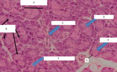
Pancreatic serous cells
Identify the cells labeled B
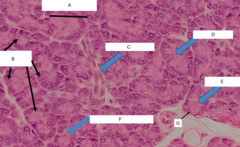
Intercalated duct
Identify the structure labeled C

Centroacinar cell
Identify the cell labeled D
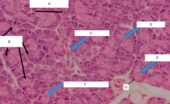
Centroacinar cell
Identify the cell labeled E
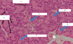
Intercalated duct
What structure is the cell labeled F the beginning of?

Serous cell
Identify the cell labeled G

Intralobular duct
Identify the structure labeled A

Intercalated duct
Identify the structure labeled B
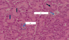
Intercalated duct
Identify the structure labeled A

Centroacinar cells
Identify the cells labeled B
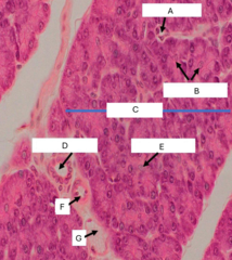
Pancreatic lobule
Identify the structure labeled C

Intralobular duct
Identify the structure labeled D

Centroacinar cell
Identify the cell labeled E

Arteriole
Identify the structure labeled F

Venule
Identify the structure labeled G

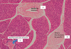
Interlobular duct
Identify the structure shown in the image

Arteriole
Identify the structure labeled B

Smooth muscle
Identify the type of tissue labeled B

Interlobular duct
Identify the structure labeled C

Pancreatic lobule
Identify the structure under the label E

Pancreatic islet (islet of Langerhans)
Identify the structure shown in the image

Alpha, beta, and delta cells
Identify the types of cells that would be found in the structure shown in the image
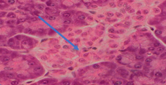
Endocrine
Identify whether the structure shown is exocrine or endocrine

Centroacinar duct cell
Identify the cell labeled A
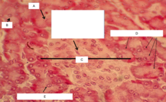
Centroacinar duct cell
Identify the cell labeled B
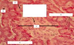
Pancreatic islet (islet of Langerhans)
Identify the structure labeled C

Pancreatic serous acinar cells
Identify the cells labeled D
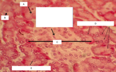
Centroacinar duct cell
Identify the cell labeled E

Intralobular duct
Identify the structure labeled A

Intramural ganglion (intrapancreatic ganglion)
Identify the structure labeled B

Pancreatic islet (Islet of Langerhans)
Identify the structure labeled C
Pacinian corpuscle
Identify A

Large interlobular duct
Identify B
