Chronic inflammation
 Call Kai
 Learn
Learn Practice Test
 Spaced Repetition
 Match
Match Flashcards
 Knowt Play
1/12

There's no tags or description
Looks like no tags are added yet.
Name | Mastery | Learn | Test | Matching | Spaced | Call with Kai | Chat |
|---|
No analytics yet
Send a link to your students to track their progress
13 Terms
what is chronic inflammation
Inflammation of prolonged duration (weeks/months/years)
Occurs when:
• The acute inflammatory response fails to remove the stimulus
• There are repeated episodes of acute inflammation
• The stimulus or microbe has unique biochemical characteristics or virulence factors which incite chronic inflammation
Mechanisms:
• Persistent or resistant infections
• Isolation (‘hiding’ from the immune response)
• Unresponsiveness to phagocytosis or enzymatic breakdown (e.g. plant material, grass awns, silica dust, asbestos fibres, some suture materials, surgical prostheses)
• Autoimmunity (response to self antigens promote chronic inflammation) or defects in leukocyte function
• Idiopathic (e.g. granulomatous meningoencephalitis)
Cytokines are a broad category of small, soluble proteins released by cells to regulate immune responses, while chemokines are a specialized subset of cytokines that specifically direct the movement and migration of white blood cells
chronic inflammation - deeper dive
The chronic inflammatory response is maintained by cytokines, chemokines and other inflammatory mediators
Incite:
• Ongoing inflammation mediated by infiltration and activation of
lymphocytes, macrophages, plasma cells and multinucleated giant
cells
• Tissue destruction (necrosis)
• Proliferation of fibroblasts and deposition of collagen
• Angiogenesis and neovascularisation (granulation tissue formation)
• Initiation of wound healing (reepithelialisation and tissue repair)

beneficial and harmful aspects of chronic inflammation
Beneficial:
• ‘Walling off’ the inciting agent or substance
Harmful:
Space occupying lesions (focal, multifocal or diffuse) can be formed resulting in:
• Displacement, replacement or obliteration of the original tissue
• Loss of function
• Clinical significance determined by the size, tissue and position
Dysplasia and malignant transformation of chronically inflamed tissue


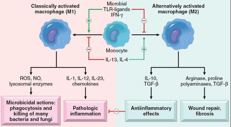
subsets of activated macrophages
there are two main types - for a) inflammation and tissue injury - fighters, b)repair - nurses
pic shows the same thing as above, just diff represnataion
you need to memorise the second pic - need to know this

fibrosis - morphology and cells of chronic inflammation
chronic inflammation leads to 3 pathwas
cytokines induce collagen deposition
growth factors
reduced matrix mellasjdajdsjsadh activity
all lead to fibrosis - result of accumulation of mature fibroblasts
starts to shrink when this tissue matures - starts to scar, dries up, depresses
blue part is fibrous connective tissue in the liver
this pic is the liver


abcess formation
Granulomatous inflammation
• Distinct type of chronic inflammation
• Dominated by cells of the monocyte-macrophage system - leading to this granular appearance
• These take the form of macrophages, epithelioid macrophages (activated
macrophages resembling epithelial cells) and multinucleated giant cells (MGCs)
The cells may be:
• Dispersed as sheets at random within the tissue = diffuse or
lepromatous granulomas
• Arranged in discrete masses or nodules = nodular or tuberculoid
granulomas


nodular (tuberculoid granulosomas) - when lymphocytes wrap around foreign substance in body
Develop with a T Helper Lymphocyte Type 1 response
May or may not have a central core of necrotic debris, which can mineralise
• Those with a necrotic core = caseating (Latin caseous = cheese)
• Those without = noncaseating
caseating-a specific type of tissue death (necrosis) where damaged tissue turns into a soft, crumbly, white substance that looks like cheese
Microscopic appearance
• Round to oval
• Numerous macrophages with variable numbers of epithelioid macrophages, MGCs
• Peripheral zone of fibroblasts, lymphocytes and plasma cells
• May form a fibrous capsule
Causes include:
• Mycobacterium bovis and Mycrobacterium tuberculosis
• Deep fungal infections
hsitology shwos the same thing
giant cell has multiple nuclei
fragment is caseous necrosis
dark material is the mineralisation of the caseating core

diffuse (lepromatous) granulomas
• Develop with a T Helper Lymphocyte Type 2 response
• Poorly delineated, forming sheets within the tissue
• Composed of numerous macrophages and few lymphocytes and plasma cells
Causes include:
• Mycobacterium avium subsp. paratuberculosis (Johne’s disease)
• Mycobacterium lapraemurium (Feline leprosy)

Eosinophilic granulomas
fibrinous not fibrous
Characterised by dense infiltration of eosinophils, macrophages and varying
numbers of lymphocytes and plasma cells
All species:
• Eosinophilic (TH2) granulomas secondary to parasitic infections
Cats:
• Eosinophilic plaque, eosinophilic granuloma, and eosinophilic
dermatitis
Dogs:
• Eosinophilic granuloma of the oral cavity of huskies and other breeds
Horses:
• Equine collagenolytic granuloma, axillary nodular necrosis and unilateral papular dermatosis
image shows parasite - the big circular thing


Pyogranulomatous inflammation
• Same cellular constituents as granulomatous inflammation
• Also contains neutrophils, plasma proteins and fibrin (acute inflammatory
response) (pyogranulomatous inflammation)
• Occurs when the inciting stimulus continues to elicit an acute inflammatory
response
• A pyogranuloma is a nodular granuloma with a central area of neutrophils

Lymphoplasmacytic inflammation
• Also termed ‘chronic inflammation’
• Composed of lymphocytes and plasma cells (lymphoplasmacytic) admixed with macrophages
• Sometimes lymphocytes and macrophages predominate over plasma cells (= lymphohistiocytic inflammation)
• Common type of inflammation often seen in the early stages of chronic inflammation
• Also seen in response to specific microbes e.g. viruses
• Seen in response to antigenic inflammation in mucosal surfaces e.g. chronic rhinitis

chronic-active inflammation
Cellular components of chronic inflammation (lymphocytes, plasma cells, macrophages)
• Also constituents of acute inflammation (neutrophils, fibrin, plasma proteins)
Occurs when the inciting cause has not been removed from the exudate of a chronic inflammatory response
• Continues to elicit an acute inflammatory response