Fracture and Dislocation Management
 Call Kai
 Learn
 Practice Test
 Spaced Repetition
 Match
 Flashcards
 Knowt Play
1/96

There's no tags or description
Looks like no tags are added yet.
97 Terms
tuning fork
compression
distraction
percussion
fracture assessment tools/techniques
deformity
open wounds
tenderness
swelling
DOTS acronym for head-to-toe assessment
transverse fracture
fracture that is perpendicular to the shaft of the bone
linear (longitudinal) fracture
fracture that splits down the length of the bone
oblique fracture
fracture with a spiral shape; occurs when one end of the bone is fixed and the other end is twisted
spiral fracture
fracture with an s-shaped separation; occurs when ends of bone are twisted in opposite directions
greenstick fracture
incomplete break of bone that has not fully ossified (children under 10)
comminuted fracture
fracture that consists of >3 fragments at the fracture site; often caused by a hard blow or fall
avulsion fracture
fracture where a ligament or tendon pulls off a piece of bone
impacted/compression fracture
fracture caused by a fall from height causing the bone to be compressed
serrated fracture
fracture where bony fragments have sawtooth fracture line
stress fracture
fracture caused by repetitive microtrauma that exceeds the stress bearing capacity of the bone
Type I
Salter-Harris fracture with complete separation of the physis with no fracture to the bone

Type II
Salter-Harris fracture with separation of the growth plate occurring along with a small portion of the metaphysis

Type III
Salter-Harris fracture that is an epiphysis fracture

Type IV
Salter-Harris fracture where portions of both the epiphysis and metaphysis fracture

Type V
Salter-Harris fracture where a crushing force causes a gross deformity without displacing the physis

signs of fracture
low chance of successful reduction
concomitant injury
1st attempt unsuccessful
children aged 6-12
suspected growth plate involvement
joint reduction contraindications
distract
disengage/recreate deformity
reposition
general steps for reducing joint
shoulder
most commonly dislocated joint
95%
% of shoulder dislocations that are anterior
anterior shoulder dislocation
shoulder dislocation that presents with abducted and externally rotated position
inferior shoulder dislocation
shoulder dislocation that presents with limited abduction
posterior shoulder dislocation
shoulder dislocation that presents in an internally rotated and adducted position
axillary artery and nerve
adjacent structures that can be damaged during shoulder dislocation
Boss-Holzach-Matter Maneuver (Davos) Technique
shoulder self-reduction technique where patient grabs their knee and leans back
77-86%
Boss-Holzach-Matter Maneuver (Davos) Technique success rate
External rotation maneuver
shoulder reduction technique where patient is supine with elbow flexed to 90 and longitudinal traction is applied while gently externally rotating
Stimson technique
shoulder reduction technique where patient lies prone with affected shoulder hanging off table in 90 forward flexion and a 5-15lb weight is attached to the wrist
28-96%
Stimson Technique success rate
~90 seconds
Boss-Holzach-Matter Maneuver (Davos) Technique relocation time
8-20 minutes
Stimson technique reduction time
spaso technique
shoulder relocation technique where patient lies supine with arm straight up, distraction force is applied while externally rotating the arm and can be aided by a second clinician stabilizing the scapula
traction-countertraction/matsen technique
shoulder relocation technique where patient lies supine; one provider provides traction on humerus while a second provider provides a counterforce along the patient’s chest
scapular manipulation technique
shoulder relocation technique where patient lies prone and longitudinal traction is applied to the humerus and another clinician applied a medial glide on the scapula
Milch maneuve
shoulder relocation technique where patient is supine with scapula fixated and longitudinal traction is applied while the arm is externally rotated and abducted
FARES method
shoulder relocation technique where patient is supine with elbow extended and forearm in neutral. Wrist is grasped with both hands and a gentle longitudinal traction is applied while oscillating the arm and slowly abducting.
Cunningham Technique
Shoulder relocation technique where patient is seated and has arm in adducted position next to the body with the elbow flexed to 90; clinician places hand between the patient’s forearm and body with the wrist/hand resting on clinician’s upper arm and gentle traction is applied while massaging the trapezius, deltoid, and biceps with the other hand.
elbow
second most commonly dislocated joint8
80%
percent of elbow dislocations that are posterior
10-19 y/o
male
wrestling, football, and gymnastics
elbow dislocation risk factors
patient seated or supine
inline traction
forearm supination
elbow flexion while placing direct pressure on tip of olecranon
elbow reduction technique
extension and valgus stress
post-reduction elbow unstable positions
dorsal DIP joint
most common finger dislocation
fracture
tendon rupture
rings present
growth plate
6-12 y/o
finger relocation contraindications
apply direct pressure over dorsal aspect of proximal phalanx with wrist in flexion
dorsal finger dislocation reduction method
apply direct pressure over volar aspect of proximal phalanx with MCP in flexion
volar finger dislocation reduction method
stabilize phalange proximal to dislocated joint and grasp dislocated segment
apply longitudinal traction and force to exaggerate dislocation
apply force to base of dislocated segment in direct opposire direction
exaggerated finger reduction method
stabilize phalange proximal to dislocated joint and grasp dislocated segment
apply axial traction to dislocated segment while pushing bone in opposite direction of dislocation
traction finger reduction method
flexed and adducted hip with axial load through flexed knee
hip dislocation MOI
90%
% of hip dislocations that are posterior
hyperextension or pivot shift mechanism
knee dislocation MOI
50%
% of knee dislocations that self reduce
male (4:1)
football
MVC
extreme sports
knee dislocation risk factors
one clinician stabilize distal femur, other grasps tibia
apply longitudinal traction to tibia
if unsuccessful, reverse dislocation of tibia
knee dislocation reduction method
30 degrees
degree to lock TROM after knee reduction
lateral
most common patellar dislocation
foot planted and internal twist while knee in valgus
patellar dislocation MOI
hip flexed
slowly extend knee while applying pressure to lateral patella
patellar dislocation reduction
31%
% of ankle dislocations that occur is ports
forceful landing on or into surface; contact
ankle dislocation MOI
posterior
most common ankle dislocation
posterior
ankle dislocation where talus is posterior to tibia; associated with fracture to lateral malleolus
anterior
ankle dislocation where talus is anterior to tibia
lateral
ankle dislocation where talus is lateral to tibia; associated with bimalleolar fracture or distal fibular fracture
superior or diastasis injury
ankle dislocation where talus is superiorly located within the mortise (separation of syndesmosis)
anatomical splint
rigid splint
soft splints
traction splints
types of splints/imobilization
anatomical splint
splint using another part of the body
soft splints
splints that do not limit as much motion and conform better to extreme injuries (ie towel, cervical collar, pillow, etc)
rigid splints
splint that limits further movement such as SAM splints, ortho-glass, and aluminum finger splints
air splints
clear, inflatable plastic splints that are inflated and provide uniform compressive force around the injured extremity; can NOT be used with displaced or open fractures
PneuSplint
splint that is held in place with quick open and close straps; work for non-displaced and angulated injuries
vaccum splint
splint composed of strong, tough plastic containing polystyrene beads
open fracture
excessive bleeding
vaccum splint potential contraindications
stabilize fracture
reduce blood loss
reduce muscle spasm
maintain distal vascular supply
traction splint benefits
2
number of people required to place traction splint
clavicle sling
sling type

desault bandage
sling type

soft splint with sling and swathe
sling type

posterior elbow splint
sling type
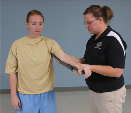
dorsolateral rigid splint elbow dislocation
sling type

sugar tong splint
sling type

Volar SAM splint
sling type

rigid volar splint
sling type
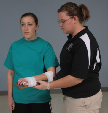
ulnar gutter splint
sling type

dorsal finger splint
sling type

SAM splint/ankle stirrup
sling type

SAM splint: cadillac
sling type

ankle soft splint
sling type

lower leg/anatomic soft splint
sling type

lower leg rigid splint
sling type

knee anatomic/soft splint
sling type
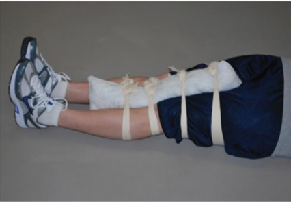
rigid straight leg splint
sling type

6 inches
distance crutches should be in front of body when fitting
2 inches
distance crutches should be at the side when fitting
~1inch (2-3 fingers)
distance between top of crutch and underarm
12-18 inches
distance crutches should be placed in front of body when moving in tripod method