UNIT 6. NUCLEIC ACID ELECTROPHORESIS
 Call Kai
 Learn
 Practice Test
 Spaced Repetition
 Match
 Flashcards
 Knowt Play
1/53

There's no tags or description
Looks like no tags are added yet.
54 Terms
electrophoresis
is the movement of molecules by an electric current.
Nucleic acids
have a negative charge so they move towards the positive pole.
tubes, slab gels, or capillaries.
Electrophoresis can be performed in
agarose gel
polyacrylamide gel
2 NA gel systems
agarose
Gel systems:
uses a _______ a polysaccharide from seaweed
smaller pores
Gel Systems:
Larger concentrations of agarose produce _________ in the gel making it harder for nucleic acids to move.
Pulse Field Gel Electrophoresis
Used for resolving very large fragments of DNA
DNA goes forward and backward, alternating, circular, and polygon, allowing better resolution of large fragments.
Pulses of current
Pulse Field Gel Electrophoresis:
_________ from alternating sides of the gel are used.
Polyacrylamide Gel
Gel systems:
Best for smaller DNA fragments.
Used for sequencing nucleic acids, mutation analyses, nuclease protection assays, and other applications requiring the resolution of nucleic acids down to the single-base level.
Acrylamide;
cross-linker methylene bisacrylamide;
polyacrylamide
____________, in combination with the _____________, polymerizes into a gel that has consistent resolution characteristics, the ____________
proteins
Polyacrylamide was originally for _____
T
T or F
Preparation of PAGE gels is more complicated than AGE gels
Capillary electrophoresis
Gel system:
A polymer is placed inside inside capillary tubes and the sample moves through there.
Sample has to be denatured.
Less sample, immediate visualization, increased sensitivity, less work, less time, but much more costly.

buffer;
electric current;
buffering capacity
In order for nucleic acids to migrate properly, the gel system must be immersed in a ______ that conducts the ________ efficiently in relation to the __________
Tris borate EDTA (TBE)
Tris phosphate EDTA (TPE)
Tris acetate EDTA (TAE)
3 Tris buffers used in electrophoresis
0.089 M Trisbase
0.089 M boric acid
0.002 M EDTA
TBE composition
0.089 M Trisbase
1.3% phosphoric acid
0.002 M EDTA
TPE composition
0.04 M Trisbase
0.005 M sodium acetate
0.002 M EDTA
TAE composition
TAE
most commonly used Trisbase
Buffer additives
__________ are often used to modify sample molecules in ways that affect their migration.
Gel loading buffer (GLB)
Samples must be first mixed with ________________
tracking dye & density agent
GLB contains (2)
Density Agent (Ficoll, sucrose, glycerol)
increases the density of the solution as compared with the electrophoresis buffer
tracking dyes (bromophenol blue or xylene cyanol green)
are used to monitor the progress of the electrophoresis run.
migrate at specific speeds in and usually run ahead of the smallest fragments.
F
T or F
Tracking dyes affect the separation of the sample DNA.
F
they only migrate along the DNA but do not bind to it
T or F
Tracking dyes help locate where the DNA is in the gel by binding onto the DNA.
Nucleic-Acid specific dyes
Used to visualize DNA/RNA
Intercalating agents: Ethidium bromide (EtBr)
SYBR green dyes
Silver stains
3 Nucleic-Acid specific dyes
Intercalating agents
3 Nucleic-Acid specific dyes
intercalate, or stack, between the nitrogen bases in double-stranded nucleic acid.
Ethidium bromide (EtBr)
3 Nucleic-Acid specific dyes
When exposed to UV light, it fluoresces (orange glow).
The more DNA present → the brighter the band.
neutrogen, carcinogen and high contamination
SYBR Green
3 Nucleic-Acid specific dyes
Binds to the minor groove or sides of DNA
Fluoresces under blue or UV light.
relatively safe, non-toxic, non-contaminating
Silver stain
3 Nucleic-Acid specific dyes
Staining solution interacts with nucleophilic groups in DNA/RNA.
Silver ions (Ag+) gets reduced and deposits directly where nucleic acids are located.
Produces dark brown/black bands.
Extremely sensitive (detects very small amounts of DNA).
Does not require UV light.
AGE of Genomic DNA
heavy, long smears with intense bright bands

ReliaPrep™ Large Volume

Maxwell® 16 LEV

Maxwell® HT 96 gDNA

Wizard® Genomic

QIAamp® DNA Blood

RNA contaminationD
smaller fragments deposited at the bottom of the gel
blurred smears at the bottom

DNA degradation
smear only with no bright bands of DNA
shearing

AGE of Plasmids
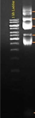
circular
linear
supercoiled
nicked
Plasmid conformations
supercoiled plasmid
AGE of Plasmids
always the band at the bottom (not RNA contamination)
Genomic DNA contamination

Open Circular / Covalently Bounded

Linear Plasmid

Supercoiled Plasmid
RNA Contamination

AGE of RNA
2 very distinct bright bands at 18S and 28S (for eukaryotes) or 16S for (prokaryotes)

2 ribosomal subunits (1 large, 1 small subunits)
AGE of RNA:
The 2 very distinct bright bands at 18S and 28S are representative of
Molecular weight markers
Consists of nucleic acid fragments of known sizes used to identify the sizes of unknown nucleic acid bands
1Kb plus DNA ladder
100 bp DNA ladder
Lambda-HindIII DNA ladder
RNA marker
different types of markers
Main application is resolve nucleic acid molecules.
DNA sequencing
Routine check for PCR products
Routine check for cloning
Routine check for enzyme digestion
Paternity testing
Application of nucleic acid gel electrophoresis