Med Term Chapter 10 - Cardiovascular, Lymphatic, Immune Systems and Blood
 Call Kai
 Learn
Learn Practice Test
 Spaced Repetition
 Match
Match Flashcards
 Knowt Play
1/138

There's no tags or description
Looks like no tags are added yet.
Name | Mastery | Learn | Test | Matching | Spaced | Call with Kai | Chat |
|---|
No analytics yet
Send a link to your students to track their progress
139 Terms

heart
muscular cone-shaped organ the size of a fist, located behind and slightly to the left of the sternum (breastbone) and between the lungs.
The pumping action of the heart circulates blood throughout the body. The heart consists of two smaller upper chambers, the right atrium and the left atrium (pl. atria), and two larger lower chambers, the right ventricle and the left ventricle (pl. ventricles).
The right atrium receives blood returning from the body through the veins and contracts to fill the right ventricle, which then pumps blood to the lungs. The left atrium receives blood from the lungs and contracts to fill the left ventricle, which then contracts to pump blood from the heart through the arteries to body tissues. The atrial septum separates the atria and the ventricular septum separates the ventricles.

atrioventricular valves
consist of the tricuspid and mitral valves, which lie between the right atrium and the right ventricle and the left atrium and left ventricle, respectively. Valves of the heart keep blood flowing in one direction.

pericardium
two-layer sac surrounding the heart, consisting of an external fibrous and an internal serous layer.
The internal serous layer is then divided into two parts: the outer layer, called the parietal pericardium, and the inner layer, called the epicardium. Between these is the pericardial space, which contains fluid produced by the serous layer that allows the heart to move without friction.

epicardium
outer lining covering the heart (also called visceral pericardium)

myocardium
middle, thick, muscular layer of the heart

endocardium
inner lining of the heart
blood vessels
tubelike structures that carry blood throughout the body
arteries
blood vessels that carry blood away from the heart.
All arteries, with the exception of the pulmonary artery, carry oxygen and other nutrients from the heart to the body cells. The pulmonary artery, in contrast, carries carbon dioxide and other waste products from the heart to the lungs.
arterioles
smallest arteries
aorta
largest artery in the body, which originates at the left ventricle, briefly ascends as the arch of the aorta, then descends through the thorax and abdomen
veins
blood vessels that carry blood back to the heart.
All veins, with the exception of the pulmonary veins, carry blood containing carbon dioxide and other waste products. The pulmonary veins carry oxygenated blood from the lungs to the heart.
venules
smallest veins

venae cavae
largest veins in the body. The inferior vena cava carries oxygen-poor blood to the right ventricle of the heart from body parts below the diaphragm, and the superior vena cava returns the blood to the right ventricle of the heart from the upper part of the body.
capillaries
microscopic blood vessels that connect arterioles with venules. Materials are passed between the blood and tissue through the capillary walls.
blood
fluid circulated through the heart, arteries, capillaries, and veins; composed of plasma and formed elements, such as erythrocytes, leukocytes, and thrombocytes (platelets)
plasma
clear, straw-colored liquid portion of blood in which cells are suspended. Plasma is approximately 90% water. The other 10% is composed of solutes (dissolved substances), which include proteins, electrolytes, and vitamins. Plasma comprises approximately 55% of the total blood volume.
serum
clear, watery fluid portion of the blood that remains after a clot has formed
blood cells (formed elements)
production of new blood cells takes place in bone marrow, the spongy tissue inside some bones

erythrocytes
red blood cells that carry oxygen

leukocytes
white blood cells that combat infection and respond to inflammation. There are five types of white blood cells.

thrombocytes
one of the formed elements in the blood that is responsible for aiding in the clotting process (also called platelets)
lymph
transparent, colorless tissue fluid; contains lymphocytes and monocytes and flows in a one-way direction toward the heart
lymphatic vessels
transport lymph from body tissues into the right and left subclavian veins, which then empty into the superior vena cava.
The lymphatic vessels begin as capillaries spread throughout the body then merge into larger tubes that eventually become ducts in the chest. They provide a one-way flow for lymph, which enters through veins into the circulatory system.
lymph nodes
small, spherical bodies composed of lymphoid tissue. They may be singular or grouped together along the path of the lymph vessels.
The nodes filter lymph to keep substances such as bacteria and other foreign agents from entering the blood. They also contain lymphocytes.

spleen
located in the left side of the abdominal cavity between the stomach and the diaphragm. In adulthood, the spleen is the largest lymphatic organ in the body. Blood, rather than lymph, flows through the spleen. Blood is cleansed of microorganisms in the spleen. The spleen stores blood and destroys worn out red blood cells.
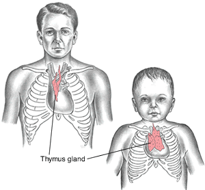
thymus gland
one of the primary lymphatic organs, it is located anterior to the ascending aorta and posterior to the sternum between the lungs. It plays an important role in the development of the body’s immune system, particularly from infancy to puberty. Around puberty the thymus gland shrinks so that most of the gland is connective tissue.
angioma
tumor composed of blood vessels
angiostenosis
narrowing of a blood vessel

aortic stenosis
narrowing, pertaining to aorta (narrowing of the aortic valve)
arteriosclerosis
hardening of the arteries
atherosclerosis
hardening of fatty plaque (deposited on the arterial wall)
bradycardia
condition of a slow heart (rate less than 60 beats per minute)
cardiomegaly
enlargement of the heart
cardiomyopathy
disease of the heart muscle
endocarditis
inflammation of the inner (lining) of the heart (particularly heart valves)
ischemia
deficiency in blood (flow); (caused by constriction or obstruction of a blood vessel)
myocarditis
inflammation of the muscle of the heart

pericarditis
inflammation (of the sac) surrounding the heart
phlebitis
inflammation of a vein
polyarteritis
inflammation of many (sites in the) arteries
tachycardia
condition of a rapid heart (rate of more than 100 beats per min)
thrombophlebitis
inflammation of a vein associated with a blood clot
valvulitis
inflammation of a valve (of the heart)
angioplasty
surgical repair of a blood vessel (also called balloon angioplasty)
atherectomy
excision of fatty plaque (from a blocked artery using a specialized catheter and a rotary cutter)
embolectomy
excision of a plug (embolus or clot, usually with a balloon catheter, inflating the balloon beyond the clot, then pulling the balloon back to the incision and bringing the plug with it)
endarterectomy
excision within the artery (excision of plaque from the arterial wall)

pericardiocentesis
surgical puncture to aspirate fluid from the sac surrounding the heart (usually to relieve cardiac tamponade and for diagnostic investigation)
phlebectomy
excision of a vein
valvuloplasty
surgical repair of a valve (cardiac or venous)
angiography
radiographic imaging of blood vessels (the procedure is named for the vessel to be studied, e.g., femoral angiography or coronary angiography)
angioscopy
visual examination (of the inside) of a blood vessel
aortogram
radiographic image of the aorta (after an injection of contrast materials)
arteriogram
radiographic image of an artery (after an injection of contrast materials)
venogram
radiographic image of a vein (after an injection of contrast materials)
echocardiogram (ECHO)
record of the heart (structure and motion) using sound (waves); (used to detect valvular disease and evaluate heart function)
electrocardiogram (ECG, EKG)
record of the electrical activity of the heart
electrocardiography
process of recording the electrical activity of the heart
cardiologist
physician who studies and treats diseases of the heart
cardiology
study of the heart (a branch of medicine that deals with diseases of the heart)
atrioventricular (AV)
pertaining to the atrium and ventricle
cardiogenic
originating in the heart
intravenous (IV)
pertaining to within the vein
acute coronary syndrome (ACS)
sudden symptoms of insufficient blood supply to the heart, indicating unstable angina or acute myocardial infarction. Rapid assessment is necessary to determine the diagnosis and treatment and to minimize heart damage.

aneurysm
ballooning of a weakened portion of an arterial wall
angina pectoris
chest pain, which may radiate to the left arm and jaw, that occurs when there is an insufficient supply of blood to the heart muscle
arrhythmia
any disturbance or abnormality in the heart’s normal rhythmic pattern
cardiac arrest
sudden cessation of cardiac output and effective circulation, which requires cardiopulmonary resuscitation (CPR)
cardiac tamponade
(KAR-dē-ak) (tam-po-NĀD)
acute compression of the heart caused by fluid accumulation in the pericardial cavity
coronary artery disease (CAD)
condition that reduces the flow of blood through the coronary arteries to the myocardium that may progress to depriving the heart tissue of sufficient oxygen and nutrients to function normally; most often caused by coronary atherosclerosis. CAD is a common cause of heart failure and myocardial infarction.
cor pulmonale
(kor) (pul-mō-NAL-ē)
enlargement of the heart’s right ventricle due to pulmonary disease
deep vein thrombosis (DVT)
formation of a blood clot in a deep vein of the body. Most often occurs in the lower extremities. A clot, or part of a clot, can break off and travel to the lungs, causing a pulmonary embolism.

fibrillation
rapid, quivering, uncoordinated contractions of the atria or ventricles, causing cardiac arrhythmia
heart failure (HF)
condition in which there is an inability of the heart to pump enough blood through the body to supply the tissues and organs with nutrients and oxygen. One common type is congestive heart failure (CHF).
hypertensive heart disease (HHD)
disorder of the heart caused by persistent high blood pressure; it may be associated with hypertrophy (abnormal thickening of the heart muscle) or dilation of the chambers of the heart (due to thinning and stretching of the heart muscle)
intermittent claudication
condition of pain, tension, and weakness in a limb that starts when walking is begun, increases until walking is no longer possible, and then completely resolves when the patient is at rest. It is caused by reversible muscle ischemia that occurs with peripheral artery disease.
mitral valve stenosis
narrowing of the mitral valve from scarring, usually caused by episodes of rheumatic fever
myocardial infarction (MI)
death (necrosis) of a portion of the myocardium caused by lack of oxygen resulting from an interrupted blood supply (also called heart attack)
peripheral artery disease (PAD)
disease of the arteries in the arms and legs, resulting in narrowing or complete obstruction of the artery.
This is caused most commonly by atherosclerosis, but occasionally by inflammatory diseases, emboli, or thrombus formation. The most common symptom of peripheral artery disease is intermittent claudication. (also called peripheral vascular disease [PVD])
rheumatic heart disease
damage to the heart muscle or heart valves caused by one or more episodes of rheumatic fever

varicose veins
distended or tortuous veins, usually found in the lower extremities

artificial cardiac pacemaker
battery-powered apparatus implanted under the skin, with leads placed on the heart or in the chamber of the heart; used to treat an abnormal heart rhythm, usually one that is too slow, due to an abnormal sinus node

automatic implantable cardiac defibrillator (AICD)
device implanted in the body that continuously monitors the heart rhythm. If life-threatening arrhythmias occur, the device delivers an electric shock to convert the arrhythmia back to a normal rhythm.

catheter ablation
procedure in which abnormal cells that trigger abnormal heart rhythms (arrhythmias) are destroyed by using a device that heats or freezes the cells

coronary artery bypass graft (CABG)
surgical technique to bring a new blood supply to heart muscle by detouring around blocked arteries

coronary stent
supportive scaffold device placed in the coronary artery; used to prevent closure of the artery after angioplasty or atherectomy; used to treat an artery occluded by plaque

femoropopliteal bypass
surgery to establish an alternate route from femoral artery to popliteal artery to bypass an obstruction

percutaneous transluminal coronary angioplasty (PTCA)
procedure in which a balloon is advanced into a coronary artery to the area where plaque has formed. When the balloon is inflated, the vessel wall expands, allowing blood to flow more freely. (also called percutaneous coronary intervention (PCI))

thrombolytic therapy
injection of a medication either intravenously or intraarterially to dissolve blood clots. It is often used in emergency departments for acute myocardial infarction.
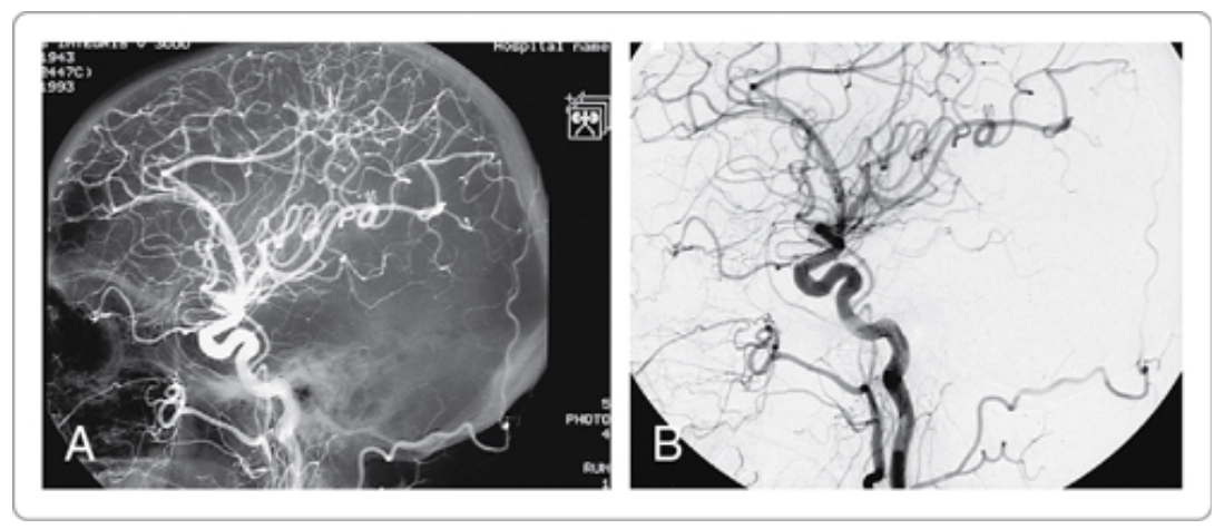
digital subtraction angiography (DSA)
process of digital radiographic imaging of the blood vessels that “subtracts” or removes structures not being studied
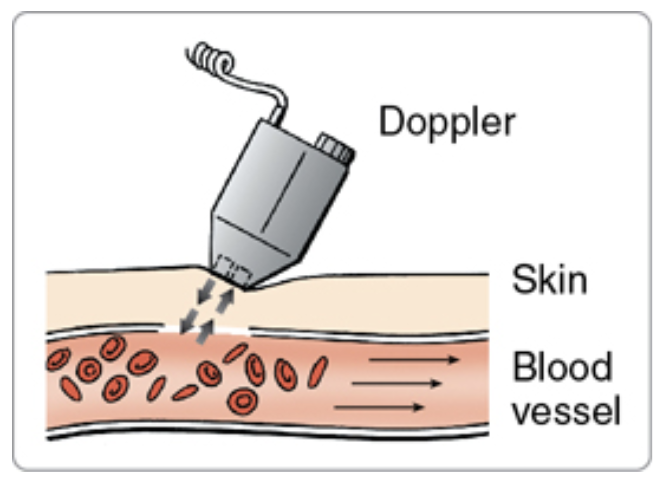
Doppler ultrasound
study that uses high-frequency sound waves for detection of blood flow within the vessels; used to assess intermittent claudication, deep vein thrombosis, and other blood flow abnormalities

sestamibi test
(ses-ta-MIB-ē) (test)
nuclear medicine test used to diagnose coronary artery disease and assess revascularization after coronary artery bypass surgery. Sestamibi, a radioactive isotope, is taken up by normal myocardial cells, but not in ischemia or infarction. These areas are identified as “cold” spots on the images produced.
single-photon emission computed tomography (SPECT)
nuclear medicine test that collects a series of images (projections) as a gamma camera rotates around the patient. These projections are then used by a computer to generate three-dimensional pictures; it also helps show the function of organs, such as coronary artery flow or active and inactive areas of the brain.
transesophageal echocardiogram (TEE)
ultrasound test that examines cardiac function and structure by using an ultrasound probe placed in the esophagus, which provides more direct views of the heart structures

cardiac catheterization
diagnostic procedure performed by passing a catheter into the heart from a blood vessel in the groin or arm to examine the condition of the heart and surrounding blood vessels; used to diagnose and treat cardiovascular conditions such as coronary artery disease
exercise stress test
study that evaluates cardiac function during physical stress by riding a bike or walking on a treadmill.
Electrocardiography is the most common method, but echocardiography and nuclear medicine scanning (diagnostic imaging tests) can also be used to measure cardiac function while exercising.
blood pressure (BP)
pressure exerted by the blood against the blood vessel walls. A blood pressure measurement written as systolic pressure (120) and diastolic pressure (80) is commonly recorded as 120/80 (blood pressure is measured in millimeters of mercury [mm Hg]).
pulse (P)
contraction of the heart, which can be felt with a fingertip. The pulse is most commonly felt over the radial artery (in the wrist); however, the pulsations can be felt over a number of sites, including the femoral (groin) and carotid (neck) arteries.
sphygmomanometer
(sfig-mō-ma-NOM-e-ter)
device used for measuring blood pressure
C-reactive protein (CRP)
blood test to measure the amount of C-reactive protein in the blood, which when elevated, indicates inflammation in the body. It is sometimes used in assessing the risk of cardiovascular disease.