Lesson 59 - Neoplastic vs Non-neoplastic Lesions
 Call Kai
 Learn
Learn Practice Test
 Spaced Repetition
 Match
Match Flashcards
 Knowt Play
1/75

There's no tags or description
Looks like no tags are added yet.
Name | Mastery | Learn | Test | Matching | Spaced | Call with Kai | Chat |
|---|
No analytics yet
Send a link to your students to track their progress
76 Terms
What lesions have low cellularity?
1. cyst
2. lipoma
3. poorly exfoliated lesions
What is an example of a poorly exfoliated lesion?
sarcoma
What are the inflammatory lesions?
1. eosinophilic
2. granulomatous
3. lymphoplasmacytic/lymphocytic
4. pyogranulomatous
5. suppurative
What are the neoplastic lesion types?
1. epithelial
2. mesenchymal
3. round
4. neuroendocrine
5. melanoma
What cells are found in eosinophilic lesions?
1. eosinophils
2. some mast cells
What cells are found in suppurative lesions?
neutrophils, often degenerate
What cells are found in lymphocytic/lymphoplasmacytic lesions?
1. lymphocytes
2. plasma cells
What cells are found in pyogranulomatous lesions?
1. neutrophils
2. macrophages
What cells are found in granulomatous lesions?
macrophages
What are the pros to cytology?
1. cheap
2. quick
3. repeatable
4. good for screening and monitoring
What are the cons to cytology?
1. less architecture
2. more operator-dependent
3. cannot always grade tumor
What are the common indications for cytology?
1. cutaneous/subcutaneous masses
2. lymphadenopathy
3. body cavity effusions
4. respiratory, urogenital, and CSF samples
5. organ masses
What are the techniques for getting samples for cytology?
1. fine-needle aspirate
2. impression smears
3. swabs
4. scrapings/brushings
What are the types of fine-needle aspirate techniques?
1. aspiration technique
2. non-aspiration technique
What are the characteristics of aspiration FNA?
1. small gauge needle (22-25) and syringe
2. negative pressure
What are the characteristics of non-aspiration FNA?
1. needle only
2. multiple redirections
What type of FNA is better for fragile or vascular lesions?
non-aspiration technique
What are impression smears used for?
1. ulcerated lesions
2. biopsies
3. surgical specimens
You want to take an impression smear, but the lesion seems to very bloody. What do you do?
blot tissue to remove blood
What are swabs best for?
1. ear
2. vagina
3. conjunctival
4. nasal
5. fistulas
How do you place a sample from a swab to a microscope slide?
roll the sample, not smear
What lesions are scrapings/brushings used for?
1. keratinized lesions
2. firm lesions
What size needle is used for FNA?
22 - 25 gauge
What size syringe is used for FNA?
5 or 10 mL
What do you not use to label your slide?
pen or sharpie
What do you do if your microscope slides have been sitting out in a drawer for a while?
clean and dry with alcohol
What is the technique for putting sample onto a slide for cytology?
draw air into the syringe and expel it out aiming it towards the slide then place two slides over top of each other and slide away
What is the prefix for change in size/shape of a cell?
aniso
What is the prefix for nucleus?
karyo
What does CHANG stand for?
1. cyst
2. hematoma
3. abscess
4. neoplasia
5. granuloma
What are the criteria for malignancy?
1. anisocytosis
2. anisokaryosis
3. prominent, multiple, and/or bizarrely shaped nucleoli
4. macrocytosis, macrokaryosis, macronuclei
5. anisonucleoliosis
6. multiple nuclei
7. increased N:C ratio
8. nuclear molding
9. coarse chromatin
10. abnormal mitotic figures
What does nuclear molding look like?
hugging nuclei
What does coarse chromatin look like?
looks "ropey"
If you have a mixed sample, how do you classify it?
based on the predominant cell type
What do epithelial lesions look like on cytology?
1. cohesive
2. polygonal
3. moderate to abundant cytoplasm
What do mesenchymal lesions look like on cytology?
1. loose sheets
2. individual
3. wispy cytoplasm
4. spindle or stellate
What do round cell lesions look like on cytology?
1. single discreet cell
2. individual
3. round in shape
What do neuroendocrine lesions look like on cytology?
1. small clusters/rosettes
2. many naked nuceli
What do melanoma lesions look like?
1. individual sometimes with clusters
2. can look like anything
What does the cytoplasm of mast cell tumors show?
purple cytoplasmic granules
What does the cytoplasm of TVT show?
discrete clear vacuoles
What are other features that may be seen with TVT?
background vacuoles
What does the cytoplasm of plasmacytoma show?
paranuclear clearing
What are other features that may be seen with plasmacytoma?
1. significant pleomorphism
2. bi/multinucleation
3. flame cells
What does the cytoplasm of histiocytoma show?
pericytoplasmic clearing
What does the cytoplasm of lymphoma show?
1. infrequently vacuolated
2. occasional paranuclear clearing
What are other features that may be seen with lymphoma?
lymphoglandular bodies
If you tap an abdomen and do cytology on the fluid and see degenerate neutrophils with bacteria, what do you suspect?
septic abdomen
What do lipoma cells show?
peripheral nuclear with large cytoplasm

What malignancy change is this cell showing?
Anisocytosis

What malignancy change is this cell showing?
Anisokaryosis
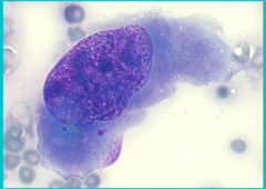
What malignancy change is this cell showing?
Prominent, bizarrely shaped nucleoli

What type of cell is this?
Mesenchymal

What malignancy change is this cell showing?
Macrocytosis, macrokaryosis, macronucleoli

What malignancy change is this cell showing?
Anisonucleoliosis

What malignancy change is this cell showing?
Multiple nuclei

What malignancy change is this cell showing?
Increased N:C ratio

What malignancy change is this cell showing?
Nuclear molding

What malignancy change is this cell showing?
Coarse chromatin

What malignancy change is this cell showing?
Abnormal mitotic figures

What kind of lesion?
Eosinophilic

What kind of lesion?
Eosinophilic

What kind of lesion?
Suppurative

What kind of lesion?
Suppurative

What lesion is this?
Lymphoma

What lesion is this?
Lymphocytic

What lesion is this?
Pyogranulomatous lesion

What lesion is this?
Granulomatous

What cell is this?
Degenerating neutrophil

What tumor cell type?
Epithelial tumor

What tumor cell type?
Mesenchymal

What tumor cell type?
Lipoma

What tumor cell type?
Mast cell tumor
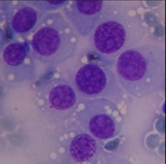
What tumor cell type?
TVT
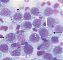
What tumor cell type?
TVT
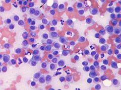
What tumor cell type?v
Plasmacytoma