Trauma
 Note
Note Call Kai
 Learn
Learn Practice Test
 Spaced Repetition
 Match
Match Flashcards
 Knowt Play
1/90

There's no tags or description
Looks like no tags are added yet.
Name | Mastery | Learn | Test | Matching | Spaced | Call with Kai | Chat |
|---|
No analytics yet
Send a link to your students to track their progress
91 Terms
Concussion
Mild TBI with GCS ≥13; may involve brief LOC, amnesia, vertigo, confusion, or delayed responses.
Coup/Contrecoup Injury
Brain hits back and forth in skull.
Post-Concussion Symptoms
Vertigo, amnesia, delayed speech, disorientation, emotional lability, incoordination.
Post-Traumatic Seizures
Occur in <5% within the first week after head injury.
Head CT Indications (Canadian Rules)
LOC with head strike, GCS <15 after 2h, skull/basilar fracture signs, ≥2 vomiting, ≥65yo, amnesia ≥30min, dangerous mechanism, neuro deficit, seizure, anticoagulant use.
Head CT Indications (New Orleans Rule)
GCS 15 + headache, vomiting, age >60, ETOH/drug use, anterograde amnesia, visible trauma above clavicle.
Head Injury Admission Criteria
GCS <15, abnormal CT, seizures, bleeding diathesis, or anticoagulant use.
Concussion treatment
Brain rest.
Head Injury Discharge Precautions
Return if confusion, worsening HA, vision change, weakness, vomiting, or seizure.
Cerebral Contusion
Focal brain bleeding; monitor and admit for observation.
Epidural Hematoma
Arterial bleed (middle meningeal artery) between skull and dura; lucid interval → rapid decline from brain herniation; needs evacuation of hematoma.
Anasacoria
Unequal pupils.
Normal ICP
<20 mmHg.
Steroids help with trauma?
No.
Epidural Hematoma Prognosis
Worse with large volume, midline shift, GCS <8.
Subdural Hematoma
Venous bleed from bridging veins between dura and arachnoid; often from falls or ETOH.
Subdural Hematoma Symptoms
LOC ± lucid interval, HA, N/V, ataxia, pupil defects.
Subdural Hematoma Treatment
Surgery or observation based on severity.
Subarachnoid Injury
Bleeding into subarachnoid space; risk of vasospasm and seizures due to brain parenchyma irritation.
Secondary Brain Injury symptoms (Intracranial)
Hemorrhage, ischemia, edema, ↑ICP, vasospasm, infection.
Secondary Brain Injury symptoms (Systemic)
Hypoxia, hyper/hypocapnia, hyperglycemia, hypotension, fever, hyponatremia.
Cerebral Blood Flow Formula
CBF = CPP − ICP; CPP = MAP − ICP.
Monroe-Kellie Hypothesis
Skull is fixed space; ↑volume in one component ↑ICP.
Increased ICP Symptoms
HA, diplopia (CN VI palsy), papilledema, bradycardia, HTN, ↓LOC.
Cushing’s Triad
Bradycardia, hypertension, irregular respirations (brainstem compression).
ICP Management
Keep ICP <20 mmHg; elevate HOB 30∘, drain CSF, mannitol, brief hyperventilation.
Fever in Brain Injury
Common; search for infection (sinusitis, UTI, PNA); cooling and antipyretics limited benefit.
Tension Pneumothorax
Clinical diagnosis: absent breath sounds, JVD, tracheal deviation, hypotension.
Tension Pneumo Chest Tube
24–28F tube, 4–5th ICS midaxillary; relieves air accumulation.
Hemothorax
Blood in pleural space; 24–28F tube at 4–5th ICS midaxillary; massive >1500 mL.
Thoracotomy Indication
Drain >20 mL/kg initially or 1500/3hrs.
Chest Tube Removal (Pneumo)
No air leak, full expansion.
Chest Tube Removal (Hemo)
<100 mL/day output, full expansion.
Traumatic Aortic Dissection
Deceleration injury tearing aortic isthmus; fatal in 80% prehospital.
Aortic Dissection Signs
Chest/back pain, L supraclavicular hematoma, widened mediastinum.
Aortic Injury CXR
Wide mediastinum (>8cm supine), apical cap, L bronchus displacement.
Aortic Injury Management
BP control (SBP <100), esmolol, CTA confirmation, endovascular repair preferred.
Cardiac Tamponade
Blood in pericardial sac; 150 mL can be fatal.
Beck’s Triad
Hypotension, JVD, muffled heart sounds.
Tamponade Signs
Tachycardia, pulsus paradoxus systolic blood pressure drops significantly during inspiration), enlarged silhouette, effusion on echo.
Tamponade Treatment
IV fluids (support preload/filling), pericardiocentesis, thoracotomy.
Primary Survey (ABCDE)
Airway, Breathing, Circulation, Disability, Exposure.
Life-Threatening Thoracic Injuries
Tension/open pneumo, massive hemothorax, tamponade, flail chest, obstruction.
Flail Chest
Paradoxical chest wall motion due to multiple broken ribs; may need fixation.
Rib Fracture Pain Control
Multimodal (NSAIDs, acetaminophen, oxycodone, methocarbamol, nerve block).
Rib fracture treatment
Symptomatic treatment only.
PIC Score
Monitors pain, incentive spirometry, cough quality.
Rib Fixation Indications
Flail chest, severe pain, failed weaning, deformity.
Intubation Indications
RR >30 or <9, SpO₂ <90%, PaO₂/FiO₂ <280, pulmonary contusion, age/poor reserve.
Ventilation Strategy
Tidal volume 6–8 mL/kg, plateau <30, use PEEP, prone/ECMO for refractory hypoxemia.
Trauma Coagulopathy (TIC)
Triad: acidosis, hypothermia, coagulopathy.
Massive bleeding Management
Whole blood or 1:1:1 transfusion, minimize crystalloids, warm fluids, early TXA, LMWH prophylaxis.
TXA in Trauma
Antifibrinolytic; give within 3 hours; not a procoagulant.
Blunt Cardiac Injury (BCI) def and complications
Myocardial trauma from MVCs; causes arrhythmia, troponin ↑, hypotension.
BCI Screening
ECG + troponin; normal results exclude BCI (100% NPV).
BCI Management
Telemetry ≥24h if abnormal; ICU if unstable or arrhythmic.
BCI Dysrhythmias
Sinus tachy, PVCs, AFib; treat per ACLS, correct electrolytes.
Commotio Cordis
Sudden VF in young athlete; R-on-T impact; no structural injury.
Myocardial Rupture
80% fatal; requires immediate thoracotomy and pericardiotomy.
Valvular Injury (Trauma)
Aortic (AR/shock), Mitral (papillary rupture), Tricuspid (RHF); surgical repair.
Coronary Artery Injury
LAD most common; causes traumatic STEMI; PCI or bypass.
Blunt Aortic Injury (BTAI)
Deceleration tear at ligamentum arteriosum.
Blunt Aortic Injury Imaging
US, CTA = gold standard (100% sens, 99.7% spec).
Blunt Aortic Injury Treatment
Esmolol, SBP <120, TEVAR preferred; open repair if arch small.
FAST Exam (EFAST)
Bedside US for pericardial, hepatorenal, splenorenal, pelvic, and pleural fluid.
FAST Sensitivity
42–100%.
Abdominal Trauma Workup
Low threshold for CT; serial exams; labs (HCG, T&S).
Renal/GU Trauma Mechanism
Usually blunt (MVCs, falls, straddle).
Renal/GU Injury Signs
Hematuria, blood at meatus/introitus = urethral injury.
Retrograde Urethrogram
Consider if blood at meatus.
GU Imaging
CT angio may miss urethral injury; US for testicular trauma.
Junctional Hemorrhage
Bleeding at neck, axilla, groin, buttocks, or perineum—difficult to compress.
Junctional Tourniquets
Abdominal aortic, combat ready clamp, SAM, junctional emergency tool.
Abdominal Aortic Tourniquet
Inflates to 80 lbs pressure; effective in small trials.
SAM Junctional Tourniquet
Fast application (<30s); approved for axillary and pelvic bleeding.
Elderly Trauma Patients
Higher morbidity due to comorbidities, meds, poor compensation.
Elderly Resuscitation Risks
Sensitive to fluid overload and under-resuscitation; fragile bones.
Crystalloid Risks
Dilutional coagulopathy, hyperchloremic acidosis, edema, hypothermia.
Effective Fluid Strategy
Permissive hypotension, hemorrhage control, avoid hypothermia, balanced transfusion.
TXA Uses
Trauma, postpartum hemorrhage, epistaxis, ICH; poor in GI bleed or angioedema.
Most deadly type of pelvic fracture
Open book.
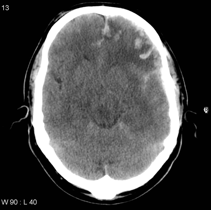
Cerebral contusion
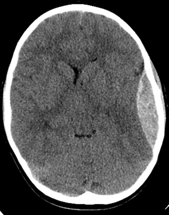
epidural hematoma
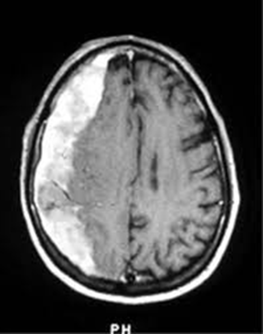
subdural hematoma
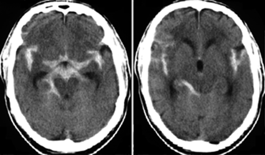
Subarachnoid hemorrhage
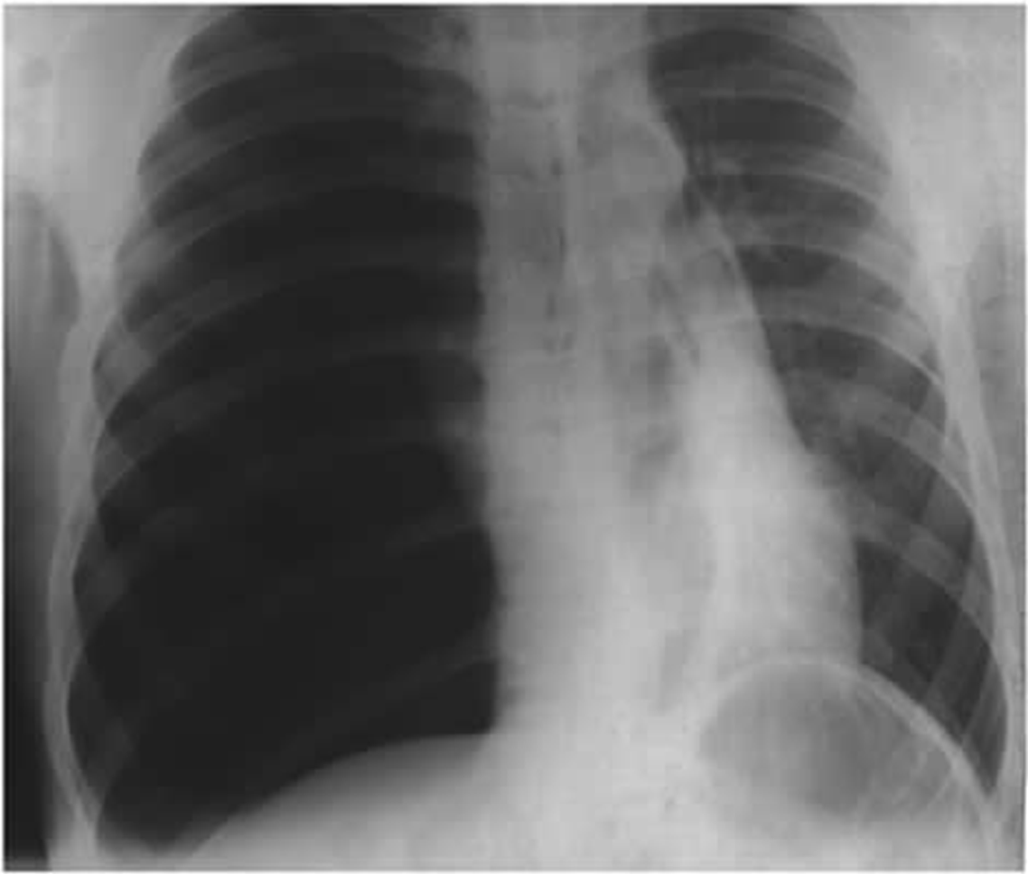
tension pnemothorax
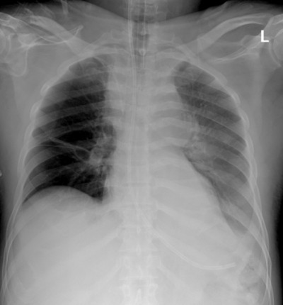
Aortic dissection
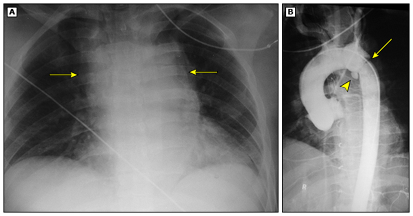
Aortic disection
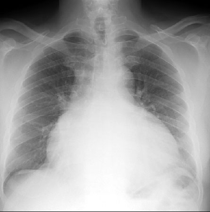
cardiac tamponade
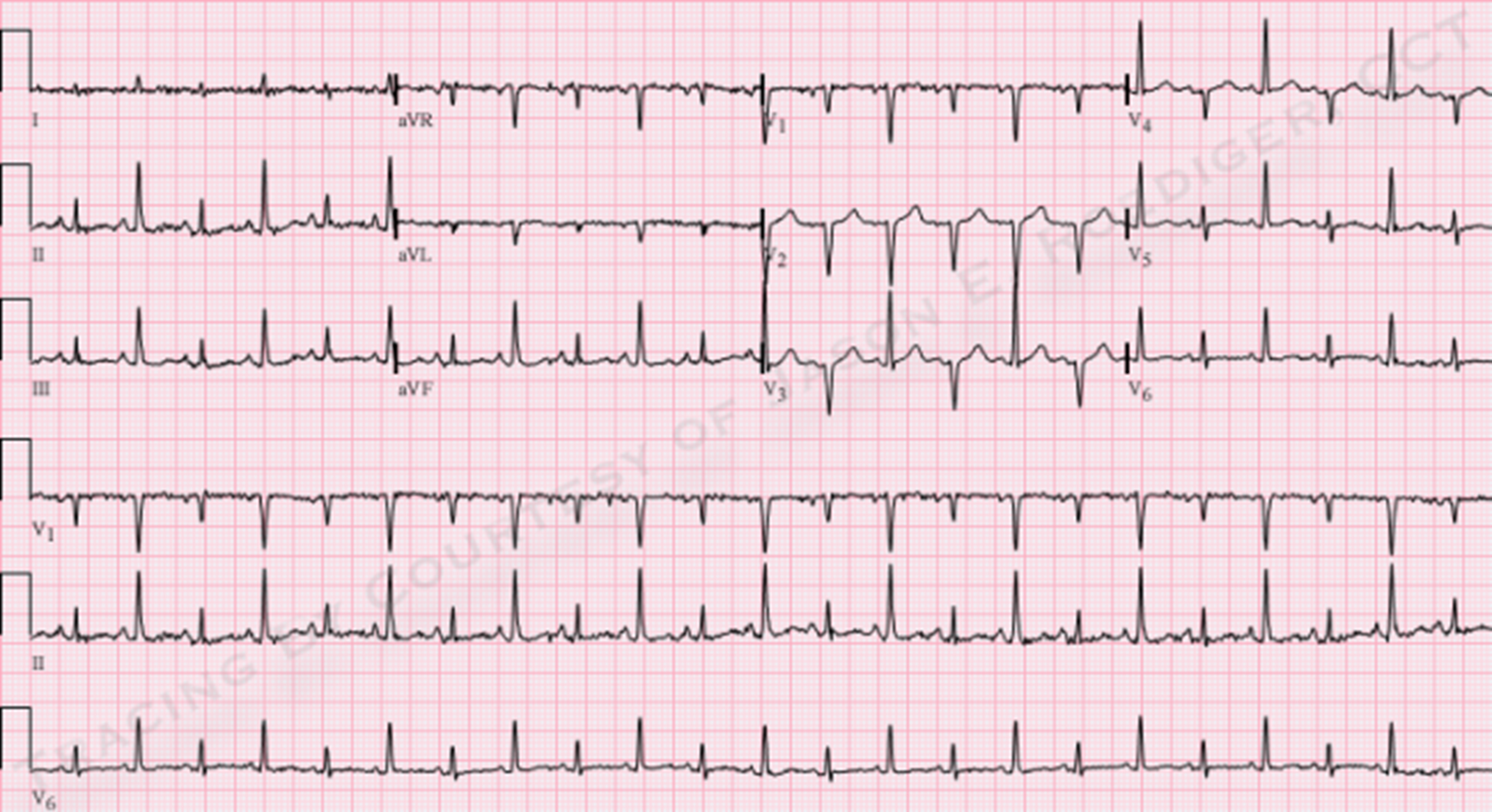
Cardiac tamponade EKG. Alternating QRS amplitude/voltage
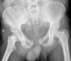
open book pelvic fracture