Haematological Malignancy 2
 Call Kai
 Learn
Learn Practice Test
 Spaced Repetition
 Match
Match Flashcards
 Knowt Play
1/23

Earn XP
Description and Tags
week 9
Name | Mastery | Learn | Test | Matching | Spaced | Call with Kai | Chat |
|---|
No analytics yet
Send a link to your students to track their progress
24 Terms
causes of leukaemia
Inherited Predispositions
Infection
Environmental Exposure
Types of inherited predispositions that can cause leukaemia
trisomy 21 (down syndrome)
fanconi anaemia
(genetic mutation that leads to defective DNA repair pathway)
Li-Frameni syndrome
(loss of p53 protein - responsible for detecting DNA damage and cell cycle arrest)
types of infections that can cause leukaemia
epstein-barr virus (EBV)
(infects B-cells, silences tumour-suppressor genes, disable apoptotic signals)
HTLV-1
(Infects CD4+ T-cells, blocks DNA repair, unregulates proliferative genes)
types of environmental exposure that can cause leukaemia
benzene and other aromatic hydrocarbons
ionising radiation
types of investigations of haematological malignancy
full blood count (FBC)
blood film
In haematological malignancies what would the FBC show for the following parameters: red cell count, platelets, MCV, white cell count
red cell count is variable - can be reduced with suppression of erythropoiesis or bleeding. or can be raised in clonal erythrocyte proliferations (e.g. polycythaemia vera)
platelets - may be reduced with myelosuppression or pooling in the spleen, raised in thrombocythaemia
MCV - usually normal; may be raised during cytotoxic therapy because of cell-cycle arrest
white cell count - variable, reduced WCC if immature cancer cells crowd out healthy production in the bone marrow, raised in lymphoma, if normal still do not rule out leukaemia
blood morphology in acute myeloid leukaemia peripheral blood film
thin cytoplasm, very large nucleus (‘scant' cytoplasm)
undifferentiated blasts
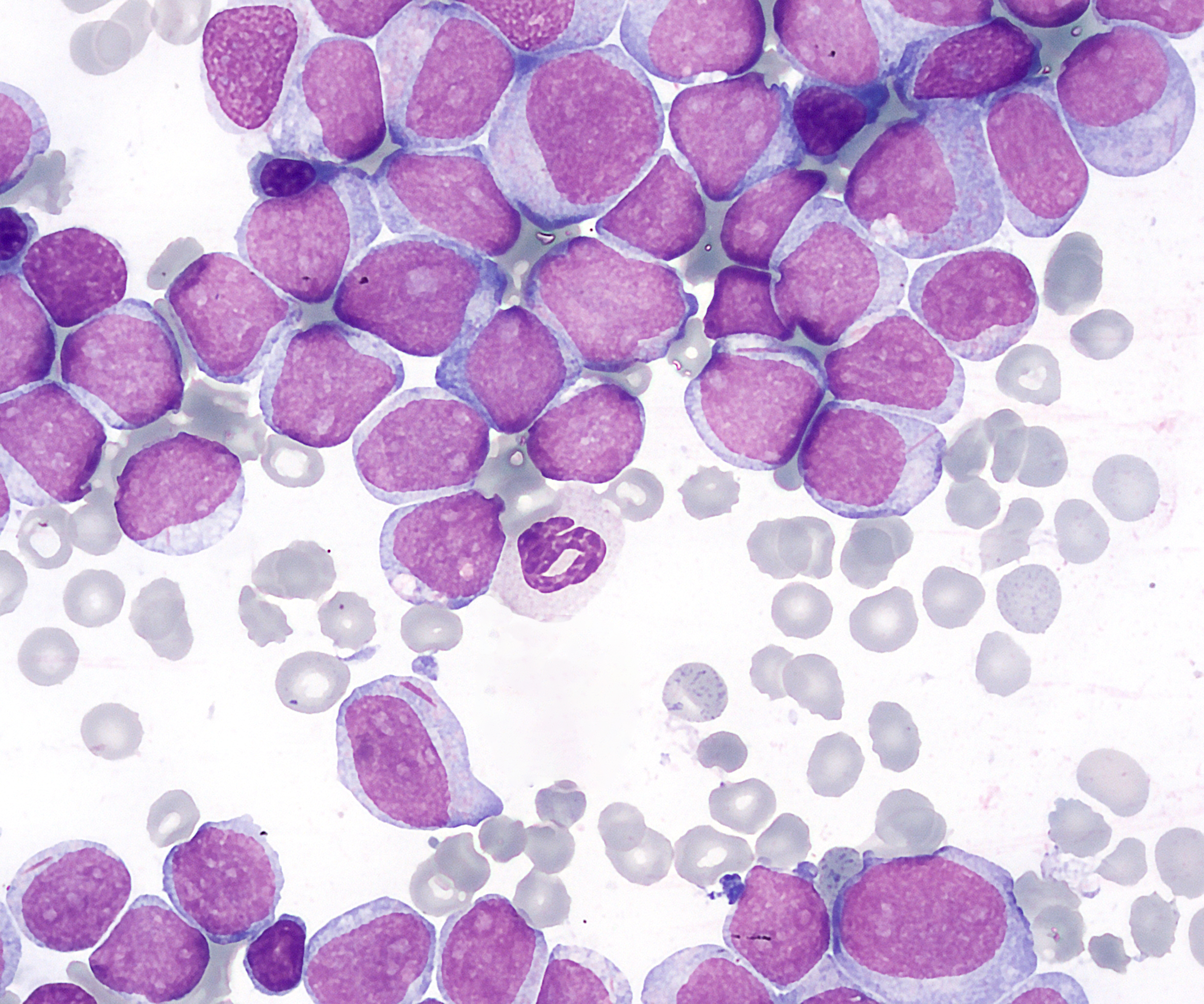
blood morphology in chronic myeloid leukaemia peripheral blood film
neutrophilic leukocytosis* - underlying genetic defect causes overproduction of neutrophils, no maturation arrest
increased basophils and eosinophils
full maturation spectrum present of other white cells (all stages of white cell development)
immature granulocytes (myeloblasts)
*leukocytosis = elevated WCC

how are white cells investigated
microscopy
nuclear structure & chromatin appearance
abnormal maturation of nucleus or cytoplasm
cytoplasmic granules
inclusion bodies and vacuoles
cell shape
presence of progenitor cells
differential count
automated analysers provide total WCC
percentage values for each cell type
3-point differentiation (lymphocytes, granulocyte, monocytes)
5-point differentiation (separates neutrophils, eosinophils, basophils, monocytes, lymphocytes)
what are auer rods
clumps of granulocytic material. only found in cells of myeloid lineage - the granulocytes to be specific!
while mostly observed in immature granulocytes, they’re not excusive to them. Have been documented in mature granulocytes too

purpose of bone marrow examination
identify abnormal cells
assess cellularity
assess degree of infiltration by leukaemic cells

what samples can be taken for bone marrow examination
lymph nodes for initial diagnosis (fine needle aspiration)
bone marrow aspirate - needle inserted into bone and marrow is pulled into a syringe
bone marrow trephine - larger needle removes a core of marrow for histology - needed in some lymphoid leukaemias

immunophenotyping
a laboratory technique used to identify and classify specific types of cells by detecting antigens on their surface or inside them
monoclonal antibodies used against cell-surface markers (CD antigens) - no single CD antigen is individually specific for malignancy, the OVERALL marker profile is interpreted
for haemopoietic investigation, what samples are used for immunophenotyping
blood
EDTA bone marrow
explain the process of immunophenotyping
cells are fixed to prevent lysis
treated to allow antibody access
fluorescent-labelled antibodies are then assessed by flow cytometry

what can immunophenotyping distinguish
distinguishes monoclonal and polyclonal populations
determines stage of maturity
distinguishes B and T cells in lymphoid population
what is CD45
a general white cell marker - assessed first in immunophenotyping to identify white cell population
the isolated population is is then examined for positive or negative populations in other markers
if more than 20% of cells show a reaction, it is considered positive
Healthy blood contains a diverse mix of cells. Leukemia creates a massive clone of identical abnormal cells, showing up as a unnaturally uniform population with the exact same surface markers
which marker is used in immunophenotyping to determine myeloid lineage
CD13, CD33, CD34
CD34 indicates blast cells
cytogenetics
the study of genetic material at the cellular level
establishes karyotype
identifies specific abnormalities in chromosome structure
**REQUIRED to classify leukaemia using WHO classifications

first step in classification of haematological malignancy
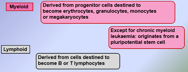
in acute leukaemia, a malignant condition is characterised by…
uncontrolled proliferation of a clonal cell population
poorly differentiated blast cells accumulate in the bone marrow and possible other tissues
how does 1976 French-American British (FAB) classification of leukaemia differ from the WHO classification

what causes symptoms of acute leukaemia
bone marrow failure
invasion of organs
metabolic disturbances

symptoms of acute lymphoblastic leukaemia
