The tongue and teeth
 Call Kai
 Learn
Learn Practice Test
 Spaced Repetition
 Match
Match Flashcards
 Knowt Play
1/48

There's no tags or description
Looks like no tags are added yet.
Name | Mastery | Learn | Test | Matching | Spaced | Call with Kai | Chat |
|---|
No analytics yet
Send a link to your students to track their progress
49 Terms
The tongue
The Organ of Deglutition (Swallowing), Drinking, Taste – Gustatory papillae, Taste
buds, Chemoreceptors
cow tongue extras
Prehension; Manipulation of food during mastication
Dog tongue extras
Thermoregulation (Panting in dogs)
Cat tongue extras
Papillae serve as a “rasp”, helping to scrape small amounts of meat off the bones and move it towards the back of the mouth
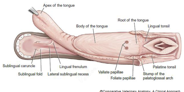
Root of the tongue
Caudal fixed part
also attached to the hyoid apparatus
Body of the tongue
Middle part: Frenulum Linguae(Lingual Frenulum) - Ventral median fold
Apex of the tongue
Rostral free part
Highly muscular nature - vigorous movements.
Median Sulcus, Sublingual Caruncles
Filiform papillae
The most numerous
Cat - Modified and highly keratinized filiform papillae on the rostral 2/3 – dorsal aspect
Conical papillae
The caudal most, oropharyngeal region of the tongue - Dog
Gustatory papillae
These papillae have taste buds associated.
➢ Fungiform papillae
➢ Vallate papillae(Circumvallate)
➢ Foliate papillae
Mucosa of the tongue
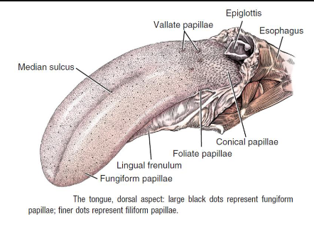
Other papillae
Marginal papillae (Mechanical) – only in young –in suckling
Lenticular Papillae (Mechanical) – Only in Ruminants
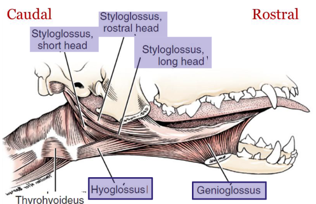
The genioglossus muscle
Depress & Protrude the tongue – Take the tongue out
The hyoglossus muscle
Retract the tongue – Pull the tongue back
The Geniohyoideus muscle
Protrude the tongue – Take the tongue out
Not a tongue muscle but action is related to movement of the tongue
Anatomy of the extrinsic muscles of the tongue

Extrinsic Muscles of the tongue
The genioglossus muscle
The styloglossus muscle
The hyoglossus muscle
The geniohyoideus muscle
Muscles of the tongue: lateral aspect
Intrinsic Muscles of the Tongue(proper lingual muscles)
The superficial longitudinal fibers
The deep longitudinal fibers
The transverse and the perpendicular fiber
Blood Supply to the tongue - Arteries

Blood Supply to the tongue - Veins

Summary of innervation to the tongue

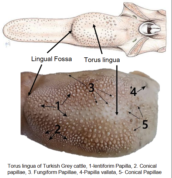
Tongue of the ruminant
The Major prehensile organ in the bovine
The lenticular papillae – Numerous Mechanical papillae on the Torus lingua
Conical papillae – larger than filiform, mainly on torus lingua
Foliate papillae are absent
Ruminant tongue pics
Tongue of the horse
Spatulated – the apex is not pointed; Not a prehensile organ
Distinct Torus lingua and fossa linguae are absent
Usual papillae present on the tongue including foliate papillae
Dorsal lingual cartilage – Hyaline cartilage accompanied by adipose tissue and muscles
Anatomy of the tongue of the horse
Microanatomy of the tongue
Stratified squamous epithelium
Dorsal – Papillae, Ventral - No papillae
Laminal propria submucosa: Lingual salivary glands(Minor)
Lyssa in the dog – fat filled, surrounded dense irregular connective tissue (fibrous rod)
Fungiform and Filiform Papillae, Tongue, Goat.
4. Filiform papilla
5. Fungiform papilla
7. Skeletal muscle

Circumvallate Papilla, Tongue, Goat
1. Core of Connective yissue,
4. Lingual salivary gland
2. papilla of Connective tissue,
5.Stratified squamous epithelium
3. Duct
6. Taste buds

Function of the teeth
Cutting and Tearing (Carnivores)
Grinding and Chewing(Herbivores)
Grasping and Holding (Omnivores and Predators)
Gnawing (Rodents and Some Mammals)
Defense and Fighting
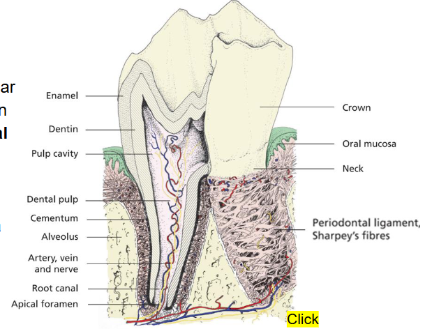
Crown
The visible part of the tooth above the gumline
Shape varies depending on tooth function (e.g., sharp incisors vs. broad molars)
Root
The part of the tooth embedded in the alveolus/socket
Number of roots varies (single, double, multiple)
Neck
The junction between the crown and the root, at the gumline.
Furcation:
The area where the roots diverge in multi-rooted teeth.
Clinical significance: common site for periodontal disease
Enamel
The hardest substance in the body; protects the tooth.
Composition: 96% mineral, mostly hydroxyapatite.
Thickness varies among species and tooth type
Dentin:
Forms the bulk of the tooth; provides support for enamel.
Composition: 70% mineral, 30% organic material and water.
Continues to form throughout life
Pulp
Contains blood vessels, nerves, and connective tissue.
Provides nutrients to the tooth and allows for sensation.
Cementum
Covers the root; attaches the tooth to the periodontal ligament.
Composition: 50% mineral, similar to bone, Important for tooth anchorage
Anatomy of a tooth
Periodontium
Includes the gum/gingiva, alveolar bone(the socket), cementum on the root, the and the periodontal ligament/ membrane → connects the two across the periodontal space
Incisor teeth
typically single-rooted with a sharp incisal edge.
Canine teeth
Large, conical teeth used for piercing and holding prey. Single-rooted with a long root for strong anchorage.
Sexual dimorphism
Premolar teeth
for grinding and shearing food, variable number of roots
Molar teeth
Large, flat teeth used for grinding food, multiple roots
Ruminants teeth
no upper incisors. Instead, the hard palate is modified as a dental pad.
The canine teeth are modified and resemble the incisors, therefore, The canine tooth of domestic ruminants has commonly been counted as a fourth incisor
Dog dental formulation

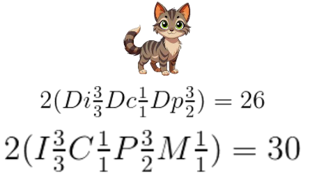
Cat dental formulation
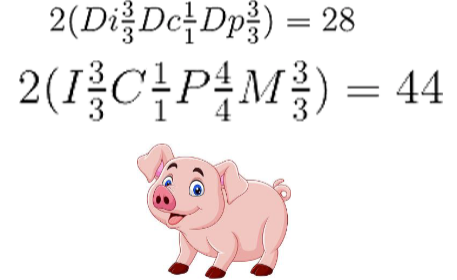
Pig dental formulation
Ruminant dental formulation
