Liver (part 2)
 Call Kai
 Learn
Learn Practice Test
 Spaced Repetition
 Match
Match Flashcards
 Knowt Play
1/31

Earn XP
Description and Tags
- congenital abnormalities & cystic disease of the liver
Name | Mastery | Learn | Test | Matching | Spaced | Call with Kai | Chat |
|---|
No analytics yet
Send a link to your students to track their progress
32 Terms
Agenesis of liver
absence of liver formation
accessory fissures of the liver
rare, caused by infolding of the peritonunem
liver vascular abnormalities
arterial anatomy variation
hepatic vein branch variation
atresia
stricture
valve formation in portal vein
Partial Situs Inversus
only abdominal contents are in reversed positions
liver is at LUQ instead of RUQ
Complete Situs Inversus
both chest and abdominal contents are in reversed positions
Liver → LUQ
Heart → right
Diaphragmatic hernia
an opening in the diaphragm that allows abdominal contents to move up into the thoracic cavity
Riedel lobe
what is it?
common in male or females?
sono appearance?
an extension of the right, inferior lobe of the liver below the lower pole of the right kidney
common with women
normal liver echotexture
normal left lobe
normal AP dimension of right lobe
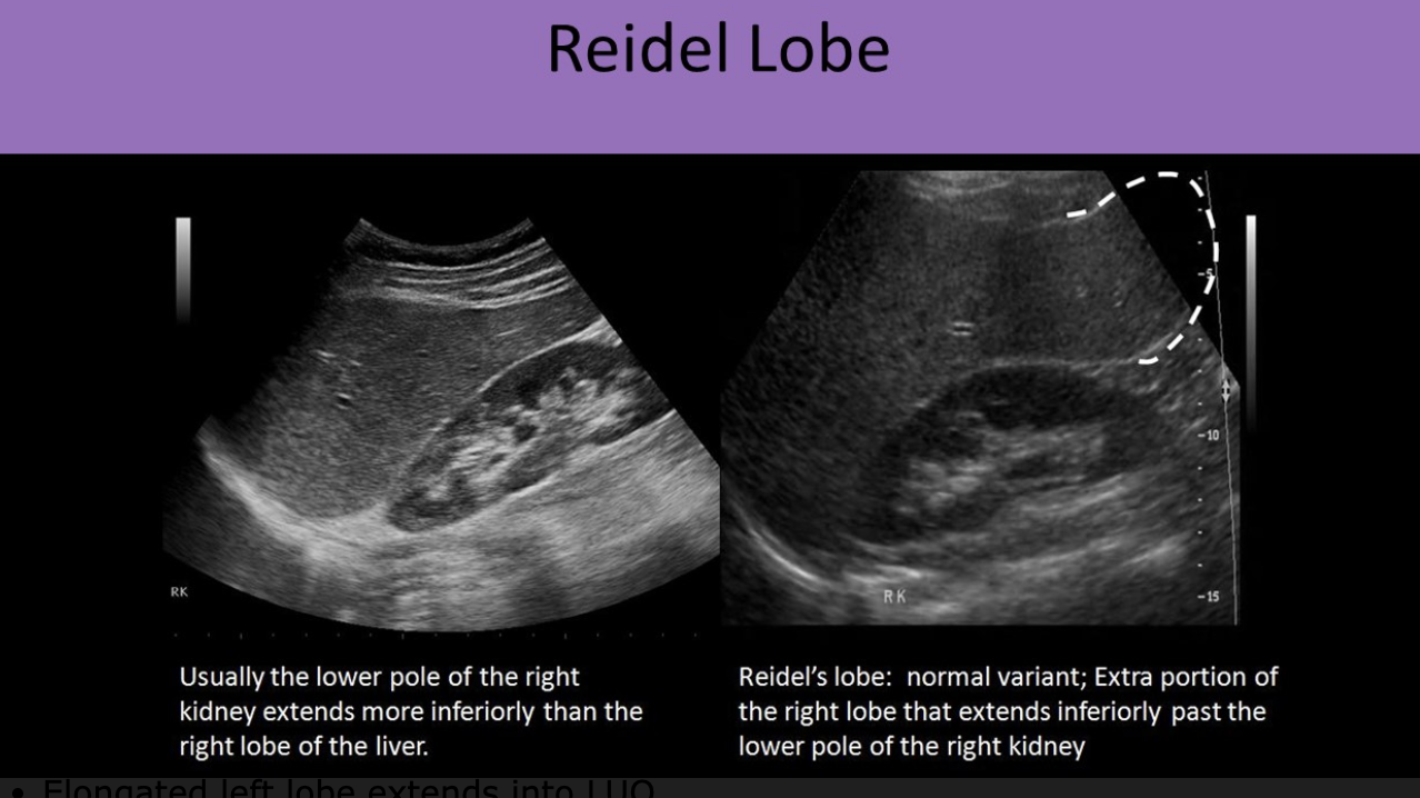
How to differentiate a Riedel Lobe with hepatomegaly?
compare the right lobe with the left lobe.
left lobe normal sized → Riedel
both left + right lobe enlarged → hepatomegaly
measure AP liver dimension of right lobe
if WNL → Riedel
if enlarged → hepatomegaly
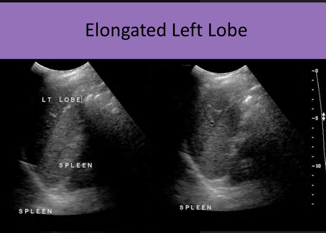
Extended left lobe
where does it extend to?
can be mistaken for what?
sono appearance
elongated left lobe that extends into the LUQ
left lobe can be seen adjacent to medial and superior spleen
left lobe extends anterolateral across AO to the LUQ
can be mistaken for hepatomegaly
normal echotexture
Liver Simple Cysts
simple, anechoic
asymptomatic
common after age 50
simple cysts before age 50
related to polycystic disease and cysts in the kidneys, pancreas and ovaries
aspiration or ablation is performed for symptomatic patients
Liver simple cyst sono appearance
smooth borders
regular, well-cricumscribed walls
anechoic, no internal echoes
posterior enhancement
through transmission

Liver hemorrhagic cysts
blood leaks into an existing cyst
thrombosis forms within the cyst
pain secondary to hemorrhage / infection
low hematocrit levels
Sono appearance of hemorrhaghic cysts
thrombus causes irregular appearnce of internal echoes
fluid/blood levels
septations

Polycystic Liver Disease (PCLD)
what is it?
are LFTs affected?
common with what age?
if pt is under 50 y/o, PCLD is associated with what?
autosomal dominant disorder (one parent has the gene)
multiple cysts that don’t communicate with biliary tree
normal LFTs
common age 30-40
simple cysts before age 50
related to polycystic disease, along with cysts in the kidneys, pancreas, spleen, ovaries
polycystic liver disease sono appearance
focal cystic areas
varied sizes
thin septations
evaluate pancreas, kidney, spleen, ovaries for cysts

biliary hamartomas (von Meyenburg complexes)
focal developmental lesions of the liver
composed of groups of dilated intrahepatic ducts within dense stroma
Biliary hamartomas sono appearance
small nodules
echogenic
causes diffused heterogeneous appearance of liver tissue
multiple, well-defined, solid nodules < 1 cm diameter
large nodules → hypoechoic
echogenic foci with comet tail / ring down artifact
twinkle artifact
associated with congenital hepatic fibrosis, autosomal dominant polycystic disease, cholangiocarcinoma

Fatty Infiltration of the Liver (steatosis)
triglyceride accumulation in the liver cells
reversible
most common cause = obesity
What are the 2 types of Fatty Liver Disease?
causes?
what can they lead to?
what LFTs are affected?
alcoholic fatty liver disease
caused by chronic alcohol intake
can lead to alcoholic hepatitis
increased AST, ALT
non-alcoholic fatty liver disease
caused by diabetes, obesity, TB, cystic fibrosis, starvation, steriod use
can lead to NASH (nonalcoholic steatohepatitis)
mild, moderate increase of AST, ALT
Distribution of Fatty Liver Disease
diffuse replacement = entire liver is fatty
focal fatty sparing = entire liver involved except localized areas
focal fatty replacement = small portion of the liver experiences fatty infiltration
Diffuse infiltration of fatty liver
mild = slight increase in fine echoes, normal visualization of diaphragm and vessel borders
moderate = moderate increase in fine echoes, impaired visualization of the diaphragm and vessel borders
severe = marked increase in fine echoes, poor or non-visualization of the diaphragm and vessel borders
Hepatomegaly > 15.5 cm
Hepatic vasculature is not displaced with fatty replacement, but flow changes can occur
hepatic veins can be narrowed due to fatty liver changes with decreased outflow, loss of pulsatility and respiratory phasicity; monophasic antegrade waveform
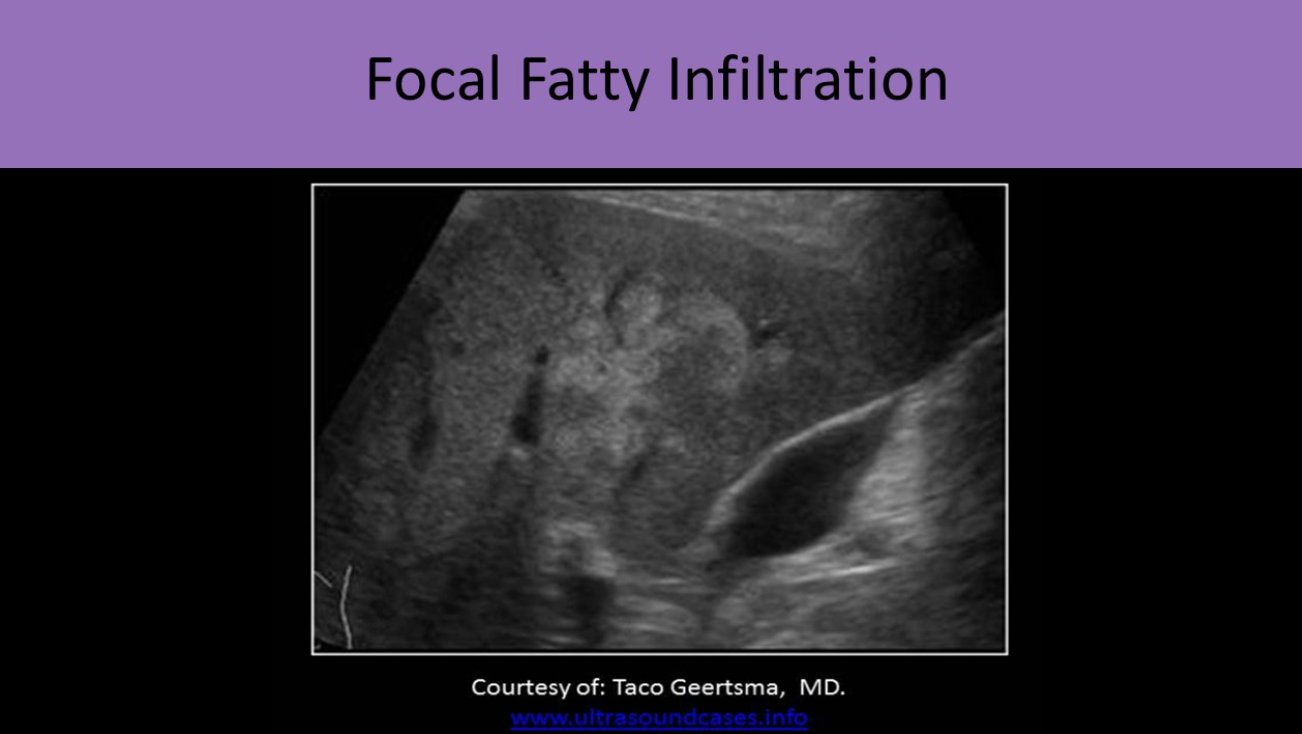
focal infiltration of fatty liver
focal area of increased echogenicity
irregular borders
adjacent to portal structures
looks like hemangiomas
does NOT displace surrounding structures
Focal sparing of fatty liver
focal area of decreased echogenicity with remaining liver tissue diffusely increased in echogenicity
irregular borders
adjacent to GB fossa
similar appearance to malignancy
does NOT displace surrounding structures

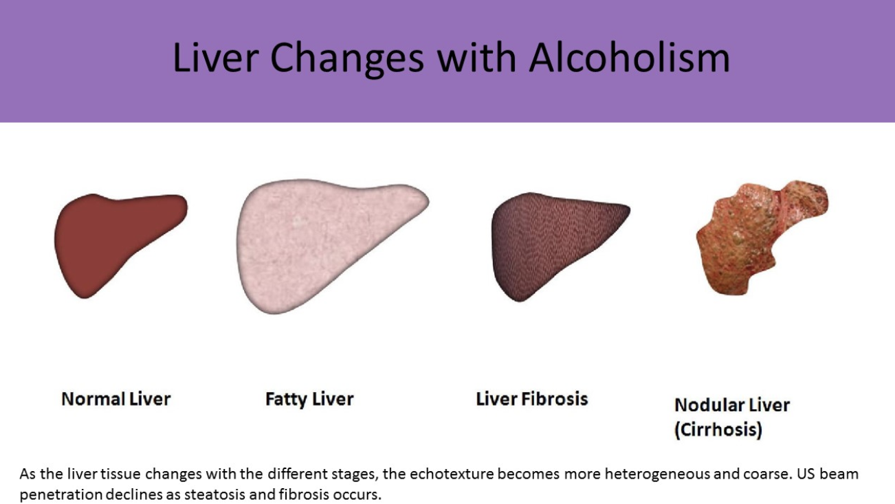
liver changes with alcoholism
normal → steatosis → fibrosis → cirrhosis
liver becomes smaller, increased heterogeneous echotexture and coarse
mild steatosis

moderate steatosis

severe steatosis

How to differentiate betweem a hemangioma and fatty infiltration?
hemangiomas will cause a mass effect and displace surrounding structures, while fatty infiltration will not affect surrounding structures.

Reye Syndrome
what is it and how does it look like on US
how does it develop?
rare disorder that affects children
develops during recovery from a viral infection like chicken pox, flu
associated with taking aspirin during viral infection
sono appearance - looks like steatosis (fatty liver)
hepatomegaly
fatty infiltration
Reye Syndrome Symptoms
what are they?
what can this syndrome result in?
symptoms:
increased cranial pressure
abnl LFTs
excessive vomiting
confusion
delirium
loss of consciousness
seizures
can result in lasting brain damage and/or death
Amyloid disease
deposition of amyloid protein within the vessel walls of various organs which leads to organ failure
Glycogen storage disease
Autosomal recessive disorder
Type 1 most common (von Gierke disease)
Affects liver and skeletal muscle
Labs = hypoglycemia, or abnl LFTs
Excessive storage of glycogen within hepatocytes - hepatomegaly, increased echogenicity, associated with adenomas
Reduced storage - hypoechogenicity
Prominent portal walls
Starry sky (similar to acute hep)