Ophthalmic Disorders
 Call Kai
 Learn
Learn Practice Test
 Spaced Repetition
 Match
Match Flashcards
 Knowt Play
1/133

There's no tags or description
Looks like no tags are added yet.
Name | Mastery | Learn | Test | Matching | Spaced | Call with Kai | Chat |
|---|
No analytics yet
Send a link to your students to track their progress
134 Terms
Strabismus: General
Ocular misalignment
Only one eye fixates on an object
Present in ~4% of children
"Ocular instability of infancy"
Strabismus: Risk Factors
family history, prematurity, refractory error, neuro disorders
esotropia
inward turning of the eye

exotropia
outward turning of the eye

hypertropia
upward deviation of one eye

hypotropia
downward deviation of one eye

-phoria
strabismus that is present only when binocular fusion is
disrupted (i.e. when one is covered)
-tropia
strabismus that is present when there is no
disruption of binocular fusion
strabismus: most common causes
CN palsy, trauma, orbital disease, hyperthyroidism
strabismus: diagnostic tests
Corneal light reflex (aka Hirschberg test)
Cover test (detects tropia)
Cover/uncover test (detects phoria)
strabismus: complications
Amblyopia (in up to half of younger children with strabismus)
Diplopia (in acquired strabismus in patients > 8 years old
strabismus: when to refer?
Constant strabismus at any age
Intermittent manifest strabismus after 4-6 months of age
Persistent esodeviations after 4 months of age
Abnormal corneal light reflex test or cover test
Deviation that changes depending on position of gaze
Diplopia or asthenopia
Parental concern about ocular alignment
strabismus: tx
Nonsurgical: glasses, contacts, occlusion therapy,
visual training exercises
Surgical: recession, resection and transposition of
extraocular muscles
amblyopia: general
Functional reduction in visual acuity caused by abnormal visual development early in life
50% of cases related to strabismus
Predominantly unilateral
Refer to ophthalmology/optometry if suspected
amblyopia: risk factors
prematurity, small for gestational age, first-degree
relative with amblyopia, neurodevelopmental delay
what is the MCC of pediatric visual impairment (1-4% of children)
amblyopia
leukoria
White pupillary reflex
leukocoria: common causes
Retinoblastoma
Cataract
Coats disease
Retinal detachment

RETINOBLASTOMA: general
Most common primary intraocular malignancy of childhood
Typically presents as leukocoria in a child < 5 years
RETINOBLASTOMA: signs and symptoms
Leukocoria
Strabismus
Red, inflamed eye
Decreased vision
RETINOBLASTOMA: tx
Deadly if untreated, but treatment is very successful! (>95% 5-year survival rate)
Treatment involves multidisciplinary approach and may include chemotherapy, surgery, cryotherapy, laser photoablation
RETINITIS PIGMENTOSA: general
degeneration and dysfunction of retina, rods then cones
May occur alone or as part of a syndrome
May be inherited or occur sporadically
what does retinitis pigmentosa cause
progressive loss of night and peripheral vision
retinitis pigmentosa: ophthalmic findings
Attenuation of retinal blood vessels
Waxy pallor of optic disc
Intraretinal pigmentation in a bone-spicule pattern

retinitis pigmentosa: tx
No great treatment currently; experimental therapies underway
conjunctivis: general
Inflammation of the mucous membrane that lines the surface of the eyeball and inner eyelids
Viral, bacterial, or allergic

conjunctivis: transmission
direct contact of contaminated fingers orobjects to other eye or other persons
conjunctivis: symptoms
Foreign body sensation
Scratching / burning
Itching
Photophobia
Sensation of fullness around eyes
viral conjunctivitis: mcc
adenovirus
viral conjunctivitis: general
May or may not be part of viral prodrome followed by adenopathy,
fever, pharyngitis, URI
Usually sequential bilateral disease
Spreads easily

viral conjunctivitis: presentation
Conjunctival injection with watery or mucoserous discharge
± preauricular lymphadenopathy
+/- follicular appearance of tarsal conjunctiva
viral conjunctivitis: tx
No specific treatment
Lasts up to 2 weeks; may get worse before it gets better
Artificial tears and cold compresses may help discomfort
bacterial conjunctivitis: mcc
staph aureus, Strep pneumo, Haemophilus species, Pseudomonas (contact lens wearers), Moraxella
bacterial conjunctivitis: general/clinical presentation
Purulent eye discharge
Eyelid matting
unilateral

bacterial conjunctivitis: tx
Usually self-limited
Lasts 10-14 days if untreated
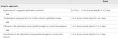
Topical antibiotics may hasten remission- Erythromycin ointment, Polytrim drops
Contact wearers- Ciprofloxacin, Ofloxacin drops
GONOCOCCAL CONJUNCTIVITIS: general
Usually acquired through genital secretions
Ophthalmologic emergency!
Corneal involvement may lead to perforation
Caused by N. gonorrhoeae
GONOCOCCAL CONJUNCTIVITIS: clinical presentation
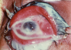
Copious purulent discharge
GONOCOCCAL CONJUNCTIVITIS: diagnosis
Confirm with stained smear and culture of discharge
GONOCOCCAL CONJUNCTIVITIS: tx
Single dose ceftriaxone IM
Also treat for chlamydia- doxycycline
Consider other STIs
CHLAMYDIAL CONJUNCTIVITIS: Trachoma general
chronic keratoconjunctivitis caused by recurrent infection with C. trachomatis
Recurrent episodes causes entropion and trichiasis leading to corneal scarring/opacity
NOT sexually transmitted

Trachoma transmission
secretions, contaminated objects, flies, NOT sexually transmitted
Trachoma treatment
SAFE: surgery, antibiotics, facial cleanliness, environmental improvement
CHLAMYDIAL CONJUNCTIVITIS: Adult inclusion conjunctivitis
Chronic conjunctivitis caused C. trachomatis
associated with genital chlamydia
Usually unilateral follicular conjunctivitis lasting weeks-months that has not responded topical antibiotics
Adult Inclusion Conjunctivitis transmission
spread by autoinoculation from genital infection
Adult Inclusion Conjunctivitis dx
Conjunctival NAAT
Adult inclusion Conjunctivitis tx
Single dose of azithromycin PO or 7 days of doxycycline
ALLERGIC CONJUNCTIVITIS: general
Usually associated with atopy (Eczema, asthma, allergic rhinitis)
May be seasonal (hay fever); usually in spring/summer
Bilateral
ALLERGIC CONJUNCTIVITIS: clinical presentation
Itching
Conjunctival hyperemia and edema (chemosis)
Watery discharge
Cobblestoning of upper tarsal conjunctiva
Bilateral

ALLERGIC CONJUNCTIVITIS: tx
Avoid known allergens, rubbing, contact lenses
Cool compresses, Artificial tears
First line- topical olopatadine or ketotifen
Oral H1 antihistamines
DACRYOCYSTITIS: general
Infection of lacrimal sac secondary to obstruction of nasolacrimal duct
sometimes seen in infants that have a membrane that blocks the duct
DACRYOCYSTITIS: pathogens
strep or staph
DACRYOCYSTITIS: clinical presentation
Tearing/discharge
Inflammation, pain, swelling, tenderness beneath medial canthal tendon in area of lacrimal sac
May be able to express purulent drainage through lacrimal puncta by applying pressure to sac

DACRYOCYSTITIS: dx
usually clinical, imaging if concerned for orbital involvement
DACRYOCYSTITIS: tx
systemic antibiotics, cannot use drops
ophthalmology referral for abscess drainage
KERATOCONJUNCTIVITIS SICCA: general
AKA dry eye disease (DED)
decreased tear production and/or increased evaporation —> unstable tear film
KERATOCONJUNCTIVITIS SICCA: symptoms
bilateral burning/gritty sensation, may find relief immediately after blinking, reflex tearing may occur
KERATOCONJUNCTIVITIS SICCA: risk factors
Advanced age
Sjogren syndrome
Contact lens wear
Certain medications
Dry environments

KERATOCONJUNCTIVITIS SICCA: tx
Artificial tears ± lubricating ointment at night
Warm compresses
Discontinue offending medications if possible
HSV KERATITIS: clinical presentation
Due to reactivation of HSV in trigeminal ganglion
unilateral, decreased corneal sensation, vesicles may occur
Dendritic (branching) corneal ulcer w fluorescein staining

HSV KERATITIS: dx
Slit lamp examination
Fluorescein stain showing branching dendritic lesion
HSV KERATITIS: tx
Topical: Trifluridine drops, ganciclovir gel
Oral: Acyclovir, valacyclovir
*Avoid topical corticosteriods (leave to ophtho)
BACTERIAL KERATITIS: mc pathogens
staph/strep; Pseudomonas aeruginosa with contact wearers
BACTERIAL KERATITIS: risk factors
Contact lens wear (especially overnight)
Corneal trauma
BACTERIAL KERATITIS: clinical presentations
painful red eye, photophobia, ± decreased vision
Central corneal ulcer
+/- hypopyon

BACTERIAL KERATITIS: dx
visual acuity + slit lamp exam + fluorescein staining
BACTERIAL KERATITIS: tx
Urgent ophtho referral, emergency!
Topical fluoroquinolones
HERPES ZOSTER OPHTHALMICUS: general
Herpes zoster frequently involves ophthalmic division of trigeminal nerve
Potentially sight-threatening
HERPES ZOSTER OPHTHALMICUS: clinical presentation
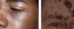
Hutchinson’s sign- vesicles on the tip, side, or root of the nose indicates increased risk of ocular involvement
Painful, unilateral vesicular rash of forehead/upper eyelid that typically does not cross midline
HERPES ZOSTER OPHTHALMICUS: tx
Urgent ophtho referral
Acyclovir, valacyclovir, famciclovir within 72 hrs of onset
PERIORBITAL CELLULITIS: general
AKA preseptal cellulitis
Infection of soft tissues anterior to orbital septum
May follow sinusitis, local skin infection, trauma, insect bite
PERIORBITAL CELLULITIS: clinical presentation
Unilateral eyelid swelling and erythema
Does NOT affect visual acuity or extraocular movements, NO proptosis

PERIORBITAL CELLULITIS: tx
Mild- Amoxicillin/clavulanate, reassess within 24-48 hrs
Consider admission in children < 1 year old, severely ill, or unsure if orbit is involved
ORBITAL CELLULITIS: general
Emergency!
Infection posterior to orbital septum (fat and extraocular muscles)
Typically caused by sinusitis
ORBITAL CELLULITIS: clinical presentation
Eyelid erythema ± Fever
Proptosis
Painful or restricted extraocular movements
Decreased visual acuity

ORBITAL CELLULITIS: dx
CT orbits and sinuses with IV contrast
ORBITAL CELLULITIS: tx
Hospitalization and Urgent broad-spectrum IV abx
Urgent optho consult
may require surgical drainage
ORBITAL CELLULITIS: complications
Cavernous sinus thrombosis
Intracranial extension
Vision loss
Orbital abscess
BLEPHARITIS: general
Chronic bilateral inflammatory condition of lid margins
Common cause of recurrent conjunctivitis
BLEPHARITIS: anterior
scales/crust at the eyelashes
associated with staph and seborrheic dermatitis
BLEPHARITIS: posterior
results from inflammation of meibomian glands (oil)
BLEPHARITIS: clinical presentation
gritty sensation, burning, itching
eyelid crusting especially on awakening
can contribute to dry eye
BLEPHARITIS: tx
First line: warm compresses, eyelid cleansing with baby shampoo,
Persistent anterior: erythromycin ointment
Refer for severe
CHALAZION: general
Sterile obstruction/inflammation of a meibomian gland
Associated with blepharitis/meibomian gland dysfunction
CHALAZION: clinical presentation
Hard, painless nodule on upper or lower lid away from lash line
May blur vision

CHALAZION: treatment
Warm compresses + gentle lid massage
Refer if persistent, optho can do steroid injection or I&C
Recurrent in same location needs to be biopsied for malignancy
HORDEOLUM: general
AKA stye
Acute infection/abscess of either meibomian gland (internal) or gland of Zeis (external)
HORDEOLUM: mc
Staph aureus
HORDEOLUM: clinical presentation
Localized red, swollen, tender eyelid nodule

HORDEOLUM: tx
Warm compresses ± gentle massage
Refer if large/persistent
CATARACTS: general
Opacities of crystalline lens
Usually bilateral
Leading cause of blindness worldwide
CATARACTS: risk factors
MCC: age
DM, smoking, steroid use, trauma, UV exposure
CATARACTS: clinical presentation
Progressive, painless blurring of vision
Flare in bright lights or night driving, faded colors, diminished red reflex

CATARACTS: tx
Mild- optimize with glasses
Surgery (improves visual acuity in 95% of cases)
Glaucoma risk factors
Older age, family hx, Black/African, Hispanic/Latino
Primary open angle glaucoma
Chronic optic neuropathy associated with elevated IOP due to reduced drainage of aqueous fluid through trabecular meshwork

Primary open angle glaucoma- clinical presentation
gradual peripheral vision loss, painless
Primary open angle glaucoma- dx
cupping of optic disc on fundoscopy
assess IOP with tonometry + gonioscopy
Primary open angle glaucoma- tx
First line- prostaglandin analog (latanoprost)
Acute angle closure glaucoma
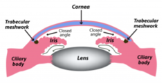
Sudden obstruction of flow of aqueous fluid into anterior chamber
Emergency!! permanent vision loss if untreated
quick pupil dilation can trigger it
Acute angle closure glaucoma- clinical presentation
severe unilateral eye pain
blurred vision, headache, N/V
poorly reactive pupil