MYCOVIRO
 Call Kai
 Learn
 Practice Test
 Spaced Repetition
 Match
 Flashcards
 Knowt Play
1/83

There's no tags or description
Looks like no tags are added yet.
84 Terms
Yeast and Molds
two types of fungi
Yeast
Single vegitative cells
smooth creamy bacteria like colony
No aerial hyphae
Budding Fission
Mode of Reproduction of Yeast
Blastoconidium
Product of budding fission in yeast
Molds
Fuzzy/ Wooly appearance
Mycelia
Other term for Molds
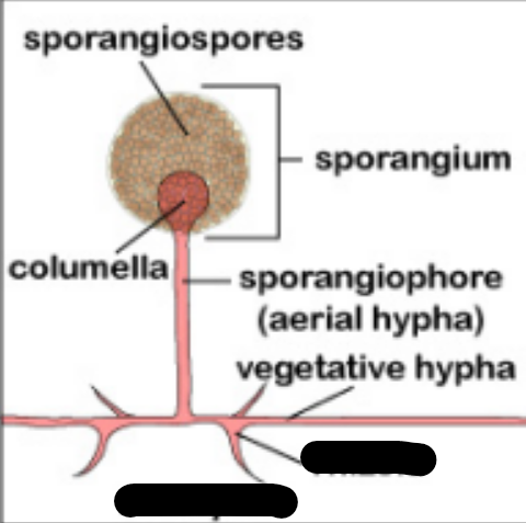
Hyphae
Branching cylindric tubules var in diameter from 2 - 10 um
Mycelium
Mass of intertwined hyphae that accumulates during active growth
Aerial Mycelia/ Hyphae
Extends to surface
Support the reproductive structures (CONIDIA)
Vegetative Mycelia/ Hyphae
Extends downwards penetrates the supporting medium
Anchors and absorbs nutrients
Ontogeny and Morphology
Most helpful phenotypic features of molds
Antler/ Hyphae/ Favic Chandelier
Branching tips
“Moose Antlers”
Racquet Hyphae
Enlarged, club shaped areas
Spiral Hyphae
tight coils of molds
Pecrinate Bodies
Comb like structures
Rhizoids
Root like structures
Septate Hyphae
Frequent cross wallss perpendicular to the outer walls of hyphae
Sparsely Septate
Few cross walls iregular intervals
Aseptate
Absence of seperations
Sparsely septate
Antler Hyphae / Favic Chandelier

Racquet Hyphae

Spiral Hyphae

Pectinate Bodies

Rhizoids
Hyaline / Moniliaceous hyphae
non-pigmented, lightly pigmented
Phaeoid / Dematiaceous hyphae
darkly pigmented
Fontana-Masson stain
Stains specifically melanin
Brown
Phaeoid hyphae on Fontana-Masson stain
DIMORPHISM
Ability of a fungi to exist in two forms depending on growth condition
Yeast phase
Spherule, Tissue phase
In vivo, or 37°C body temperature, increased CO2
Growth condition yeast phase
Mold phase
Growth condition: 22° - 25°C room temperature, ambient air
POLYMORPHISM
Both yeast and mold form in the same culture
SUPERFICIAL MYCOSES
Confined to outermost layer of skin or hair No tissue response, No inflammatory reaction
MALASSEZIA FURFUR
Chronic mild superficial infection
Patchy, scaling lesions
Varying pigmentation
Sites of infection: chest, upper back, face arms, trunks, abdomen
Pityriasis versicolor / Tinea versicolor Folliculitis, Seborrheic dermatitis (Dandruff)
Disease of M. furfur
Calcofluor white
Staining agent for M. furfur
M. furfur
Moist, smooth, cream colored colonies
M. furfur

PIEDRAIA HORTAE
Nodular infection on the hair of the scalp; Hard, brown or black gritty nodules
Black Piedra
Disease of Piedraia hortae
TRICHOSPORON SPP.
Larger, softer, yellowish nodules on the hairs. May also occur on, beard, pubic region and axillary
White Piedra
Disease for Trchosporon spp.
LPCB stain
Stain for Trichosporon spp.
TRICHOSPORON SPP.
White Piedra
Straw to cream colored, smooth or wrinkled, moist, creamy or velvety
TRICHOSPORON SPP.
White Piedra
Carbohydrate non-fermenter
Potassium nitrate utilizer
Urease positive
HORTAEA WERNECKII
Lesions appear as a dark (brown to black) discoloration often on the palm.
Tinea nigra/ Tinea nigra palmaris
Hortaea weneckii disease
HORTAEA WERNECKII
Branched, septate hyphae and budding yeast cells with melanized cell walls
Shiny, moist, yeast-like colonies, start with brownish coloration olive greenish black
Malassezia furfur
Piedraia hortae
Trichosporon spp.
Hortaea werneckii
Superficial Mycoses
Cutaenous Mycoses
Dermatophytosis fungal disease of the keratinized tissue of humans
keratinophilic
Geophilic
Soil causative agent Cutaenous Mycoses
Zoophilc
Animal causative agent Cutaenous Mycoses
Anthropophilic
Humans causative agent Cutaenous Mycoses
Trichophyton
infects hair, skin and nails
Microsporum
infects hair and skin
Epidermophyton
infects nails and skin
2 weeks at 25°C on Sabouraud’s dextrose agar
Growth conditions Cutaneous Mycoses
MICROSPORUM SPP.
Multicellular macroconidia with echinulate walls
Infect only hair and skin.
MICROSPORUM CANIS
(Zoophilic) Colony with a white cottony surface and a deep yellow color on the reverse

MICROSPORUM GYPSEUM
(Geophilic) Tan, powdery colony; Most common geophilic species causing human infections.

Microcondidia, Microsporum canis
Abundant, maintained in cultures even after serial transfer
Macroconidia, Microsporum canis
Spindle shaped, echinulate, tapered ends, Thick walled
Microcondidia, Microsporum gypseum
Clavate, sessile
Macrocondidia, Microsporum gypseum
Cigar-shaped, rounded ends Thin walled
MICROSPORUM AUDOUINII
Anthropophilic
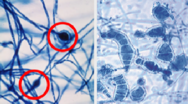
Microconidia Microsporum audouinii
Rare ▪ Chlamydospores
Macroconidia Microsporum audouinii
Odd shaped, chlamydoconidia-like swellings
EPIDERMOPHYTON FLOCCOSUM
Infects nails and skin; colonies are usually flat and velvety with a tan to olive-green tinge

Macroconidia Epidermophyton floccosum
Smooth-walled, club shaped
2-4 celled;
Singly or in clusters
TRICHOPHYTON SPP
May infect hair, skin, or nails.
Cylindric, smooth-walled macroconidia and characteristic microconidia
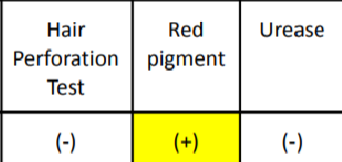
In vitro hair perforation
Laboratory Diagnosis for Trichopython spp.
T. rubrum
Pencil Shaped -- Macroconidia
Tear shaped - Microconidia
T. mentagrophytes
Cigar shaped -- Macroconidia
Grape like cluster - Microconidia

TRICHOPHYTON TONSURANS
Flat, powdery to velvety colony on the surface that becomes reddish brown on reverse

Microconidia, Tricophyton tonsurans
Abundant; mostly elongated
Tear-shaped or Balloon-shaped
TRICHOPHYTON VERRUCOSUM
Glabrous to velvety white colonies; rare strains produce yellow- brown color

Macroconidia, TRICHOPHYTON VERRUCOSUM
3-5 celled with rat-tail end
TRICHOPHYTON MENTAGROPHYTES
Cottony to granular
Microconidia TRICHOPHYTON MENTAGROPHYTES
Globose, teardrop shaped ▪ Grape-like clusters
TRICHOPHYTON RUBRUM
Deep red, nondiffusible pigment when viewed from the reverse side of the colony.
Trichophyton, Epidermophyton
Causative agent for Tinea Pedis “Athlete’s Foot”
Tinea Pedis “Athlete’s Foot” / Moccasin Foot
MOT: Contact with infected skin scales
Affected areas: Soles of the foot, toe web
Tinea Unguium (Onychomycosis)
Infection of the nails caused by dermatophytes. The most difficult to treat (oral antibiotics or surgical removal of the nail)