Lab 3 Blood supply
 Call Kai
 Learn
Learn Practice Test
 Spaced Repetition
 Match
Match Flashcards
 Knowt Play
1/53

There's no tags or description
Looks like no tags are added yet.
Name | Mastery | Learn | Test | Matching | Spaced | Call with Kai | Chat |
|---|
No analytics yet
Send a link to your students to track their progress
54 Terms

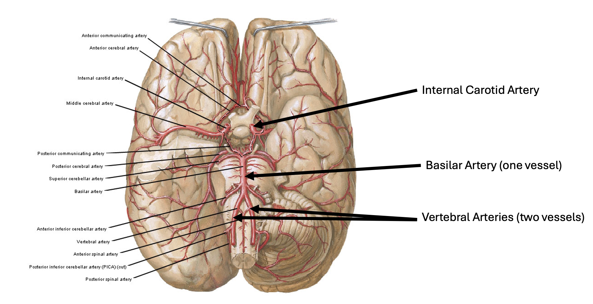
Blue rectangle structure?
Internal carotid artery


Structures top to bottom
Basilar artery
Left and right vertebral artery
Internal carotid artery


Structures top to bottom
Internal carotid artery
Basilar artery
Vertebral arteries

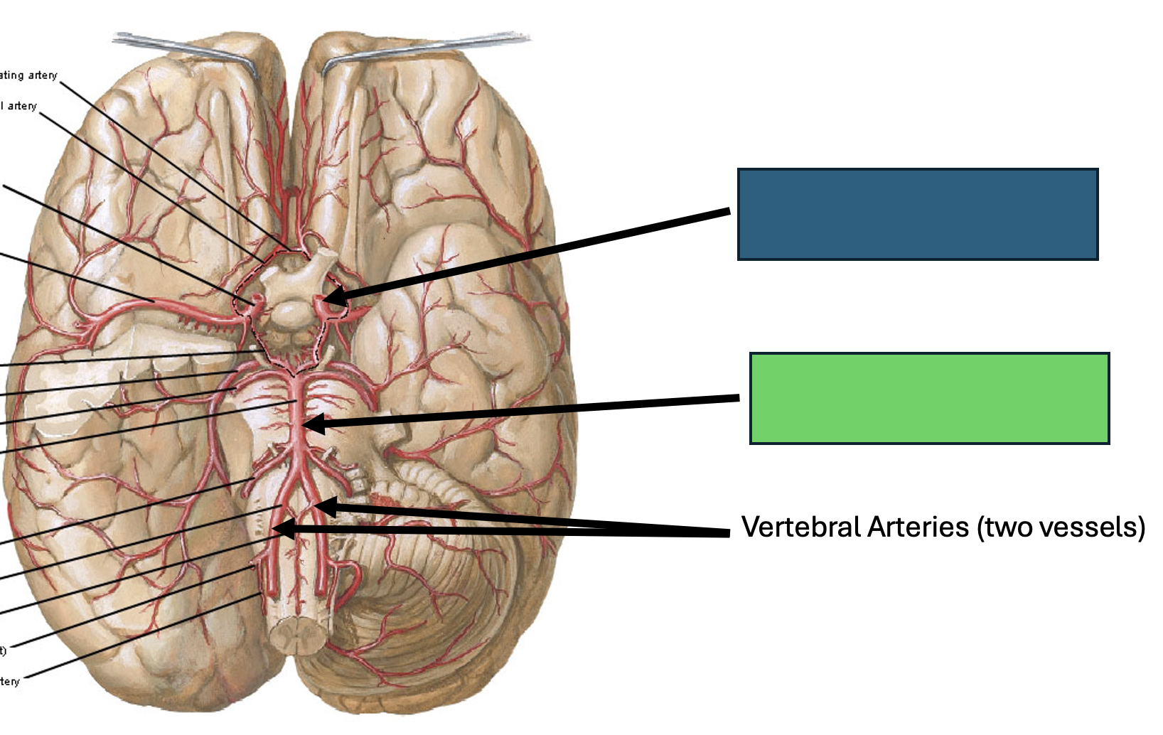
Green rectangle structure?
Basilar artery


Orange rectange structure?
Vertebral arteries (2 vessels)
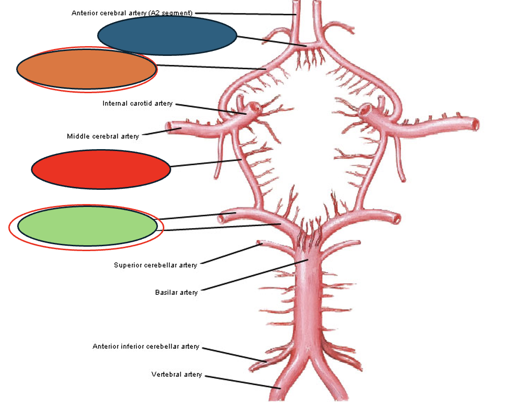
Circle of Willis structures: Blue circle
Anterior communicating artery


Orange circle Circle structure
Anterior cerebral artery (A1 segment)


Red Circle structure
Posterior communicating artery
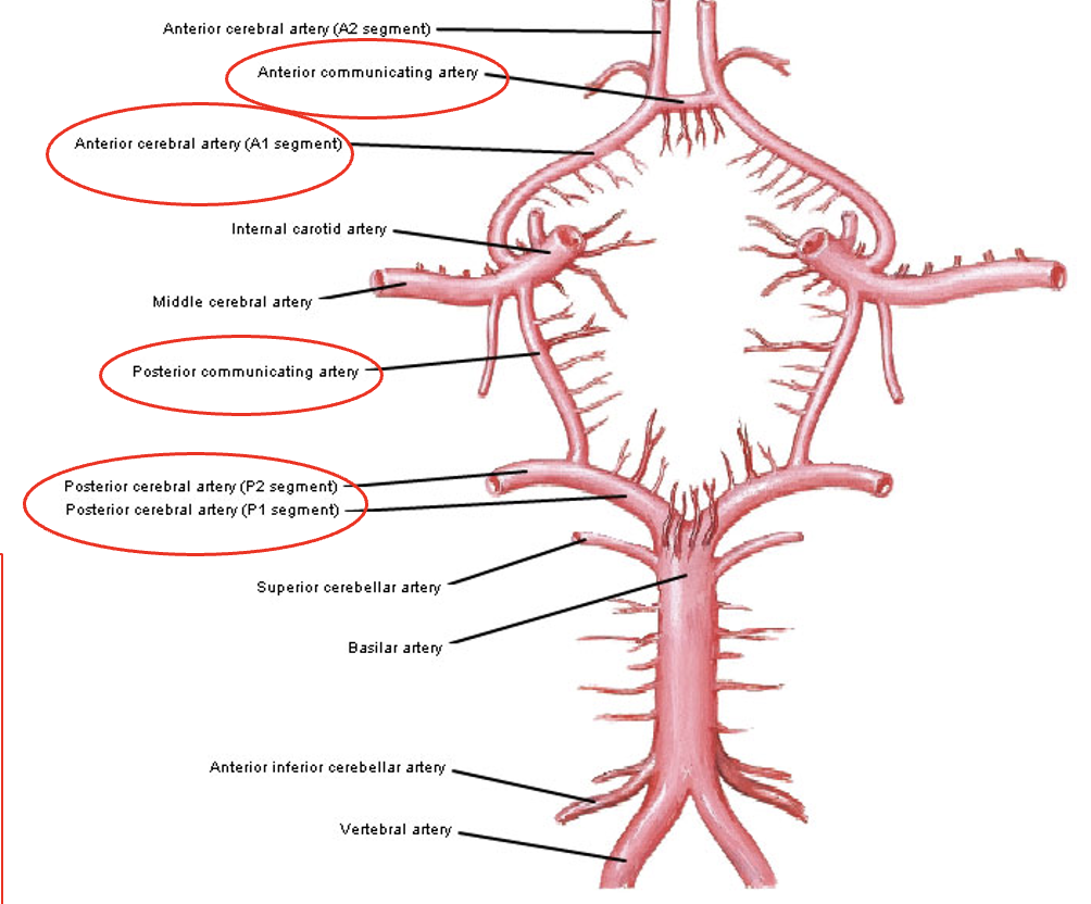

Green Circle structure(s)
Posterior cerebral artery (P2 segment)
Posterior cerebral artery (P1 segment)


Branches of…blank, label structures 1 - 5
Internal Carotid Artery Branches
Opthalmic artery
Anterior Cerebral artery
Anterior communicating artery
Middle cerebral artery
Posterior communicating artery
Opthalmic artery funciton
small branch to the orbit of the eye

Anterior cerebral artery supplies…
medial frontal/parietal lobes

Anterior Communicating artery connects…
both anterior cerebral arteries

Middle cerebral artery supplies out…
lateral frontal, parietal, and superior portions of temporal lobe

Posterior Communicating artery connects which 2 structures to each other
Posterior Communicating artery connects internal carotid artery to posterior cerebral artery (#5)

MOTOR & SENSORY homunclus (Pre + Post Central Gyrus) is supplied by…
Lateral side (Lower extrem): middle cerebral artery
Medial side (Face + trunk + Upper extrem): anterior cerebral artery


Overlap/small arteriolar connections that link the terminal branches of the major cerebral arteries
leptomeningeal anastomosis (Watershed region)

Structures 1 - 2
Anterior inferior cerebellar artery
Labyrinthe artery
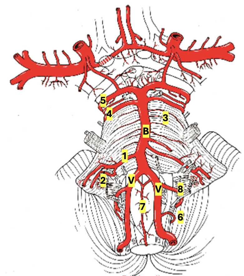
Structures 3 - 4, B
Pontine arteries
Superior cerebellar artery
Basilar artery
Structures 5 - 6, V
Posterior Cerebral artery (connected to anterior circulation via Pcomm a.)
Posterior Spinal artery
2 vertebral arteries

Structures 7 - 8
Anterior Spinal artery (single vessel)
Post. Inferior Cerebellar artery:
Anterior inferior cerebellar artery function
supplies ventral cerebellum and dorsolateral mid-lower pons
Labyrinthe artery function
Difficult to find, can be a branch off the AICA, serves inner ear
Pontine branches (paramedian, short/long circumferential)
Blood supply to the midline pons via paramedian branches, and the lateral/dorsal pons via the short/long circumferential aa.
Superior cerebellar artery function
supplies superior cerebellum, midbrain structures, superior & middle cerebellar peduncles

Posterior Cerebral artery function (#5)
Supplies cerebral hemispheres: serves midbrain, hypothalamus, thalamus, occipital lobe (visual cortex)/inferior temporal gyrus
Connected to anterior circulation via posterior communicating artery


Posterior spinal artery (2) function (#6)
blood supply to dorsolateral aspect of spinal cord and dorsal caudal medulla
supplies posterior of spinal cord
Each spinal artery supplies 1/6 of spinal cord, with both spinal arteries, supplies 1/3 of spinal cord

Anterior Spinal artery #7
supplies anterior 2/3s of spinal cord and ventral medulla

Posterior Inferior Cerebellar artery #8
Supplies inferior cerebellum and lateral aspect of medulla
How many total pairs of spinal nerves?
31 pairs of spinal nerves
How many pairs of cervical nerves
8 cervical pairs
How many pairs of thoracic nerves
12 thoracic pairs
How many pairs of lumbar nerves
5 lumbar
How many pairs of sacral nerves
5 sacral
How many pairs of coccygeal nerves
1 pair
conus medullaris
tapered end of the spinal cord, L1-2 level
Cauda equina
bundle of spinal nerves which exit the conus
Dural sac
Central canal
Dorsal horn
Ventral horn

intermediate gray
Lateral horn

Label A - F
A: dorsal root
B: dorsal root ganglion
C: Ventral root
D: spinal nerve
E: dorsal rami
F: ventral rami
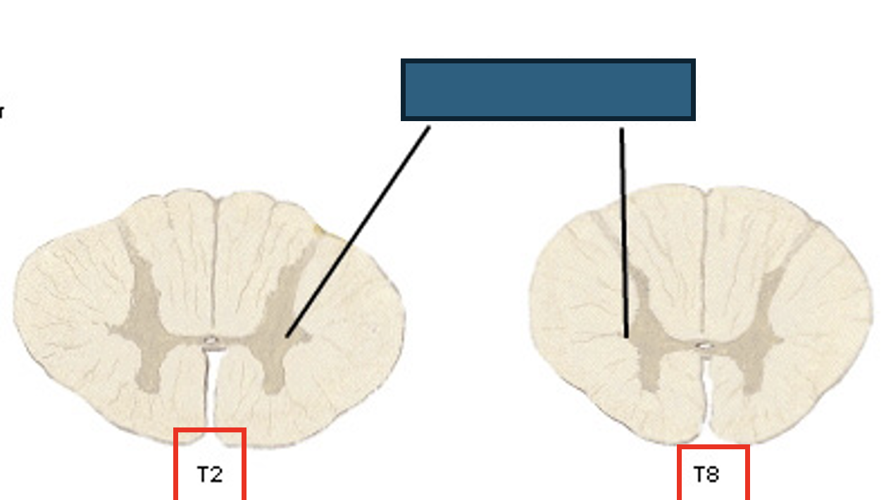
Intermediate Zone
Intermediolateral cell column: a group of neuron cell bodies in the spinal cord that controls the sympathetic and parasympathetic (autonomic) nervous systems
Interneurons
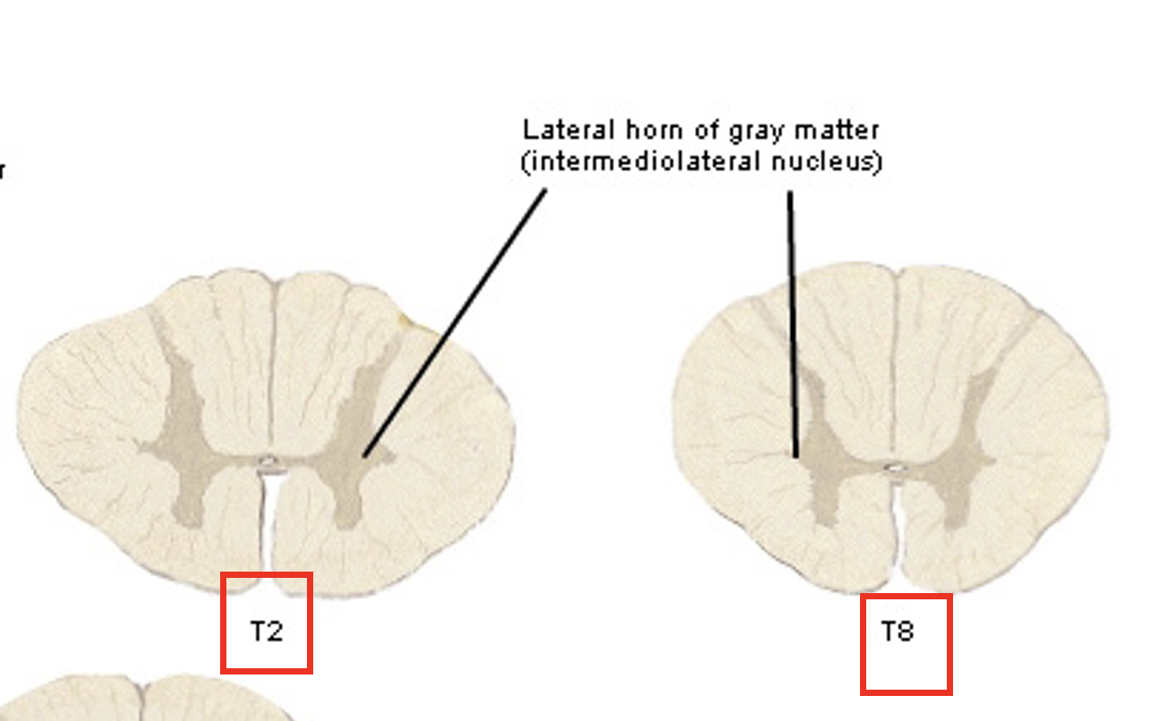
Ventral Horn
Nuclei Motor Nuclei (Medial/Lateral)
Interneurons

Structures of blue, green, and red?
Blue: dorsal column
Green: lateral column
Red: ventral column


Blue and red structure?
Blue = Gracile fasciculus: carries LE sensory
Red = Cuneate fasciculus: carries UE sensory

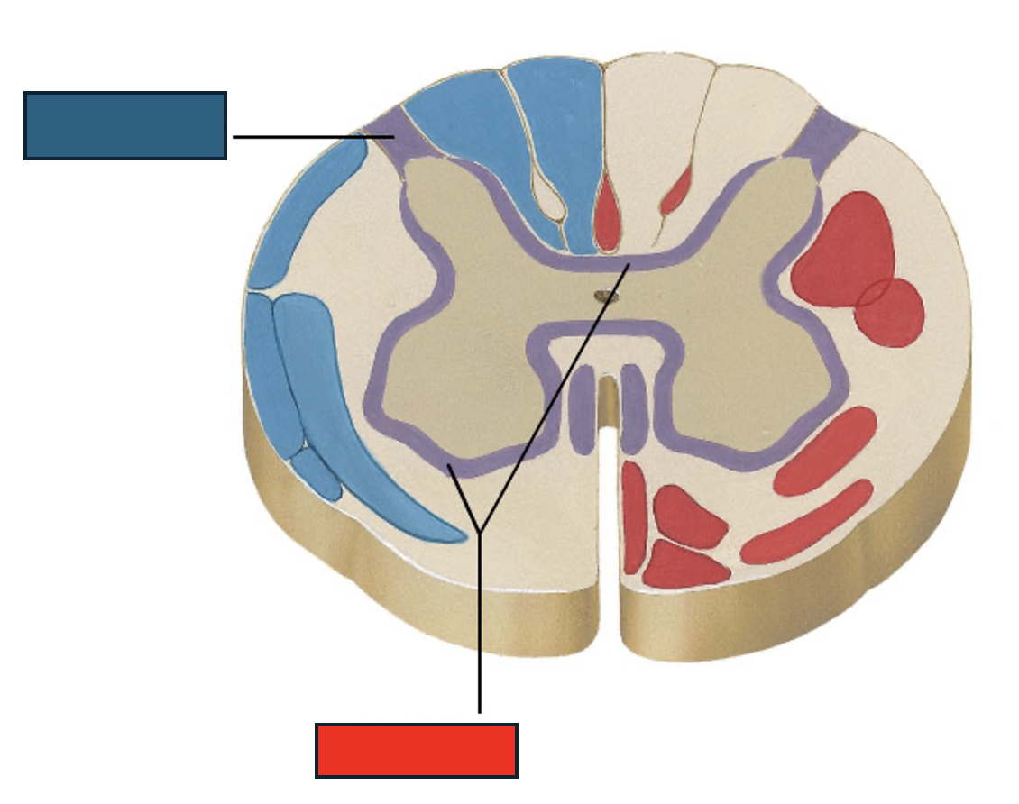
Blue and red structures
Blue: Lissauer’s tract: sensory fibers, pain and temp, ascend or descend 1-2 level of the cord before synapsing in the dorsal horn gray matter.
Red: Propriospinal tract: surrounds gray matter, connects & communicates between spinal cord levels.


Label the number of these Cranial Nerves

Cranial nerves 1 - 3
Olfactory nerve
Optic nerve
Oculomotor nerve
Cranial Nerves 4- 6
Trochlear nerve
Trigeminal nerve
Abducens
Cranial nerves 7 - 9
Facial motor to muscles
Vestibulomotor
Glossopharyngeal:
Cranial nerves 10 - 12
Vagus nerve
Spinal accessory nerve
Hypoglossal nerve