Case 9: John Fumer - COPD
 Call Kai
 Learn
Learn Practice Test
 Spaced Repetition
 Match
Match Flashcards
 Knowt Play
1/38

There's no tags or description
Looks like no tags are added yet.
Name | Mastery | Learn | Test | Matching | Spaced | Call with Kai | Chat |
|---|
No analytics yet
Send a link to your students to track their progress
39 Terms
COPD: Description
Lung condition with chronic resp symptoms and airflow obstruction from airway and alveoli abnormalities
COPD: Types
Chronic Bronchitis (Blue Bloater)
Emphysema (Pink Puffer)
Alpha-1 Antitrypsin Deficiency (AATD)
COPD: Chronic Bronchitis
Airway inflammation + occlusion from mucus
Chronic productive cough
Hypoxemia, hypercapnia, cor pulmonale (right ventricle failure from lung pathology)
COPD: Emphysema
Alveolar destruction decreasing elastic lung recoil
Increase ventilation, normal PO2
COPD: AATD
Genetic predisposition to COPD
AAT: Protein from liver inhibiting elastase in lungs
COPD: Epidemiology
Risk Factors:
Particle exposure (smoking, pollution)
Genetics and fam history
Older age
Low SES
Infections
COPD: Etiology
Main: Exposure to smoke and pollution
Epigenetic factors
Exacerbations from infection
COPD: Pathogenesis
Increase inflammatory cells in airways, lung parenchyma, and pulmonary vessels = Induce chemotaxis, inflammation, and structural changes
Structural Changes:
Smooth muscle hyperplasia = Small airway + pulmonary vasculature narrowing
Proteases/elastases destroy elastin
ROS destroy alveolar attachments
Oxidative stress and proteases in lungs = Exacerbate inflammation
Impact lung function
Airway narrowing = Decrease airflow
Decrease elastic recoil = Lung hyperinflation
Alveolar destruction + Blood vessel remodelling = Decrease gas exchange
Pulmonary vasculature narrowing = Pulmonary hypertension
Progression
Inflammatory mucus occlude small airways
Inflammatory infiltrates in small airway walls (edema)
COPD: Investigations
Spirometry:
FEV1/FVC < 70%
Low FEV1
Normal/low FVC
PFT:
Body Plethysmography: High TLC, FRC, RV
Diffusing Capacity: Low DLCO
Serum AAT Level: Screening
Resp failure tests
COPD: Clinical Manifestations
Chronic productive cough
Dyspnea
Pursed lip breathing (emphysema)
Tachypnea
Long expiratory phase
Wheezing
Cyanosis (hypoxemia)
COPD: Treatment/Management
Decrease exposure to risk factors (smoking cessation)
Inhaled bronchodilators
GOLD severity
LABA, LABA + LAMA, + ICS
COPD Effects on Alveoli Physiology
Emphysema:
Alveolar wall destruction increases compliance (low elastic recoil) = Decrease expiratory force = Air trapping + hyperinflation
Supporting lung parenchyma destruction = Airway collapse

COPD Effects on Immune System
Innate:
Increase alveolar macrophages and neutrophils
Decrease phagocytosis
Increase pro-inflammatory phenotype
Epithelial and dendritic cells release cytokines and chemokines
Adaptive:
Increase T-cells
Increase B-cell production of autoantibodies against lung antigens
Gas Transfer
Gas movement/diffusion across alveolar-capillary membrane
Depend on:
Pressure gradient (O2 and CO2 move high to low pressure)
Membrane SA and thickness (Large SA and thin membrane = Easier)
V/Q match
COPD Effects on Gas Transfer
Emphysema: Decrease membrane SA and increase thickness
Alveolar wall and capillary destruction = Decrease SA
Chronic Bronchitis and Emphysema: V/Q mismatch
Non-uniform decrease in ventilation (low and high V/Q in lungs = Heterogenous regions)
Low V/Q (low V) from mucus plug and edema
High V/Q (low Q) from capillary destruction
Dead Space
Ventilated air not participating in gas exchange (not reaching perfused alveoli)
Anatomic: Volume in conducting airways
Alveolar: Volume in alveoli ventilated but not perfused (embolism)
Physiological: Anatomic + alveolar
Bohr Enghoff equation
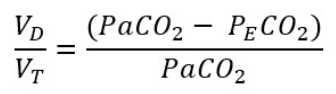
Dead Space: Bohr Enghoff Equation
Calculate physiological dead space (VD) relative to tidal volume (VT)
PECO2: CO2 in expired air
COPD Effects on Dead Space
Decrease expiration (air trapping) = Increase dead space = Less perfusion (no gas exchange) = High V/Q
Emphysema:
Alveolar-capillary unit and small airway destruction = Ventilated air not perfused (no capillaries) = Increase dead space
Chronic Bronchitis:
Airway narrowing + destruction = Air trapping + uneven ventilation = Increase dead space
Venous Admixture
Deoxygenated blood mixing with oxygenated blood in pulmonary circulation to decrease arterial PO2
CO passing through lungs without gas exchange (shunt)
Increase A-a ratio
Low V/Q
Cause hypoxemia

COPD Effects on Venous Admixture
Increased from V/Q mismatch
Airway + vascular changes = Poor ventilation, normal perfusion = Physiological shunt
Spirometry
Measure exhaled volume vs time during forced exhalation
Spirometry: FEV1
Forced expiratory volume in 1 sec
Spirometry: FVC
Forced vital capacity
Spirometry in COPD
Decreased expiratory force = Low FEV1/FVC < 0.7
Lung Volumes
Measure volume in lungs before and after expiration
Lung Volumes: VC
Vital capacity
Measured with spirometry
Lung Volumes: TLC, RV, FRC
Total lung capacity, residual volume, functional residual capacity
Measured with dilution tests and body plethysmography
Lung Volumes: Dilution Tests
Known volume + conc of inert gas inhaled
Inert gas diluted by gas in lungs
Conc of exhaled gas = Initial volume in lungs
Lung Volumes: Body Plethysmography
Compress and expand gas in thorax in airtight box
Boyle’s law calculates gas volumes
Lung Volumes in COPD
Gas trapping + Hyperinflation = Increase RV and FRC (+ TLC in emphysema), Decrease VC

Diffusing Capacity (DLCO)
Measure gas exchange rate from alveolus to Hb in capillary
Small [CO] inhaled → Measure [CO] during expiration
DLCO in COPD
Emphysema:
Decrease gas exchange units = Decrease DLCO
Chronic Bronchitis:
Airway changes (no gas exchange unit changes) = Normal DLCO
Resp Muscle Strength Tests
Measure inspiratory and expiratory pressures
Max inspiratory pressure (MIP)
Max expiratory pressure (MEP)
Resp Muscle Strength: MIP
Inspiratory muscle strength
Exhale to RV and suck on tube at max effort
Record greatest neg pressure
Resp Muscle Strength: MEP
Expiratory muscle strength
Max inspiration and exhale in tube at max effort
Record greatest pos pressure
Resp Muscle Strength in COPD
Hyperinflation pushes muscles into suboptimal force-generating positions = Decrease muscle contraction force
Ex: Chronic diaphragm depression
Smoking Pack-Year
Measure lifetime smoking exposure
# packs per day x # years smoked
Smoking Health Effects
Impair resp ciliary function
Shorten cilia + decrease beating frequency
Mucus buildup
Increase bronchial mucus glands (size + number)
Excess number
Decrease airway lumen diameter
Impair resp immune system
Increase inflammatory cells and mediators
Inhibit macrophages
Inhibit anti-protease/elastase
Release ROS
Oxidative stress damage tissues
Increase CO
High Hb affinity = Shift dissociation curve left = Prevent O2 delivery
Smoking Cessation (5 A’s)
Help up to 67%
Ask: About tobacco use
Advise: Encourage to quit
Assess: Willingness to quit + dependence
Assist: Help users quit (plan, pharm, behaviour support)
Arrange: Follow-up care and support