Module #9 Intro to the Mesoderm
 Call Kai
 Learn
 Practice Test
 Spaced Repetition
 Match
 Flashcards
 Knowt Play
1/53

There's no tags or description
Looks like no tags are added yet.
54 Terms
What is a very particular characteristic of Xenopus gastrulation regarding the germ layers?
This particular kind of gastrulation puts the mesoderm in very close contact with the ectodermal layer.
How do Xenopus embryos compare to human development, and what is a more frequently used model organism?
Xenopus embryos are not as similar to us. One of the most frequently used model organisms (that is more similar) is the chicken.
In the context of the chicken embryo, what are the primary components of the egg and the "beginning of the beginning" of the embryo?
The entire structure is the cell, containing the shell, the yolk, and the blastodisc. The blastodisc represents the initial division of the cytoplasm of the embryo.
How does the rotation of the egg in the oviduct affect the blastodisc and embryo orientation?
The eggs continuously rotate in the oviduct, causing the blastodisc to always be tilted of rotation. The area towards the ventral side is what will become the posterior
Describe the initial cleavage in chicken embryos and the resulting primitive structure.
Chickens undergo meroblastic discoidal cleavage, which occurs only at the top of the embryo. This process forms the blastoderm (the most primitive layer)
The space between the yolk and the blastoderm is the
subgerminal space

Distinguish between the Area pellucida and the Area opaca in the blastodisc.
The inside area; it is transparent because it is formed by only two layers of cells.
Area opaca: The outer, darker layer; it has many more cells and more physical integrity.

What is the epiblast, and how does the hypoblast relate to it during early development?
The epiblast is the surface layer of cells. In contraposition is the hypoblast, which consists of cells that divide and fill the gap between the yolk proper and the surface
What is Koller’s sickle, and what does its location determine at the time of laying?
Koller’s sickle is a darker region with a high concentration of cells. Its appearance defines the posterior side of the embryo's axis

What is the function of the primitive streak, and what is the specific mechanism of cell movement called?
The primitive streak is the site where cells move inside the embryo to begin gastrulation. This process of cells entering the embryo is called ingression.

Do the three germ layers (ectoderm, mesoderm, endoderm) derive from the hypoblast or the epiblast?
All three germ layers come from the epiblast. Cells that do not enter the embryo remain ectoderm, while those that ingress through the primitive streak become the future mesoderm and future endoderm.
Are the hypoblast and endoblast permanent structures that give rise to adult tissues?
No. The hypoblast and endoblast are transient structures; they do not give rise to the final endoderm
In the chicken embryo, which signaling molecules are expressed in the marginal zone, and where is their expression strongest?
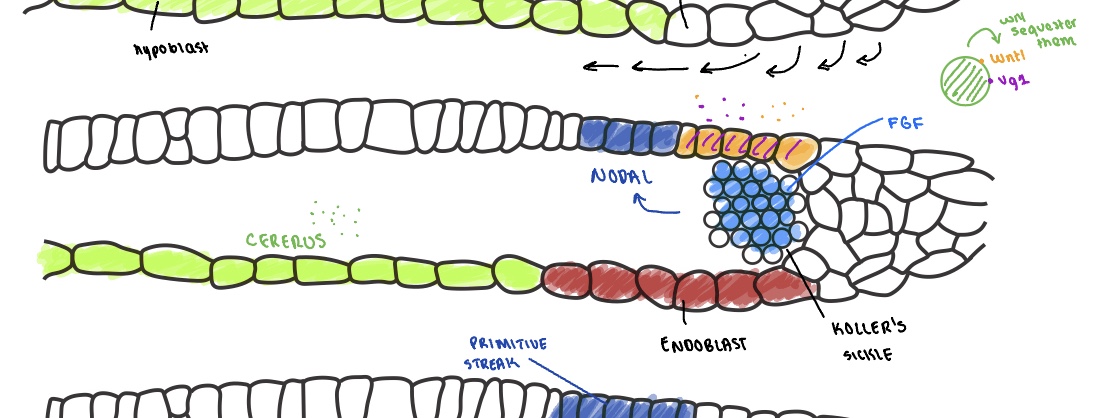
Wnt8C (a Wnt ligand) and Vg1 (a TGF-beta signaling ligand). While Wnt8C is expressed across the marginal area of the epiblast, both are particularly strongly expressed in the posterior side.

What is Cerberus, and how does it interact with Wnt8C and Vg1?
Cerberus is a master inhibitor expressed in the hypoblast. It acts as a protein trap that sequesters both Wnt and Vg1, preventing them from signaling to the overlying cells.

How is the endoblast formed, and what is its significance regarding Cerberus?
Cells from the Area opaca and marginal area proliferate and invade the hypoblast, forming Koller’s sickle. These cells push the Cerberus-expressing hypoblast cells away (toward the midline), creating the endoblast, which does not express Cerberus.
Which three signals must combine in the absence of Cerberus to allow the expression of Nodal?
Vg1
2. Wnt
3. FGF (expressed by the cells of Koller’s sickle).

What process is triggered by Nodal expression, and what role does the resulting primitive streak play?
Nodal expression causes cells to organize and undergo ingression. This area becomes the primitive streak, which acts as an organizer to define the anterior-posterior path of development.
Describe the movement of the primitive streak and the fate of the epiblast cells involved.
The primitive streak moves from the most posterior to the most anterior side. As it moves, cells from the epiblast migrate toward the line and enter the embryo through ingression, giving rise to the mesoderm and endoderm.

What is the specific name for the most anterior side of the primitive streak, and what is its function?
It is called Hensen’s node. It serves as one of the embryo's signaling centers.
Describe the two-step movement of the primitive streak during development.
Migration: The streak moves from the posterior to the anterior side, where cells (mesoprogenitors) enter through ingression.
2. Regression: The entire streak then regresses from the anterior back to the posterior side.
What structure is "left behind" inside the embryo as the primitive streak regresses, and what is its composition?
The notochord is left behind. It is a mesodermal rod formed during the process of ingression and streak regression.
How does the formation of the notochord relate to the development of the neural plate?
The progenitor of the notochord must signal and release Chordin and Noggin. These signals are required for the neural plate to form from the epiblast.
What is the developmental significance of the organizer and the primitive streak (labeled by Nodal)?
The organizer is both needed and sufficient to organize the axis of the embryo. The primitive streak, labeled by Nodal, is a very important element in this organizational process.
Describe the interaction between Wnt and DKK-1 (Dickkopf-1) in the early embryo and its developmental significance.
Wnt is a dominant posterior signal. It is contrasted by DKK-1, a Wnt antagonist emerging from the most anterior region. DKK-1 is required to suppress Wnt signaling anteriorly to specify the head; mutants for DKK-1 show head malformations.

How does Cerberus contribute to the signaling environment of the anterior region?
Cerberus, expressed in the hypoblast, reinforces the anterior environment by inhibiting both BMP and Wnt. This ensures the most anterior region is devoid of Wnt signaling.
What is the stem zone, and which signal drives its posterior program?
The stem zone is a middle region with high proliferation that provides cells for both anterior and posterior structures. Wnt (where not inhibited by DKK-1) induces the expression of FGF8, which forms a gradient from the posterior to the anterior.

How does FGF8 influence the expression of Wnt8C?
FGF8 acts as a morphogen. In areas of low FGF8 expression, it activates Wnt8C, creating a border region between the most posterior and anterior domains.

What is the relationship between Wnt8C, RALDH2, and Retinoic Acid (RA)?
Wnt8C stimulates cells to produce the enzyme RALDH2. This enzyme is essential for the production and release of Retinoic Acid (RA), which forms a signaling gradient, particularly strong in the area of the somites.

True or False: All morphogens are proteins. Explain using Retinoic Acid (RA) as an example.
False. RA is a morphogen, but it is a small molecule (not a protein) derived from another molecule. It is a diffusible signal that induces a dose-dependent response in receiving cells.
Describe the inhibitory "fight" between FGF8 and Retinoic Acid (RA) that establishes tissue domains.
The two molecules mutually inhibit each other:
FGF8 inhibits the expression of RALDH2 (blocking RA production).
Retinoic Acid blocks the expression of FGF8. This creates distinct domains where only one of the two signals dominates.

Besides the posterior gradient, where is another "future source" of FGF8 located in the embryo?
There is an area where the heart is going to develop that serves as a future source of FGF8.
How is the expression of Retinoic Acid eventually positioned in the embryo?
It becomes "sandwiched" in the area where the somites are located.
What is the precursor for Retinoic Acid (RA), and how is it transported into cells?
The precursor is Retinol (Vitamin A), which must be obtained through the diet. Because it cannot move freely in the blood, it is bound by retinol binding proteins and enters cells via a retinol transporter.
Describe the two-step cytoplasmic process that transforms Retinol into the active form, Retinoic Acid (RA).
Step 1: Retinol is catalyzed by retinol dehydrogenase to produce retinaldehyde.
Step 2: Retinaldehyde is transformed by the enzyme RALDH2 (retinaldehyde dehydrogenase) to produce the active form, Retinoic Acid (RA).
How is Retinoic Acid (RA) signaling limited or eliminated within specific tissue domains?
RA is degraded by enzymes of the Cytochrome P450 family, most notably Cyp26. Expressing Cyp26 is a primary way for the embryo to eliminate RA and restrict its signaling domain.
Once Retinoic Acid (RA) enters a target cell, what specific families of receptors/transcription factors does it bind to?
It binds to two main families, each coming in alpha, beta and gamma
RAR (Retinoic Acid Receptors)
RXR (Retinoid Receptors)Where do the RA-receptor complexes bind within the cell to regulate gene expression?
Where do the RA-receptor complexes bind within the cell to regulate gene expression?
They migrate to the nucleus and bind to specific sequences in the promoter regions of DNA called RARE (Retinoic Acid Response Elements).
Contrast the state of target gene expression in the presence versus the absence of Retinoic Acid (RA).
With RA: Receptors bound to RA at the RARE domains promote the transcription of target genes.
Without RA: The transcription factors are either absent from the nucleus or remain bound to the promoter to inhibit gene expression.
What is the axial mesoderm, and what are its primary derivatives?
Also known as chorda-mesoderm, it gives rise to the notochord. While the notochord is a transient structure, it remains in the adult as part of the intervertebral discs.

Into which two main regions does the paraxial mesoderm differentiate?
It divides into the dermomyotome (the region closest to the ectoderm) and the sclerotome (a more ventral structure).

What tissues originate from the dermomyotome?
It gives rise to skeletal muscle, the dermis, and certain skeletal elements.

List the developmental fates of the sclerotome.
The sclerotome forms:
Tendons
Vertebral structures (e.g., proximal ribs)
Endothelial cells and large vessels, including the aorta

What specific organ systems are derived from the intermediate mesoderm?
It is the source of the kidneys and the gonads.

Distinguish between the fates of the somatic and splanchnic layers of the lateral plate mesoderm.
Somatic layer: Gives rise to the pelvis and the limb bones.
Splanchnic layer: Gives rise to the heart, as well as blood vessels and blood.

Describe the temporal relationship between gastrulation and neurulation along the embryo's axis.
Development follows an Anterior-to-Posterior gradient; while the anterior region has already completed gastrulation and formed brain vesicles (neurulation), the posterior region is still active at Hensen’s node and undergoing gastrulation. This "everything everywhere all at once" state means the head is developmentally much older/more advanced than the tail.
Describe the early cleavage of a mammalian embryo and the resulting structure.
Mammals have isolecithal eggs (very little yolk) that undergo complete division. This forms a solid ball of cells called the morula.

What morphological process follows the morula stage in mammals, and what structure does it create?
The process is called cavitation. It creates a fluid-filled cavity called the blastocoel within a structure known as the blastocyst (the mammalian version of a blastula).

Name and describe the two distinct cell populations found in the mammalian blastocyst.
Trophoblast: The most external layer of cells.
Inner Cell Mass (ICM): The internal cluster of cells, also known as embryonic stem cells.

According to the text, which cell is the only truly totipotent cell in development?
The zygote is the only totipotent cell, as it is the only one that gives rise to both the inner cell mass and the trophoblast.
What does the trophoblast give rise to, and what are its two differentiated cell layers?
The trophoblast gives rise to the placenta. Its differentiated layers are the syncytiotrophoblast and the cytotrophoblast.

Describe the structure and function of the syncytiotrophoblast during implantation.
Structure: It is syncytial, meaning it is one massive cell composed of multiple nuclei sharing a single cytoplasm with no physical separation.
Function: It actively participates in forming the placenta by invading the endometrium. This process requires maternal immunosuppression so the embryo is not rejected.

What two lineages does the Inner Cell Mass (ICM) divide into?
1. Epiblast
2. Hypoblast (also known as primitive "endoderm")

Is the mammalian hypoblast the source of the true endoderm? What is its primary role?
No, it is not the true endoderm. It is a transient structure that forms the yolk sac before eventually disappearing.
What are the primary derivatives of the mammalian epiblast?
It is the source of the embryo proper.
It gives rise to extra-embryonic mesoderm, which helps form structures for nutrient absorption.
It is the only part of these early lineages that remains in the adult.
