development of the ear
 Call Kai
 Learn
 Practice Test
 Spaced Repetition
 Match
 Flashcards
 Knowt Play
1/23

There's no tags or description
Looks like no tags are added yet.
24 Terms
how does the inner ear form
forms first
ectoderm thickens into otic placodes
roll inwards to form otic pit
pinch off to form otic vesicle - octic vesicle will form membranous labyrinth (sensory organ)
how does the middle ear form
forms from an outgrowth of the 1st pharyngeal pouch
the inside of the embryo’s throat
endoderm
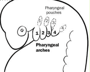
how does the external ear space form
from the 1st pharyngeal groove
outside of the embyro’s neck
ecotderm
where is the ear drum and ear bones formed
also known as the tympanic membrane
formed where the pouch and groove touch
ear bones develop endochondrally from neural crest cells inbetween
describe the outer ear
outside of the skull
the pinna (auricle) - made of skin and hyaline cartilage
auricular muscles - control position of ear
ear canal (externa auditory meatus)
ends at ear drum (tympanic membrane)
direct and transmits sound to middle ear

describe the function of the pinna
funnel shape to collect and direct sound
highly mobile, moved by auricular muscles (locate sounds & communication function)
domestication syndrome - weakened ear cartilage, floppy ears
thermoregulation - surface area and blood supply
innervated by CNVII (facial nerve)
terminology used to describe pinna
helix - the long side
apex - the tip
tragus - base of ear in center (targi hairs protecting entrance to meatus)
annular cartilage - right at the base

describe the external auditory meatus
lined by skin - contains ceruminous glands
cerumen (earwax) traps particles that might damage delicate tympanic membrane
in carnivores/pigs - ear canal has a bend (vertical canal, horizontal canal)
describe the tympanic membrane
separates external and middle ear
stretched like drumskin over tympanic ring (incomplete dorsally)
first ossicle (malleus) attaches to it - visible as stria malleris
sound waves cause membrane to vibrate - vibration transmitted along the chain of ossicles
describe the middle ear
housed inside temporal bone of skull - includes a bulbous expansion of temporal bone (tympanic bulla)
lined by simple squamous epithelium
middle ear communicates with pharynx via auditory tube - useful for pressure equalisation and drainage of gland secretions
describe the middle ear ossicles shapes
malleus - hammer shape
incus - anvil shape
stapes - stirrup shape

functions of the middle ear ossicles
transmit vibrations
connect tympanic membrane to oval window of inner ear
amplify vibrations - tympanic membrane much larger than oval window
muscles attaching to ossicles alter sensitivity
describe the guttural pouches in horses
airfilled cavity with structures that run through it
outpouchings of auditory tubes - may function in brain cooling
divided into two compartments by stylohyoid bone - medial and lateral
blood vessels and nerves pass through this pouch
infections/disorders common can lead to severe complications
what does the inner ear consist of
membranous labyrinth - series of interconnected ducts and chambers, filled with endolymph
bony labyrinth - hollow cavity in temporal bone of skull
perilymph fills the space between osseous and membranous labyrinths

functions of the inner ear
hearing - cochlea
balance - vestibular system
describe the cochlea and hearing
divided into three parts - cochlear duct, scala vestibuli, scala tympani
two scala communicate at the tip of the cochlea’s spiral

describe the process of hearing to brain
as the stapes vibrates in the oval window, it compresses the perilymph in the scala vestibuli
pressure waves are transmitted to the endolymph in the cochlear duct via reissners membrane
movement of the endolymph causes pressure on the tectorial membrane inside the cochlear duct
tectorial membrane puts pressure on hair cells, which sends impulse along vestibulocochlear nerve to brain (as hair cells bend because tectorial membrane doesn’t move)

describe the organ of corti - auditory senses
sits on dorsal surface of basilar membrane
contains acoustically sensitive hair cells that have stereocilia
support cells
tectorial membrane - extends from spiral limbus, in contact with stereocilia of hair cells
vibration in tectorial membrane displaces the stereocilia leading to signal

describe the inner ear and the vestibular system and balance
vestibular part of inner ear is responsible for sensing balance and movement - 3 semicircular canals and vestibule - saccule and utricle
information is carried by vestibular part of vestibulocochlear nerve to the brain
usually sensory input is balanced, if there is in-balance, the brain interprets this as drift towards the side with reduced sensation
what do semicircular canals detect
rotations of the head
what does the saccule and utricle detect
accelerations
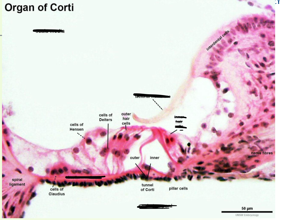
label histological photo of organ of corti

describe the semicircular canals
three canals perpendicular to one another
each detects rotation around one directional axis - pitch, roll, yaw
rotations cause endolymph to move inside canal, moving hair cells in the ampulla of the canal

describe the saccule and utricle
expansions of the membranous labyrinth
detect linear acceleration
contain hair cells in thickened patches (maculae) - covered with gel like layer contain crystals
endolymph movement bends the hairs and information about movement is transmitted via the vestibulocochlear nerve