Quiz 5 (L17-L18)
 Call Kai
 Learn
 Practice Test
 Spaced Repetition
 Match
 Flashcards
 Knowt Play
1/23

There's no tags or description
Looks like no tags are added yet.
24 Terms
Construct a Go vs Progress of Reaction diagram for a given reaction mechanism (i.e., number of substrates and products, number and order of intermediates) and information about rate-limiting step, relative Gibbs free energy of states, relative rates of steps, etc.
Greater concentration (favorable/unfavorable)
Faster Formation (Favorable/unfavorable)
Dissociation Constant > or < 1 (Favorable/Unfavorable)
Stable (Favorable/Unfavorable)
The greater the x concentration the more favorable that x is (so lower Go)
The faster the formation = The more favorable(so lower Go)
Dissociation Constant < 1 Binding more favored(I,e ES)
Dissociation Constant > 1 Dissociation more favored(I.e E+S)
More Stable S or P is the lower G on graph
Overall TS is the highest G on graph

For chymotrypsin reaction pathway: why ES is higher energy than E+S
Enzymes drive the reaction, so it directly acts on the transition state and binding tightly.
The substrate 6 peptide bond planar shape doesn’t fit well in the active site so it’s a higher energy ES(Tetrahedral intermediate is more favorable)
For chymotrypsin reaction pathway: what happens at step 1, given the blue boxes that precede and follow the step
1) Chymotrypsin Free Enzyme: No catalytic reaction occurs. Peptide planer/bond(substrate) comes in, but it doesn’t fit well to the active site.
Hydrophobic packet (specificity pocket) which offers favorable interaction.

For chymotrypsin reaction pathway: what happens at step 2, given the blue boxes that precede and follow the step
2) ES Complex: First tetrahedral intermediate formed where Histidine acts as a base taking H+ from OH group of Ser (rate-determining step and the overall TS) Followed by, Serine195 oxygen performs nucleophilic attack on the carbonyl
Oxyanion hole that stabilizes the tetrahedral intermediate.
“histidine takes H⁺ → serine attacks → tetrahedral intermediate forms.”

For chymotrypsin reaction pathway: what happens at step 3, given the blue boxes that precede and follow the step
3) Short-lived Intermediate: The first tetrahedral intermediate collapses, releasing product 1(via the help of the HIS being an acid and giving it's H+ to the departing N) and forming the acyl-enzyme intermediate.

For chymotrypsin reaction pathway: what happens at step 4, given the blue boxes that precede and follow the step
4) Acyl Enzyme intermediate(R-C=O-): Water molecule comes in the active site.

For chymotrypsin reaction pathway: what happens at step 5, given the blue boxes that precede and follow the step
5) Acyl Enzyme intermediate(R-C=O-): The water performs a nucleophilic attack on the acyl-enzyme intermediate & forms second tetrahedral intermediate via Histidine acting as a base by accepting H from water
Oxyanion hole that stabilizes the tetrahedral intermediate.

For chymotrypsin reaction pathway: what happens at step 6, given the blue boxes that precede and follow the step
6) Short-lived Intermediate: The collapse of the tetrahedral intermediate, meaning the oxyanion hole is disrupted. Histidine acts as an acid(giving its H back to ser).

For chymotrypsin reaction pathway: what happens at step 7, given the blue boxes that precede and follow the step
7) EP Complex: Peptide product released from enzyme + enzyme regenerated

What group & amino acids form the oxyanion hole
Backbone amide N–H groups of Gly193 and Ser195.
In what steps are His acts as a base to form the tetrahedral intermediates
His acts as base in step 2&5 => forms tetrahedral intermediate (first forms-step 2, second forms-step 5)
In what steps are His acts as an acid to form Acyl enzyme intermediate
His acts as acid in step 3&6 => forms Acyl-enzyme intermediate (only form-step 3); step 3 release first product and step 6 is release of 2nd product
In which step does water perform nucleophilic attack on Acyl Enzyme intermediate
Step 5 water perform nucleophilic attack on Acyl-enzyme intermediate
In which step does ser perform nucleophilic attack
Step 2
For chymotrypsin reaction pathway, what is chymotrypsin
Chymotrypsin: the serine protease (e.g. elastase, trypsin, chymotrypsin, thrombin, plasmin) => break down protein
A digestive enzyme produced in pancreas helping break down proteins in food to smaller peptides/amino acids
Contributes to peptide bond cleavage that targets carboxyl end of amino acid
What are the catalytic triad of the chymotrypsin reaction pathway & other enzyme that have the triad as well
Histidine (57), Aspartate (102), and Serine (195) are catalytic triad at active site that lowers the G° of every step of catalysis that involves H+ transfer and TS binding
Asp positions His and helps stabilize its protonated form; His transfers protons; Ser attacks the substrate. Their precise arrangement makes catalysis possible.
serine protease (e.g. elastase, trypsin, chymotrypsin, thrombin, plasmin)
Understand the “limited chemistry” of amino acids (2 of them)
Most electropositive atom
Redox reactions reversibility
Amino acids have no more electropositive atoms than NH3+
Amino acids have little ability for reversible redox except for Cys-S-S-Cys disulfide, and this could cause enzyme structure to change
For these and other reasons, enzymes use certain small molecules and ions.
Case example: Carboxypeptidase A needs Zn+2
Definitions of Non-AA components of Enzymes:
Coenzyme
Vitamin
Metal ion
most abundant cofactor metal
Coenzyme: Organic compound required for enzymatic function. It is regenerated. (some: covalent bond while others: reversible enzyme binding)
Vitamin: Organic compound required in small amounts in the diet.
i.e Vitamin B3 = Niacin = Nicotonic Acid
Metal ions: important roles as enzyme cofactors (~⅓ of all enzymes require metal ions)
Maintain proper 3D structure
Help bind substrate
Participate in catalysis (e.g. redox)
iron or Fe2+
Categories of enzyme control
Is enzyme there?
4 ways of Regulation of synthesis (transcription, splicing, RNA degradation, translation)
Overview: Determination of RNA polymerase structure by Roger Kornberg’s laboratory work that won him Nobel Prize in 2006
4 ways of regulating/controlling protein synthesis
Control transcription: Synthesize more or less mRNA
Control splicing of mRNA: pre-mRNA enters as “RNA processing machine”, the spliceosome (most complex protein-RNA structure known: 5 small RNAs and ~200 different proteins bound together)
Control of RNA degradation: RNA lifetime determined by degradation rate of mRNA molecule that is broken down into nucleotide fragments
Regulation of protein synthesis on the ribosome (translation)
Categories of enzyme control
Is enzyme there?
Regulation of degradation (e.g., ubiquitin-mediated proteasomal degradation)
Proteins lifetime(histone, Hb, Mitotic cyclin)
Principle
Mechanism in lysosome & cytosol
Why is it important to remove no longer needed protein
Protein lifetimes (vary a lot): Histones ~ life of cell; Hb months; mitotic cyclin ~2 min
Principles: Protein that partially unfolds or chemically reacts (e.g. oxidation) is recognized and then hydrolyzed. Healthy proteins can also be hydrolyzed!
Hydrolysis through fasting, infection, cell death (apoptosis) - Nobel Prize award in 2016 to Yoshinori Ohsumi for apoptosis discovery, cell cycle
Mechanisms:
a) In lysosome, proteins and whole organelles degrade by various proteases
b) In cytosol, the proteasome has the major role as the “protein degradation machine” consisting of 4 rows of 7 subunits
Ubiquitin: 76 amino acid protein, found in all cells as marker for proteolysis
Why is it important to remove proteins no longer needed?
Steps of cell division/DNA repair only when needed/destroy only defective newly synthesized proteins/immune activity turned off/transcription regulation (e.g. degradation of transcription repressor to turn on specific genes)
Categories of enzyme control
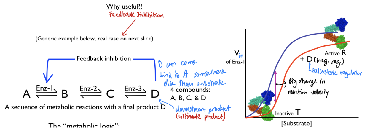
Regulation of enzyme activity overview: R vs T state
Overview: Enzymes have two main states →
(1) active(R)
(2) less active meaning enzymes perform to have an “off-state” (T)
Categories of enzyme control
Regulation of enzyme activity
Reversible(4) & Irreversible Covalent modification (many types, e.g. phosphorylation, proteolytic cleavage)
Reversible through other enzymes, controlled by signals, via removal of chemical modification
Addition of (a) phosphate group, (b) AMP, (C) Methyl group, (d) 100s of others
Irreversible covalent modification
Cleavage of peptide bond (proteolytic cleavage) through chymotrypsin
Categories of enzyme control
Regulation of enzyme activity
Allosteric regulation (Example of allosteric control: ATCase[AKA Aspartate transcarbamoylase] (negatively regulated by CTP, positively regulated by ATP and why this makes sense, how these regulators shift Vin vs [S] curve; R T model)
Allosteric Control: Binding at a site different from active site that is reversible between T and R transitions of quaternary structure + reversible allosteric regulation of monomer possible (e.g. human glucokinase)
Covalent modification can also be allosteric (not mutually exclusive)
Key point: binds to another site and changes the shape of the active site, either activates or inhibits the protein function
ATCase: first enzyme for characteristic identification of allosteric regulation
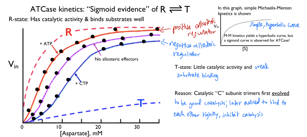
ATP: Positive allosteric effector, favors R-state(Left)
Substrates (e.g. carbamoyl phosphate, and Asp) are also positive allosteric effectors
CTP: Negative allosteric effector, favors T-state (Right)
[S] a concentration unit like M
Vin a concentration unit / time
Be able to plot (or recognize) Vin vs [S] to show sigmoidal curve and impact of positive and negative allosteric regulators; where populations of R vs T state are high/low on this plot.
Key takeaway:
R state: looks like myoglobin curve
No allosteric effectors: somewhat of sigmoidal graph
Positive allosteric effectors: shifts the graph to the left
Negative allosteric effectors: shifts the graph to the right
T State: linear line(lowest activity)
They all have different Vmax