SDSU Chem 365 Exam 2
 Call Kai
 Learn
 Practice Test
 Spaced Repetition
 Match
 Flashcards
 Knowt Play
1/259

There's no tags or description
Looks like no tags are added yet.
260 Terms
*** _____ are the principal biomolecules of life.
Proteins
*** What are 5 examples of protein functions?
1) Enzymes
2) Sensors
3) Pumps
4) Structural support
5) Regulators
*** Proteins are generally longer than ___ amino acids in length.
40
*** Most proteins are ___ to ___ amino acids in length.
100 to 1000
*** What is the largest known protein?
How long it is?
Titin; 34,350 amino acids long
*** Name 3 characteristics of proteins.
1) Stable fold
2) Functional capability
3) Likely an upper limit (mistakes increase with size)
*** Table 5-1: Compositions of Some Proteins

*** Common Protein Expression Systems
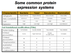
Name 4 types of assays. What are the properties of a useful assay?
Types of assays
1) Binding assay
2) Enzyme assay
3) Immunological assay
4) Biological assay
Properties of a useful assay
1) Specific
2) Sensitive
3) Easy to perform
*** Comparison of Protein Purification Procedures
Salting Out: Solubility
Ion exchange: Ionic charge
Affinity: Binding specificity
SDS-PAGE: Size/Molecular mass
Hydrophobic: Polarity

*** What can a biological assay be used to find?
- Biological assay for the presence of NGF ???

*** What is a type of assay that combines two types of assays?
Enzyme-linked immunosorbent assay (ELISA)
*** Describe the 5-Step procedure for ELISA.
Enzyme-linked immunosorbent assay (ELISA)
1) Immobilize first antibody on solid support
2) Incubate with protein-containing sample
3) Add a second antibody that is covalently linked to an assayable enzyme
4) Wash and assay enzyme activity.
5) Determine the amount of protein present; - Based on the amount of substrate converted to product

*** Protein purification:
What is the first step of protein purification?
What are the 2 ways this can be done?
First step: Homogenize the starting material
1) Blender/Grinder/Scissors
2) Lysis of cells from culture
*** What are 3 ways that cells can be lysed from culture?
1) Sanitation
2) Shear force (French press, glass beads)
3) Chemical lysis (detergent, enzyme degradation)
*** Protein Purification:
What is the second step?
What are 3 ways this can be done?
Second step: Fractionate the mixture of biomolecules by some physical/chemical means
1) Centrifugation
2) Salting out
3) Chromatography
*** Isoelectric Point (pI)
- The pH at which the net charge on a molecule is zero
- In other words, the pH at which the molecule would not move under the influence of en electric field
- This can be found by averaging the two pKa values that border the +1 ==> 0 ==> -1 transition
- Example: The amino acid Arg has three pKa values (2, 9, 12.5). What is the pI of Arg? (Answer: 10.75)
- The isoelectric point (pI) can be calculated for any molecule including whole proteins
*** Describe the solubility of a protein at its isoelectric point.
- Proteins are LEAST SOLUBLE at their isoelectric point (pI)

*** "Salting Out":
What is the purpose?
What are 3 ways this can be achieved?
Purpose: Obtain optimum solubility of a protein(s) in a protein mixture
- Vary the salt concentration, pH or temperature
*** What is the most common salt used in "Salting out"? What are 2 other options?
Most common:
- Ammonium sulfate (NH4)2SO4
Less common (More specific for RNA/DNA):
- Streptomycin sulfate
- Polyethyleneimine
*** The solubility of most globular proteins is markedly influenced by ____ and _____.
1) pH
2) Ionic strength (i.e. various salt concentrations)
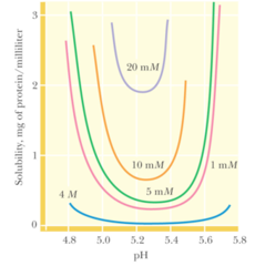
***Describe "salting out".
- The manipulation of 1 or more variables to selectively precipitate certain proteins, while others remain soluble
1) Concentration of dissolved salts
2) Polarity of the solvent
3) pH
4) temperature
- Protein solubility increases at low ion concentration increases as salt is added ("salting in"). Eventually, as more salt is added, protein solubility decreases ("salting out").
- The solubility begins to due to competition between added salt ions and other dissolves solutes for molecules of solvent.
- At very high salt concentrations, so many added ions are solvated that there is less solvent available to dissolve other substances, such as proteins.
- B/c each protein has a different ionic and hydrophobic composition, they precipitate at different salt concentrations.
- In order to precipitate the unwanted proteins from solution, the salt concentration must be adjusted to just below the precipitation point of the protein to be purified
- The unwanted proteins are removed via filtration or centrifugation
- In order to precipitate the desired protein from solution, the salt concentration of the remaining solution is increased
- Result: Significant purification and concentration of large quantities of protein
- Most commonly used reagent: Ammonium sulfate, or (NH4)2SO4, b/c its high solubility (3.9 M in water at 0oC) allows the preparation of solutions with high ionic strength
- pH may be adjusted to approximate the isoelectric point (pI) of the desired protein b/c a protein is least soluble when net charge = 0
Use the phenomenon of "salting out" as a means to fractionate protein mixtures.
1) The salt of choice, usually ammonium sulfate, is added to a solution of macromolecules to a concentration just below the precipitation pain too the protein o interest
2) After centrifugation, the unwanted precipitated proteins (red spheres) are discarded and more salt is added to the supernatant to a concentration sufficient to salt out the desired proteins (green spheres)
3) After a second centrifugation, the protein is recovered as a precipitate, and the supernatant is discarded

*** Define "Column Chromatography".
- Any procedure that separates individual components of a mixture ("mobile phase") due to their unique interactions with a matrix contained in a column ("stationary phase").
*** Column Chromatography: What are the 3 Types? What are they based on?
1) Ion exchange chromatography
- Separation based on variable electrostatic interactions between proteins and charged matrix
- Ex. Anion or cation exchange chromatography
2) Size exclusion chromatography
- Separation based on exclusion of larger proteins from pores in matrix
- Aka: "Gel Filtration" or "Molecular Sieve" chromatography
3) Affinity chromatography
- Separation based on affinity of target for some component on the matrix.
- Ex. Immunoaffinity, Ni-affinity, Glutathione-affinity
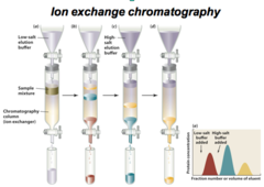
Describe protein purification by ion exchange chromatography
Purpose:
- Separation of anions and cations in order to precipitate the protein of interest
Facts:
- Oppositely charged molecules bind to oppositely charged groups that are chemically linked to a matrix, such as cellulose or agarose
- Anions bind to cationic groups on anion exchangers
- Cations bind to anionic groups on cation exchangers
- Polyelectrolytes (have both + and - charges) can bind to both cation and anion exchangers
- A particular protein's binding affinity depends on the presence of other ions that compete with the protein for: 1) Binding to the ion exchanger and 2) pH of the solution, both of which influence the net charge of the protein
Steps:
1) A mixture of proteins are dissolved in a small volume of buffer with an appropriate pH and salt concentration, which is applied to the top of the matrix in the column containing the ion exchanger
2) Column is washed with the buffer
3) As elution progresses, the proteins separate into discrete bands as a result of their different affinities for the ion exchanger
- Low affinity = Move faster
- High affinity = Move slower
4) Column effluent is collected in a series of fractions
5) Proteins that bind tightly to the ion exchanger can be eluted (washed through the column) by adding a buffer, called the "eluant". The eluant has either a higher salt concentration or pH, which works to reduce the affinity with which the matrix binds to the protein.
- Ex. The first protein (red) passes through the column more quickly and is isolated as a separate fraction.
6) The salt concentration of the eluant is increased to elute the remaining proteins.
- Ex. The other proteins pass through the column more slowly (c and d)
7) Test for the protein of interest:
- Column effluent: Measure its absorbance at 280 nm.
- Eluted fractions: Using a more specific assay
8) An elution diagram can be used to view the protein mixture from the column.
- X axis: Fraction number or volume of fluent
- Y axis: Protein concentration
Describe protein purification by size exclusion chromatography
Purpose: Separate according to size and shape
Facts:
- Stationary phase: Gel beads with pores that are narrow in size range. Pore size is determined by the extent of cross-linking between the polymers of the gel material.
- If an aqueous solution of molecule son various sizes is passed through a column containing such "molecular sieves" or "pores", the molecules that are too large to pass through the pores are excluded from the solvent volume inside the gel beads.
- Large molecules: Move through column more rapidly; Often have access to fewer pores and thus elute faster
- Small molecules: Move through column more slowly b/c they must pass through the pores; Often have access to more pores and thus elute slower b/c they have access to more of the gel's interior volume
- If a gel filtration column is calibrated with several proteins of known molecular mass, the mass of an unknown protein can be estimated by its elution position
Steps:
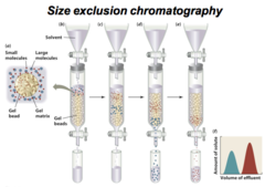
a) A gel bead (tan spheres), which consists of a few matrix, encloses an internal solvent space
- Small molecules (red): Freely enter the internal space of the gel bead
- Large molecules (blue): Cannot penetrate the gel pores
b) The sample solution is applied to the top of the column
c) The small molecules can penetrate the gel, and thus, migrate through the column more slowly than the large molecules that are excluded from the gel
4) The large molecules elute first and are collected as fractions
5) The smaller molecules elute later, and require larger volume of solvent to elute
6) An elution diagram, or chromatogram, can be used to view the complete separation of the two components
- X axis: Volume of effluent
- Y axis Amount of solute
Describe protein purification by affinity chromatography
Purpose: Exploits specific binding behavior
Facts:
- Many proteins can bind to specific molecules tightly, but noncovalently. This property is used to purify proteins by affinity chromatography.
- Ligand: Molecule that specifically binds to the protein of interest, and is covalently attached to an inert matrix
- Protein of interest: A nonreactive analog of an enzyme's substrate
- The ligand must have an affinity high enough to capture the protein of interest, but not so high as to prevent the protein' subsequent release without denaturing it
Steps:
1) Construct an affinity chromatography column by chemically attaching small molecules or proteins to a chromatographic matrix
2) A ligand (black dots) is immobilized by covalently binding it to the chromatographic matrix
3) An impure protein solution, composed of many macromolecule proteins (orange, blue, and green) is passed through the chromatographic material. Each protein has unique cutout regions that symbolize their ligand-binding sites. These differ from one another based on the unique shape/size of their ligand binding site.
4) Only the specific protein of interest (orange) bind to the matrix-anchored ligand
5) The other macromolecules (blue and green) are washed through the column with the buffer
6) Protein of interest can be recovered in highly purified form by changing the elution conditions to release the protein from the matrix.
- Ex. Washing the column with a solution containing a high concentration of free ligand
- Ex. Washing the column with a solution of different pH or ionic strength
Advantages:
- Able to exploit the desired protein's unique biochemical properties, rather than the small differences in physiochemical properties between proteins
- Greater separation power for a specific protein

*** Contrast Preparative vs. Analytical Techniques
- Chromatography and other techniques differ in scale and purpose. They are either:
1) Preparative
- Large scale
- Purpose: make material that will be used in experiments
2) Analytical
- Small scale
- Purpose: Monitor the quality of/make measurements on the material
*** What does SDS-PAGE stand for?
Sodium dodecyl sulfate polyacrylamide gel electrophoresis; NOTE: 11 carbons!
*** Is SDS-PAGE an analytical or preparative technique?
Analytical
*** What is the structure of SDS

*** What are the four steps of SDS-PAGE?
1) Treat the protein mixture with SDS (a detergent), reducing agent, and heat
2) The proteins denature (unfold). This gives all proteins a net negative charge.
3) Separate proteins in a polyacrylamide gel "cage" under the influence of an electric field (electrophoresis)
4) Visualized through stained gel with bands that correspond to different sized proteins

*** SDS-PAGE separates proteins by _______.
MASS
*** In SDS-PAGE:
As _____ decreases, _____ increases.
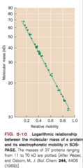
As MOLECULAR MASS decreases, RELATIVE MOBILITY increases.

Describe how SDS-PAGE works and what information it gives
- "SDS-PAGE" = Sodium Dodecyl Sulfate Polyacrylamide Gel Electrophoresis
- Purpose: Protein separation/purification by gel filtration effects, that is, according to molecular mass.
- Steps (2):
1) Treat the protein mixture with SDS, a detergent that denatures proteins and renders them negatively charged (reducing agent), and heat.
- This denatures (unfolds) the protein and gives them all a net negative charge.
2) Separate proteins in a polyacrylamide gel "cage" by electrophoresis, which puts the proteins under the influence of an electric field.
- Note: Normalized shape and charge-migration is due ONLY to size of protein
Result: Stained gel with bands corresponding to different sized proteins.
Graph: The SDS-PAGE method separates proteins by mass.
1) X-Axis: Relative mobility
2) Y-Axis: Molecular Mass (kD)
3) Trend: Negative linear slope; As molecular mass decreases, relative mobility increases.

Use the Beer-Lambert law to determine protein concentration
Equation: A = εcl
- A = absorbance (logI0/I) at a specific wavelength (280 nm for protein)
- ε = molar extinction coefficient (M-1cm-1)
- c = concentration (M)
- l = path length (cm)
Note: ε can be estimated for a protein of known sequence by assuming the native protein absorbs as the sum of its Trp, Tyr and Cys residues

*** What biological macromolecule can absorb ultraviolet radiation? At what wavelength?
Proteins and Nucleic acids
- 260 nm absorbance signal
- Reserved for purified nucleic acids
- Can be used to quantitate DNA and RNA
- A typical purified sample of protein will exhibit an A280/A260 ratio of approximately 2

*** How can scientists trace evolutionary relationships between proteins?
Amino acid sequence comparison
Note: Amount of evolution varies significantly from protein to protein as their amino acid sequences are complex and unique to that protein
*** What 3 ways can proteins evolve?
1) Point mutations
2) Gene duplication
3) Domain shuffling
*** What are the different types of global proteins and how did they evolve?

*** Phylogenetic tree of Cytochrome C

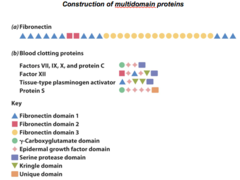
*** Name 2 types of multi domain proteins. What are they composed of?
1) Fibronectin
2) Blood clotting proteins

** Compare the sequence divergence rates between the following:
1) Fibrinopeptides
2) Hemoglobin
3) Cytochrome C
4) Histone H4
Graph:
- X axis: Millions of years since divergence
(0-1000)
- Y axis: Number of amino acid changes/100 sites
(0-140)
- Rate of evolution:
Fibrinopeptides > Hemoglobin > Cytochrome C > Histone H4
Describe some conclusions about molecular evolution that arise from protein sequence comparisons
1) Protein sequences reveal evolutionary relationships
- Ex. Cytochrome c is a protein found in nearly all eukaryotes, from yeast to humans. The cytochromes c of all species - whether "primitive" or "advanced" - have evolved to about the same extent. This is illustrated in the Figure 5-22, in which the evolutionary distances from all modern cytochromes c to the lowest point, the earliest common ancestor producing this protein, are approx. the same.
2) Proteins evolve by the duplication of genes or gene segments.
- Domain: Evolutionarily conserved sequences of 40-200 residues; Can be grouped into 1000-1400 families, but ~50% of all known domains fall into just 200 families.
- 10-20% of proteins appear to be unique
- Domains with 40% sequence identity usually have the same function
- Domains with less than 25% sequence identity usually perform different roles
3) Protein families can arise through gene duplication
- Proteins with similar functions = Similar sequences = Shared, common ancestor
- Homolgous proteins with the same function in different species (ex. cytochromes c) are said to be "orthologous"
- Two independently evolving genes that are derived from a gene duplication event are said to be "paralogous"
- Gene duplication: Genetic recombination event; One member of a chromosome pair acquires both copies of the primordial gene
- Following duplication, sequences may diverge as mutations occur over time
- Gene duplication = Efficient mode of evolution b/c one copy of the gene evolves a new function via natural selection, while the other gene directs the synthesis of the original protein
- Many protein domains have been duplicated: Prokaryotes-60%; Eukaryotes-90%; & Humans-98%
- Example of evolution via gene duplication: Globin family. Hemoglobin (transports O2 from lungs/gills/skin --> tissues) has a and B subunits that are similar to each other and to the sequence of the protein myoglobin (oxygen diffusion through muscle tissue).
- Gene duplication allowed one global to evolve into a monomeric hemoglobin a chain. Duplication of the a chain --> paralogous gene for the B chain.
- "Pseudogenes": Globen genes that are not expressed
- A duplicated gene has only a limited time to evolve a new function that provides a selective advantage before it is inactivated via mutation, that is, becomes a "pseudogene"
3) The rate of sequence divergence varies
- Protein mutations accumulate at a constant rate over a geological time scale (i.e. Linear slope for the graph of Protein evolution rate)
- However, the rate at which mutations are accepted into a protein varies among proteins
- The rate at which a protein evolves is largely influenced by the effect of amino acid changes on the protein's function. For example, Histone H4 is so finely tuned to its function of packaging DNA in cells that it is extremely intolerant of any mutations. Cytochrome c is only slightly more tolerant. Thus, any changes in its amino acid sequence must be compatible with all its binding partners.
- Rate of protein evolution also depends on the protein's structural stability. For example, a mutation that slowed the rate in which a polypeptide chain folded into its functional 3-D shape may affect cell survival, although the protein's function remains unchanged.
- Genes for highly expressed proteins evolve more slowly than genes for rarely expressed proteins.
4) BOTH proteins AND DNA sequences that control the expression of proteins (when, where, how much) are subject to mutational changes.
- Although the proteins (cytochromes c) of humans and chimps are > 99% identical, they are grouped into different families due to significant anatomical/behavioral differences
5) Many proteins control domains that occur in other proteins
- Most of the proteins involved in blood clotting are composed of sets of smaller domains
- Sequence identity between homologous domains is imperfect since each domain evolved independently
- Individual domain functions: catalyzing ac chemical reaction, binding a particular molecule, spacers, scaffolding for other domains, unknown, etc.
- New proteins can be generated via gene duplication and domain shuffling
- Domain shuffling is a much faster process than the duplication of gene, followed by its evolution of a new functionality.
- Proteins evolve through changes in primary structure. Protein sequences can be compared to construct phylogenetic trees and identify essential amino acid residues.
- New proteins arise as a result of the duplication of genes or gene segments specifying protein domains, followed by their divergence
*** What determines a protein's ability to function?
Its overall 3-D shape or conformation
Define protein structural hierarchy as primary, secondary, tertiary, and quaternary
1) Primary: A protein's amino acid sequence, which facilitates the formation of secondary and tertiary structure.
- Ex. Lys-Ala-His-....
2) Secondary: Results from local hydrogen bonding patterns of amino acids.
- Ex. Alpha helix or Beta pleated sheets
3) Tertiary: Results from the arrangement of secondary structure elements into a core domain
- Ex. One complete protein chain, such as a Beta chain of hemoglobin
4) Quaternary: When multiple subunits interact to form the active protein (not found in all proteins)
- Ex. The 4 separate chains of hemoglobin assembled into an oligomeric protein

Define protein structural hierarchy as "primary",
- Sequence of amino acids in a protein, which is a unique characteristic of every protein
- Encoded by the nucleotide sequence of DNA
- Thus, it serves as a form of genetic information
- Read from amino-terminus --> carboxyl terminus
Define protein structural hierarchy as "secondary".
- Secondary structure motifs form stably folded protein domains
- These include: a-helices + B-sheets
Define protein structural hierarchy as "tertiary".
Define protein structural hierarchy as "quaternary".
***Describe the fundamental structural pattern in proteins: What is the rxn? What is the bond?
Condensation reaction (loss of H2O)
- 1 H atom comes from the amino terminus (NH3+) of an amino acid
- 2 O atoms come from the carboxyl terminus (COO-) of another amino acid
C and N are linked by a peptide bond
The linkage of 2 amino acids from a peptide bond is called a "dipeptide"

Describe the unique chemical properties of the peptide bond
- Peptide bonds have a partial double bond character through conjugation with the lone pair of electrons on the O of the C=O (carbonyl)
- Trans conformation (usually)
- Partial (40%) double bond character
- Approx. 0.133 nm (1.33 Å) long
- Due to the double bond character, the six atoms of the peptide bond group are always planar!
- N = partially positive
- O = partially negative

*** How is peptide bond length confirmed?
- Peptide bond length confirm partial double bond character

*** Describe the coplanar nature of the peptide bond.
"Six atoms on a plane" (SOAP) ???

*** Describe the consequences of partial double bond character of the peptide bond.
1) Rotation around peptide bond is restricted
- A pure double bond between C and O would permit free rotation around the C-N bond
2) Atoms associated with the peptide bond are coplanar
- Prohibits C-N bond rotation, but would place too great a charge on O and N
3) The amide N is partially positively charged and the carbonyl is partially negatively charged. Therefore, the peptide bond has a permanent dipole.
- The true electron density is intermediate. The barrier to the C-N bond rotation of about 88 kj/mol is enough to keep the amide group planar

Explain how peptide bond planarity limits bond rotations and protein folding
1) Two degrees of freedom per residue for the peptide chain
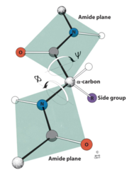
- The angle about the Cα-N bond is denoted phi (φ) and the angle about the Cα-C bond is denoted psi (ψ)
- If all the angles phi and psi are specified then the entire path of the peptide backbone is known
- Some values of phi and psi are more likely than others
*** Contrast the angles "phi" vs. "psi".
Notes:
- The peptide bond planes are joined by the tetrahedral bonds of the a-carbon
- The rotation parameters are f and y
- In this conformation, f = 180° and y = 180°
- Positive values of f and y correspond to clockwise rotation as viewed from Cα
- Starting from 0°, a rotation of 180° in the clockwise direction (+180°) is equivalent to a rotation of 180° in the counterclockwise direction (-180°).
Illustrate the previous point by Ramachandran analysis
G. N. Ramachandran was the first to demonstrate the convenience of plotting phi and psi combinations from known protein structures
The sterically favorable combinations are the basis for preferred secondary structures
- Dark area: "low energy", or favored regions for particular combinations of F (phi) and Y (psi) angles)

*** Steric constraints on Phi and Psi
Unfavorable orbital overlap precludes some combinations of phi and psi (3 examples)
1) Unfavorable: phi = 0°, psi = 180°
2) Unfavorable: phi = 180°, psi = 0°
3) Unfavorable: phi = 0°, psi = 0°
List the elements of protein secondary structure and describe the forces that drive their formation
All secondary structures are localized and stabilized by hydrogen bonds
1) Alpha helix
2) Beta sheets (composed of "beta strands")
3) Tight turns (aka beta turns or beta bends)
*** Describe the alpha helix.
- First proposed by Linus Pauling and Robert Corey in 1951
- Identified in keratin by Max Perutz
- A nearly ubiquitous component of proteins
- Stabilized by H bonds
- Residues per turn: 3.6
- Rise per residue: 1.5 Ångstroms
(Å; 0.1 nm)
- Rise per turn (pitch)
3.6 x 1.5Å = 5.4 Å
- This information can be used as a "ruler" to give you an idea of the size of a protein

*** How does an alpha helix have a net dipole?
- The alpha helix has a LARGE net dipole
- This is due to the arrangement of N-H and C=O groups (each with an individual dipole moment) along the helix axis

*** What do the numbers on an alpha helix represent?
Indicate fractional charges on respective atoms

*** Name two examples of proteins whose 3-D structures contain substantial amounts of alpha helices in their structures.
1) B-hemoglobin subunit
- O2-carrying protein in vertebrates
2) Myohemerythrin
- O2-carrying protein in certain invertebrates, such as "Sipunculids", a phylum of marine worm
Note: The helices are represented by the regularly coiled sections of the ribbon drawings

*** Describe the Beta sheet.
- Pleated
- Composed of beta strands
- First postulated by Pauling and Corey, 1951
- Rise per residue:
3.47 Ångstroms for antiparallel strands
3.25 Ångstroms for parallel strands
Each strand of a beta sheet may be pictured as a helix with two residues per turn
- Strands may be parallel or antiparallel

*** Describe a Beta sheet (Parallel)

*** Describe a Beta sheet (Antiparallel)

** Describe a mixed beta-sheet protein with alpha helices. What is an example?
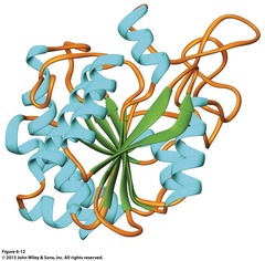
- Example: Bovine Carboxypeptidase
*** Describe the Beta Turn.
- Aka: "Beta bend" or "Tight turn"
- Allows the peptide chain to reverse direction
- Carbonyl C of one residue is H-bonded to the amide proton of a residue three residues away
- Proline and glycine are prevalent in beta turns
- Can be described as "connecting" + "adjacent"

*** Describe reverse turns in polypeptide chains.

*** What are the 3 basic classes of proteins?
1) Fibrous
2) Globular
3) Membrane

Classify folded proteins fibrous, globular, or membrane
1) Fibrous: Structural role; Mechanically strong; Usually insoluble
- Ex. Usually structural proteins, such as collagen and keratin
2) Globular: Spherical shape; Soluble in cell cytosol (an aqueous environment)
- Ex. myoglobin, serum albumin
3) Membrane: Associated with membranes; Insoluble in water; Solubilized by detergents
- Ex. Glucose transporter, Ca2+-pump ATPase, Bacteriorbodopsin

*** In fibrous proteins, how is most of a polypeptide chain organized?
Approx. parallel to a single axis
*** Describe the structure of a-Keratin and how it relates to its function?
- Sequence consists of 311-314 residue alpha helical rod segments capped with non-helical N- and C-termini
- Primary structure of helical rods consists of 7-residue repeats: (a-b-c-d-e-f-g)n
- In the above sequence, a and d are NONPOLAR --> ASSOCIATION OF HELICES (thus, its structural role and mechanical strength)
*** What is the name of the interaction that takes place between side groups of neighboring a-helices?
- Coiled coil interactions

Describe the primary and secondary structures of the fibrous proteins α-keratin and collagen
Collagen
1) Primary
- Nearly 1 of 3 residues is Gly
- Proline content is unusually high
2) Secondary
- Triple helix (thus, unique aa composition)
- Three intertwined, left-handed polypeptide chains (1000 residues each)
- MW = 285,000
- 300 nm long, 1.4 nm diameter
- Rise per residue of 2.9 Ångstroms (Å)
- 3.3 residues per turn
- Long stretches of Gly-Pro-Pro/HyP
- Much more extended than α-helix
α-keratin
1) Primary
- Helical rods
- 7-residue repeats: (a-b-c-d-e-f-g)n
- a and d are non polar --> Helices association
2) Secondary
- Alpha helical rod with 311-314 residues
- Each residue is capped with non-helical N- and C-termini
*** Where is Collagen vs. α-Keratin found?
Collagen
- Hair, Horns
- Fingernails, Claws
- Beaks
- Processed to make gelatin!
Keratin
- Tendons, Cartilage
- Bones, Teeth
- Lip injections
*** What are the 3 higher order structures of α-Keratin?
1) Dimer: Coiled alpha helical coil rod with non-helical N and C-terminal tails
2) Protofilament: 2 dimers
3) Microfibril: Many protofilaments
*** What 4 things are most collagen-based amino acids made of? (4) Name three examples.
1) Glycine - 33% (1 in every 3 residues)
2) Proline and Hypro - 30%
3) Enzyme prolyl hydroxylase
4) Cofactor Vitamin C (deficiency --> scurvy!)
Examples:
- 4-hydroxyproline
- 3-hydroxyproline
- 5-hydroxylysine
*** What reaction do proline residues undergo?
Hydroxylation
*** What enzyme catalyzes the hydroxylation of proline residues?
The reaction requires. The Hyl residues are often covalently linked to carbohydrates and participate in covalent crosslinking of collagen fibers
Prolyl Hydroxylase
*** Besides Prolyl Hydroxylase, what two other things are needed for the hydoxlation of proline residues?
1) α-ketoglutarate
2) Ascorbic acid (vitamin C)
*** How are the Hyl residues linked?
- Covalently linked to carbohydrates
- Covalently cross-linked to collagen fibers
*** Name an example of a collagen-like protein.
Poly(Gly-Pro-Pro)
- Right-handed triple helix with three left-handed helical chains

Describe how the properties of secondary structure and the application of the principles of thermodynamics give rise to tertiary structures known as "domains"
- Proteins fold to make the most stable structures
- In order to make the most stable structure, there needs to be minimal solvent contact with hydrophobic side chains
- Structural stability is supported by
1) Hydrogen bonds
2) Electrostatic interactions
3) Van der Waals bonds
- Elements of secondary structure combine to form stable folded regions within a protein that remain ordered even when removed from the protein (this is called the "domain concept")
*** Describe the structure of globular proteins.
- Polar proteins: Face the exterior; Interact with solvent
- Hydrophobic residues: Bury on the interior; Interact with each other
- Packing of residues is close (Van der Waals interactions)
- Structures of globular proteins are not static
- Various elements and domains of protein move to different degrees
- Some segments of proteins are very flexible and "disordered
- Their fold is described by the secondary structure elements that they contain or by their homology to other proteins.
- Well defined domains can occur in within different contexts among proteins of diverse function
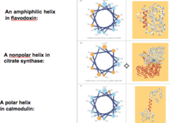
*** What are the 3 types of helices? Name an example of each.
1) Amphiphilic Helix
- Ex. Flavodoxin
2) Nonpolar Helix
- Ex. Citrate synthase
3) Polar Helix
- Ex. Calmodulin
*** How can you improve the quality of electron density in maps?
Increased resolution data

*** Examine the structures of three types of common domains

*** Examine the structures of three types of 8-stranded beta-barrels
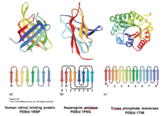
*** What is an example of a two-domain protein?
GAP Dehydrogenase

*** Describe quaternary structure.
- Quaternary structure: Multiple subunits associate to form active protein
- Noncovalent interactions only (usually)
- Typical KD for two subunits: 10-8 to 10-16 M. - - These values correspond to energies of 50-100 kJ/mol at 37° C
- Unfavorable: Entropy loss due to association
- Favorable: Entropy gain due to burying of hydrophobic groups
- Example: Hemoglobin

*** What are the 4 Structural/Functional Advantages Driving Quaternary Association?
1) Stability: reduction of surface to volume ratio
2) Genetic economy and efficiency-viruses
3) Bringing catalytic sites together- Metabolism
4) Allostery/Cooperativity (Ch. 7)
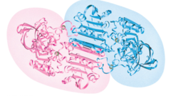
Classify the quaternary structure of a particular protein on the basis of its subunit organization
Example: Liver Alcohol Dehydrogenase
- Each subunit = 6-stranded parallel sheet
- Between each subunit = Antiparallel sheet
- Center point = C2 symmetry axis
*** Where are polar vs. non polar amino acids found? (i.e. "The Hydrophobic Effect")
Nonpolar/Hydrophobic amino acid: Interior
- Bury on the interior of a protein
Polar/Hydrophilic amino acid: Exterior
- Decorate the exterior of a protein
*** What are the 4 forces that dictate and stabilize folding?
1) Hydrophobic effect
2) Noncovalent Interactions
- Van der Waals
- Hydrogen bonds
- Ionic bonds
3) Hydrophobic interactions
4) Disulfide bonds
*** Table 6-3: Hydropathy Scale for Amino Acid Side Chains

*** Table 6-3.5: Hydropathy Plot of Chymotrypsinogen
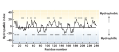
*** All of the information required to ensure proper folding of a peptide chain into its native structure is contained in what structure?
The primary amino acid structure of the peptide