LBBBIO8
 Call Kai
 Learn
Learn Practice Test
 Spaced Repetition
 Match
Match Flashcards
 Knowt Play
1/77

There's no tags or description
Looks like no tags are added yet.
Name | Mastery | Learn | Test | Matching | Spaced | Call with Kai | Chat |
|---|
No analytics yet
Send a link to your students to track their progress
78 Terms
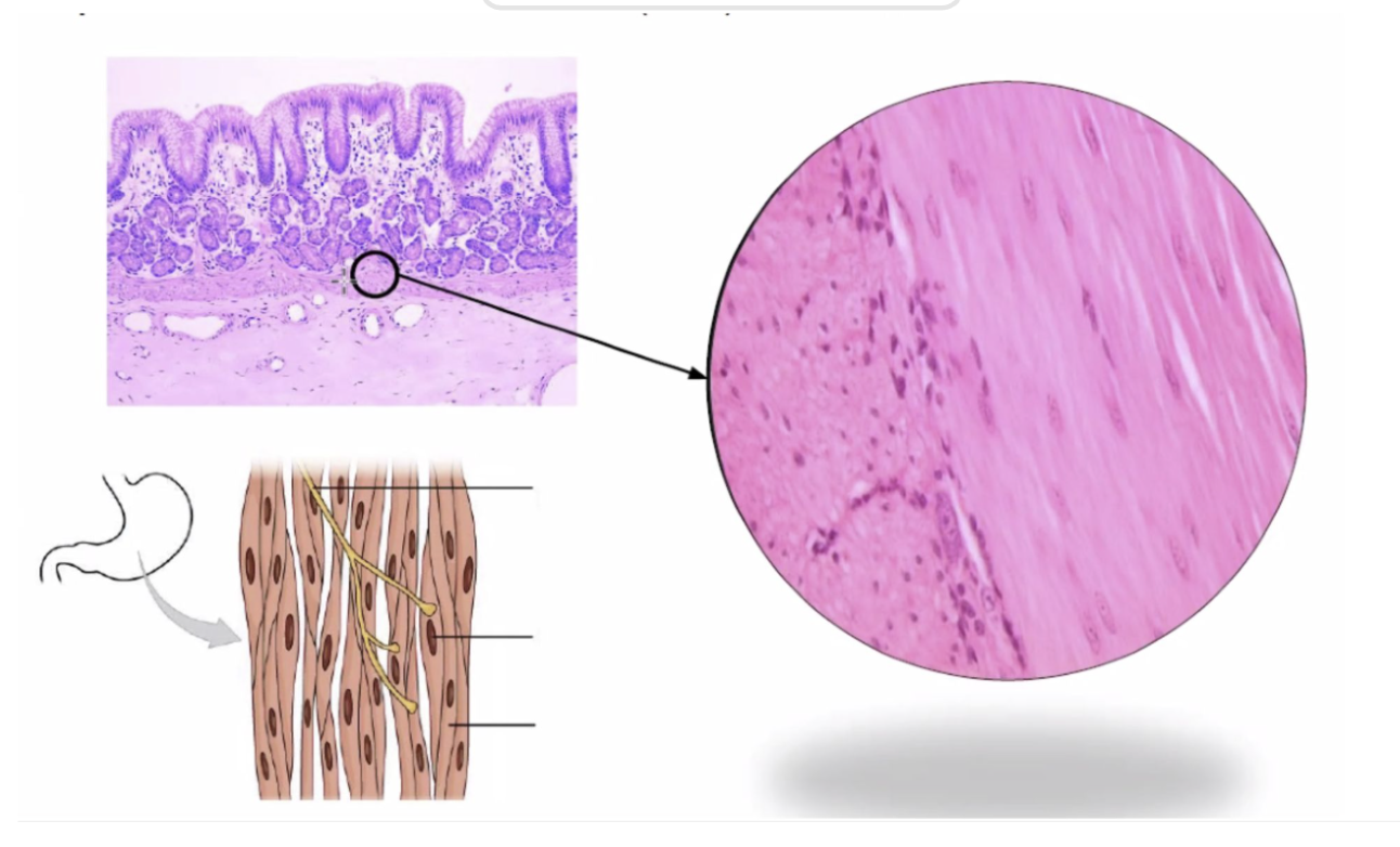
What specimen is this and what is the shape?
Frog stomach and Fusiform or Spindle-shaped cell

Shape of skeletal muscle fiber
cylindrical, striated
cardiac mucos shape of fiber
short and branched
Smooth muscle shape of the fiber
spindle-shaped
Number and location of nucleus in skeletal muscle
multinucleated; located in the periphery
Number and location of nucleus in cardiac muscle
1 or 2; located at the central region of the cell
Number and location of nucleus in smootj muscle
singular, located at the central region of the cell
Presence of striations
Only no striations in the smooth muscle
Presence of intercalated disks
Only present on cardiac muscle
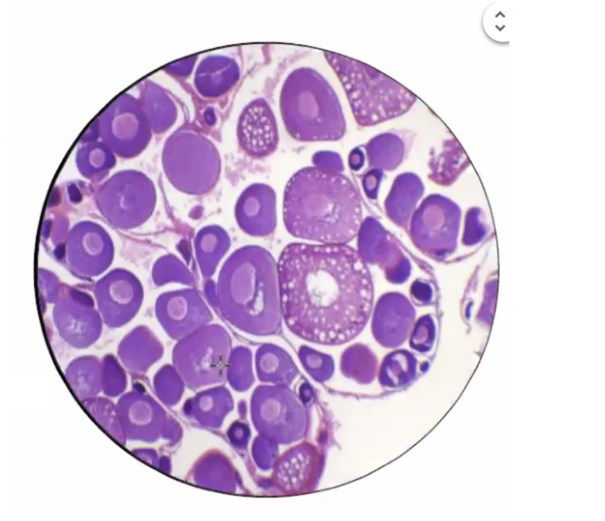
What specimen is this and what is the shape?
Frog ovary and spherical shape cell

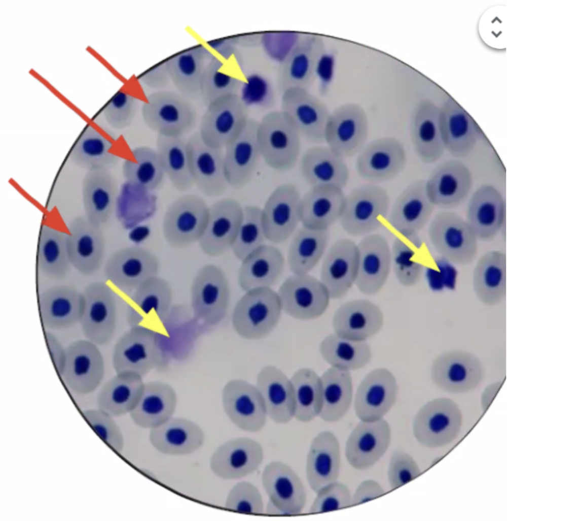
What specimen is this and what is the shape?
Frog blood smear and Oval and Amorphous

Enumerate the major stages of embroyonic development
Fertilization, cleavage, blastulation, gastrulation, neurulation, organogenesis

What specimen is this and what is the shape?
Frog Testis and Whiplike flagella


what is the specimen?
Human cheek cells


what is the specimen?
Frog early cleavage

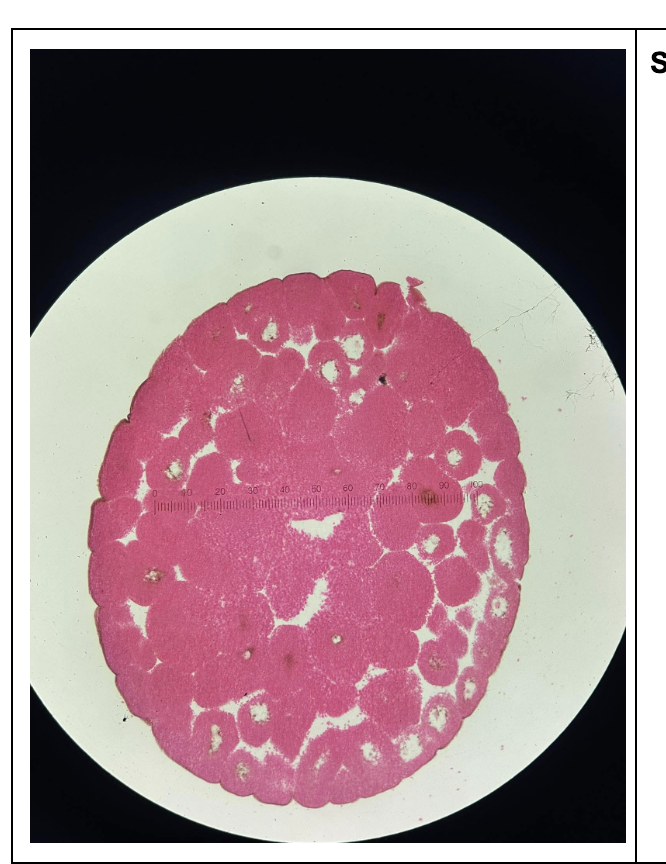
what is the specimen?
late cleavage


what is the specimen?
early blastula


what is the specimen?
late blastula


what is the specimen?
late gastrula


what is the specimen?
frog neural plate


what is the specimen?
frog neural fold


what is the specimen?
frog neural tube


What specimen is this and what is the tissue type?
simple squamous epithelial tissue
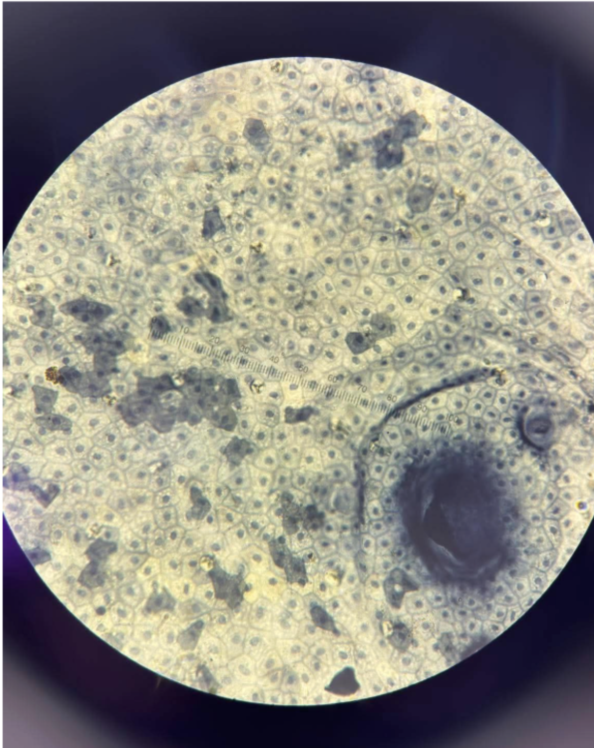

What specimen is this and what is the tissue type?
frog’s ovary and simple cuboidal epithelial tissue


What specimen is this and what is the tissue type?
frog’s intestine and simple columnar epithelial tissue

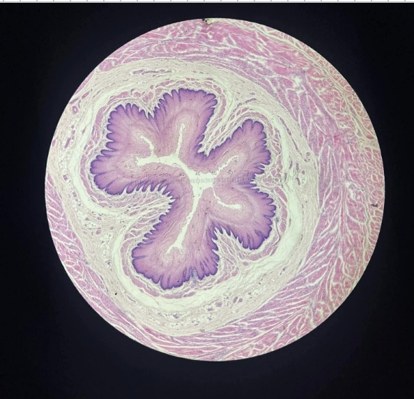
What specimen is this and what is the tissue type?
mammal’s trachea and ciliated pseudostratified epithelium


What specimen is this and what is the tissue type?
frog skin and stratified squamous epithelial tissue


What specimen is this and what is the tissue type?
human urinary bladder and transitional epithelium


What specimen is this and what is the tissue type?
mammal and areolar/loose connective tissue


What specimen is this and what is the tissue type?
dense connective tissue - human tendon


What specimen is this and what is the tissue type?
frog blood smear


What specimen is this and what is the tissue type?
human trachea and hyaline cartilage
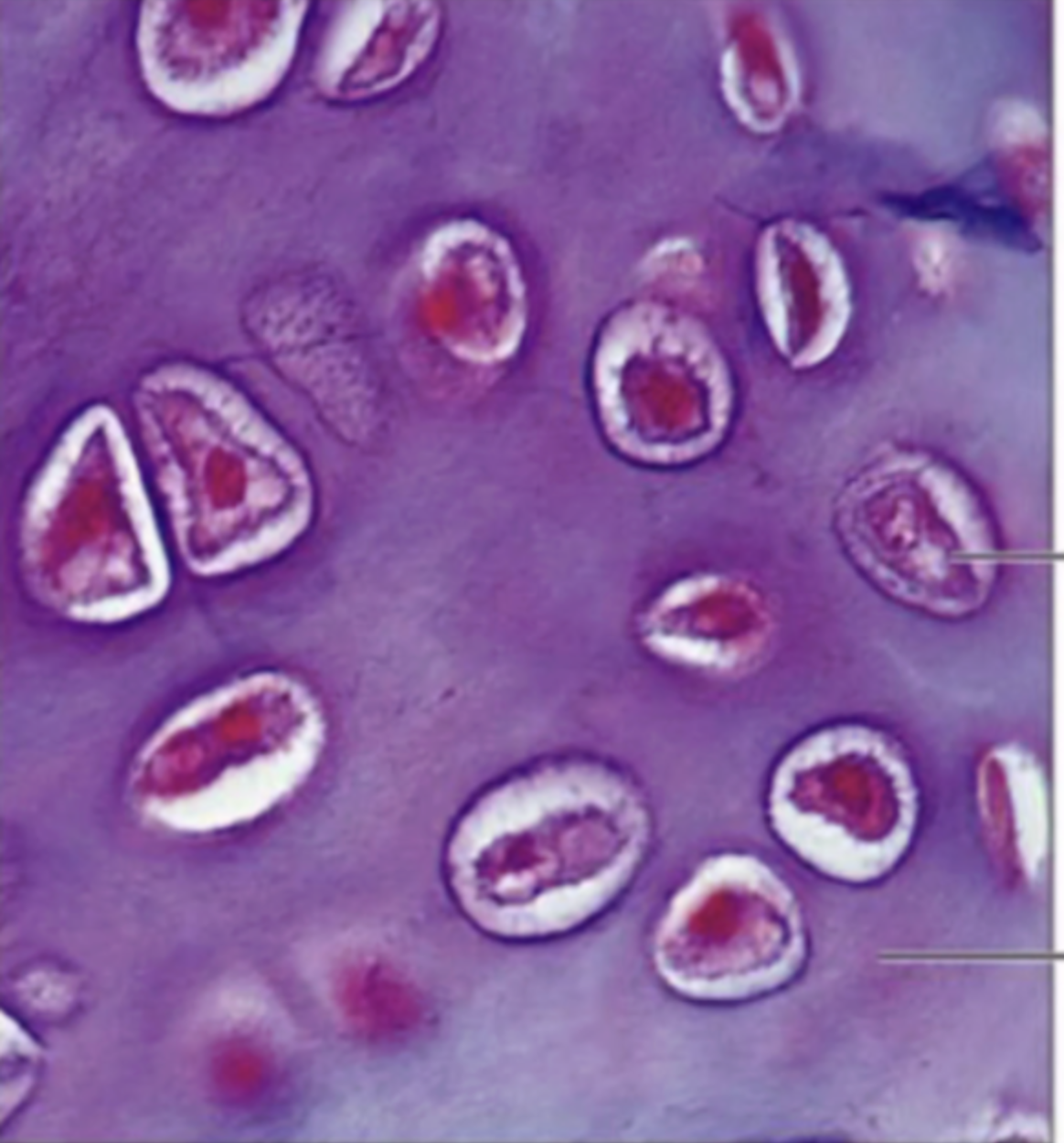

What specimen is this and what is the tissue type?
mammal and elastic cartilage


What specimen is this and what is the tissue type?
fibrocartilage


What specimen is this and what is the tissue type?
human bone and decalcified
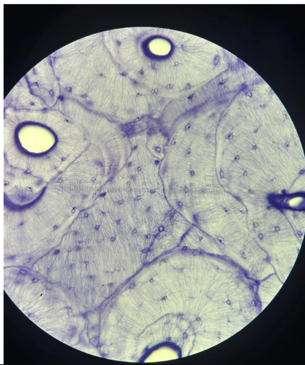

What part of cell division is this?
Interphase
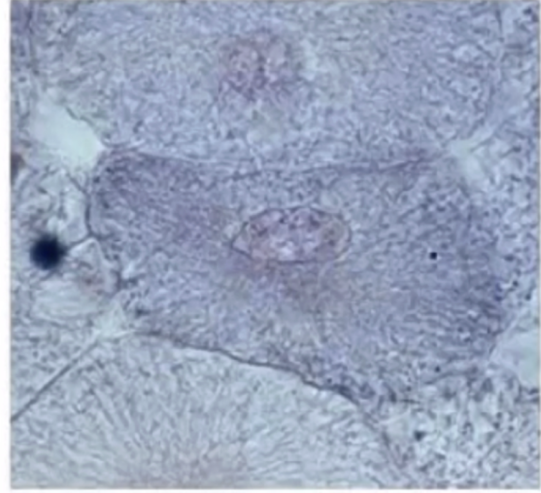

What part of cell division is this?
Interphase


What part of cell division is this?
prophase


What part of cell division is this?
prophase


What part of cell division is this?
metaphase


What part of cell division is this?
metaphase


What part of cell division is this?
metaphase
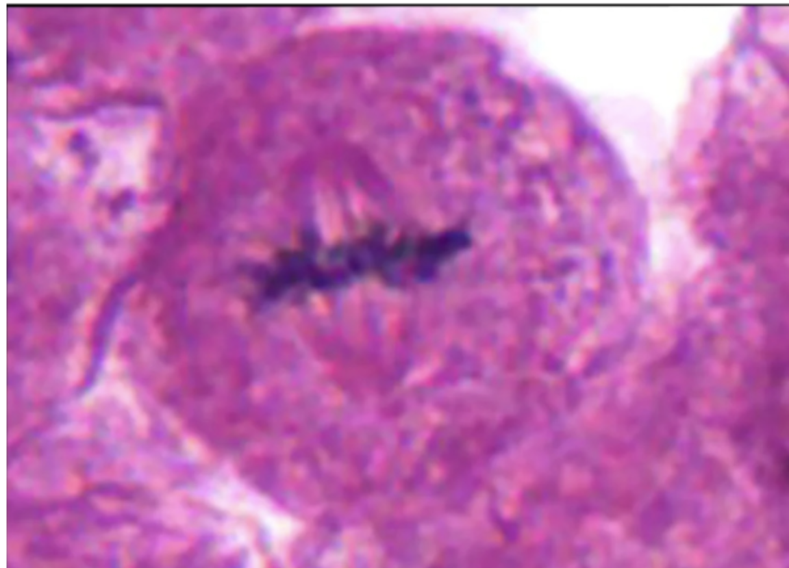

What part of cell division is this?
anaphase
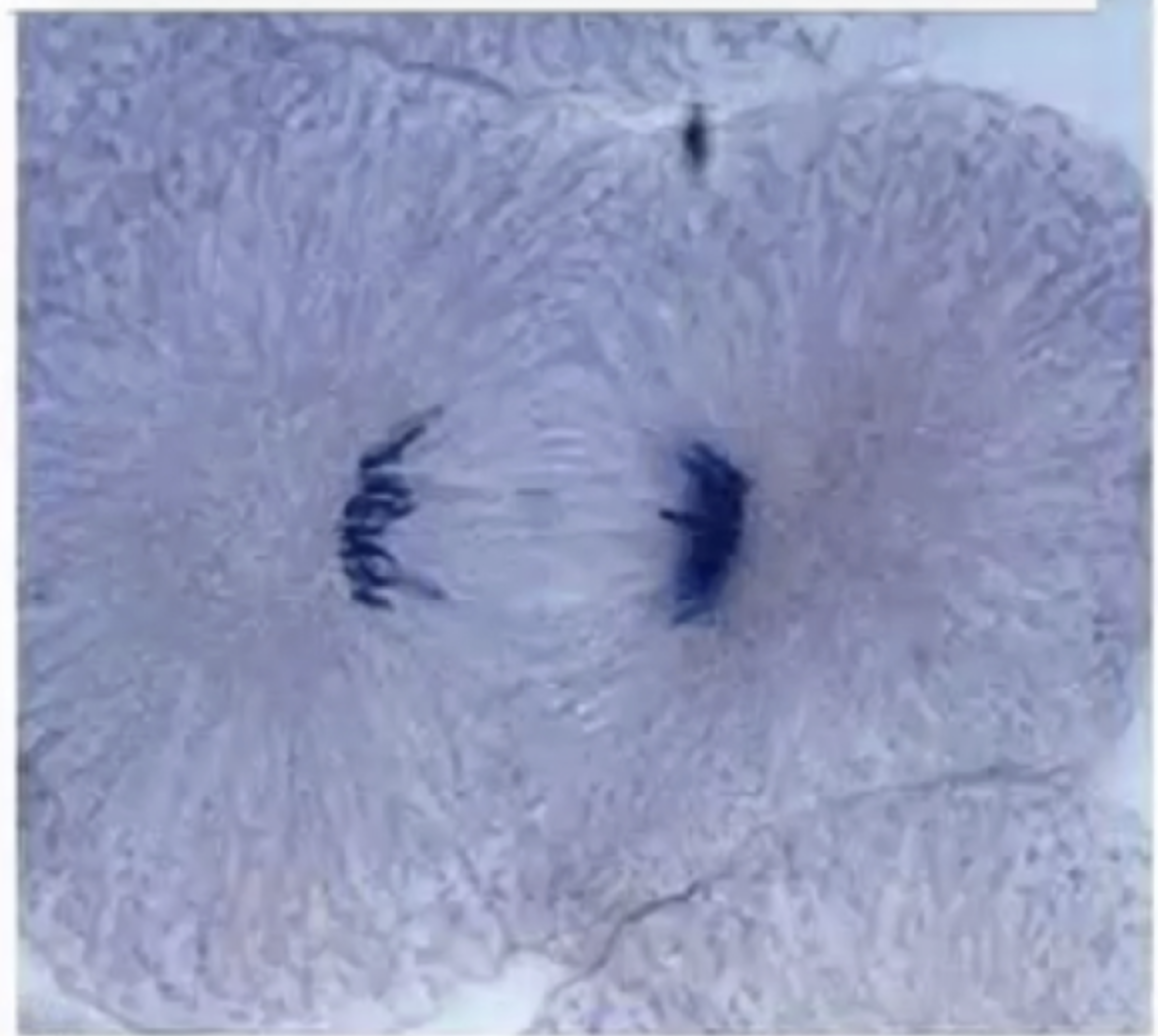

What part of cell division is this?
anaphase


What part of cell division is this?
telophase


What part of cell division is this?
cytokinesis


What part of cell division is this?
cytokinesis

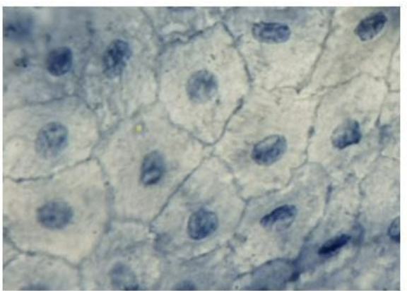
What is this
Stratified squamous epithelial tissue and Frog


what is this
simple cuboidal epithelium tissue and thyroid follicle

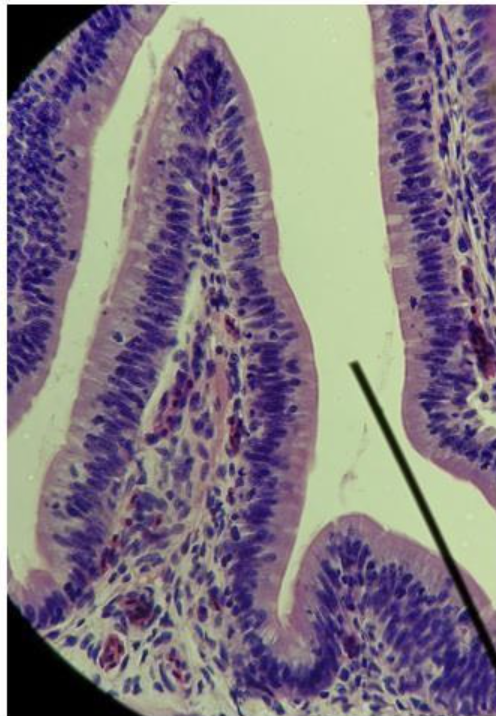
what is this
simple columnar epithelial tissue and frog intestine


what is this
stratified squamous epithelial tissue - frog skin


what is this
transitional epithelial tissue and urinary bladder
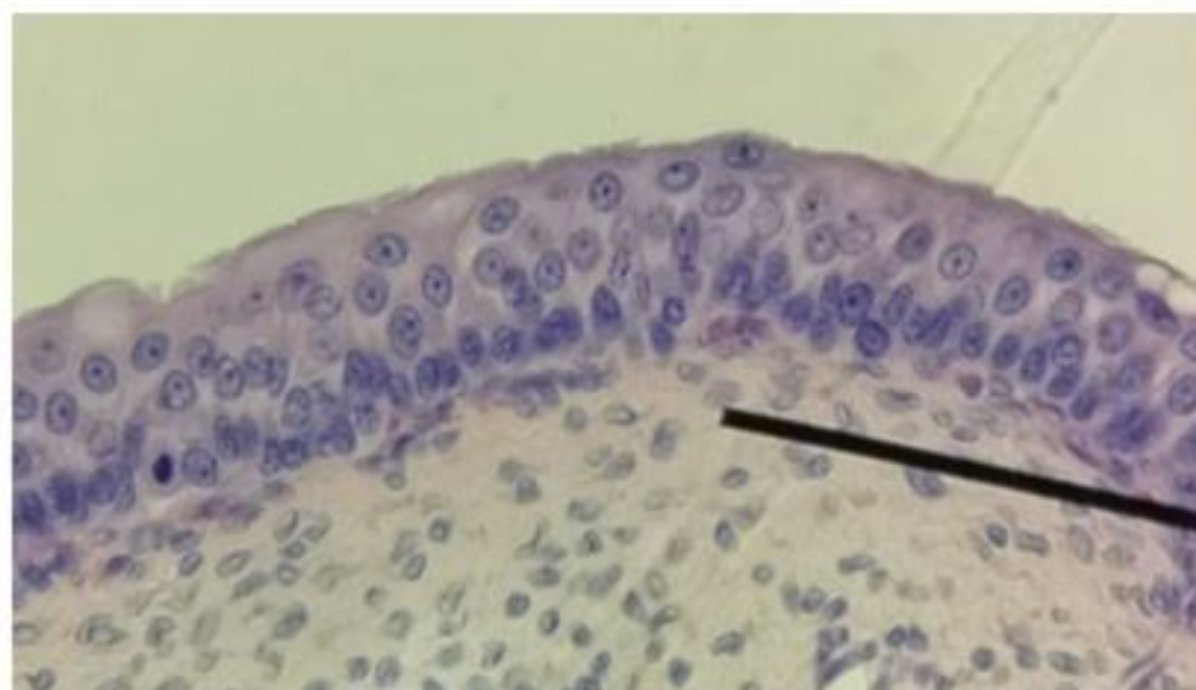

what is this
artery


what is this
frog spinal cord


what is this
ciliated pseudostratified columnar epithelial tissue and human trachea


what is this
loose connective tissue or areolar tissue


what is this
dense connective tissue and human tendon


What is this
adipose connective tissue and subcutaneous layer


what is this
blood smear


what is this
bone tissue


what is this
skeletal muscle
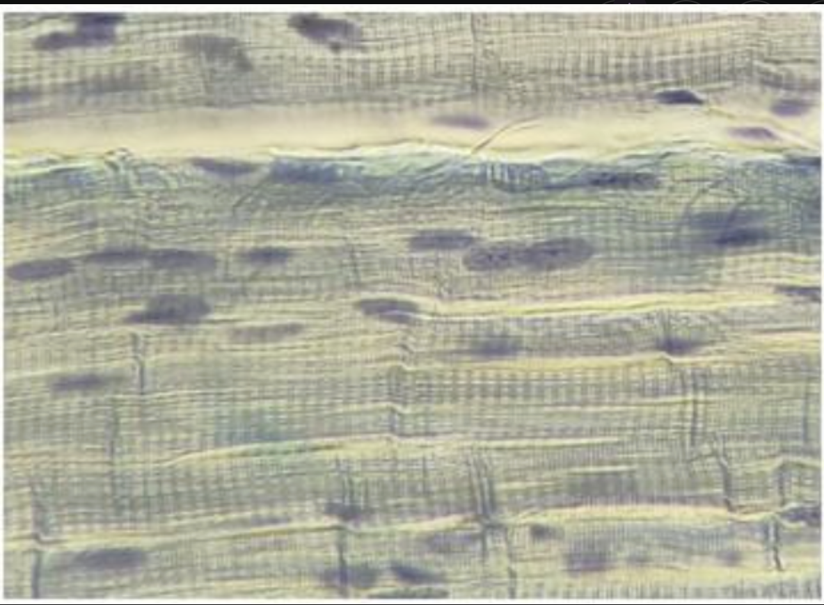

what is this
cardiac muscle

What are the advantage of staining the cheek cells?
specimen is clearer and more observable
Relate the shape of frog's red blood cells and frog's egg cell with their respective
function.
RBC is oval its helps transport oxygen to through the frog’s body. WBC is amorphous it helps stores nutrients for growth and developement
Enumerate the shapes of cells studied and indicate the organ where they are found.
Fusiform or spindle shaped - frog stomach ( smooth muscle)
Spherical - frog ovary
Amorphous - wbc
oval - rbc
Whiplike flagella - frog testis
What is the magnification of a compound microscope?
40x to 100x
What is the magnification of dissecting microscope?
5x to 50x
What is the dimension of a compound microscope
2 dimension
What is the dimension of a dissecting microscope
3 dimensional
Inversiom of image of a compound microscope?
Inverted
Inversion of dissecting microscope
Upright direction
Direction of movement of a compound microscope
Opposite direction
Direction of movemen of a dissectinf microscope
Same direction
How does one properly carry the microscope
Hold it with two hands, one at the arm, and one on the base
How does one improve contrast when using a dissecting microscope?
Shift the light source, make it dimmer or brighter, and properly adjust the iris diaphragm
Differentiate chromatin, chromatids, and chromosomes
chromatin are thin, thread like DNA material found in the nucleus when the cell is not dividing. Chromosomes are condensed and coiled chromatin visible during cell division, and chromatids are thick, identical copies of a chromosome joined at the centromere
why is white fish blastula used to demonstrate the different stages of the cell cycle?
the cells divide rapidly