Non-Malignant Granulocyte Disorders
 Call Kai
 Learn
Learn Practice Test
 Spaced Repetition
 Match
Match Flashcards
 Knowt Play
1/46

There's no tags or description
Looks like no tags are added yet.
Name | Mastery | Learn | Test | Matching | Spaced | Call with Kai | Chat |
|---|
No analytics yet
Send a link to your students to track their progress
47 Terms
WBC Reference Range
3.5-10.5 × 10⁹/L
Neutrophil Reference Range
1.7-7.0 × 10⁹/L
Lymphocyte Reference Range
0.9-2.9 x 10⁹/L
Monocyte Reference Range
0.3-0.9 x 10⁹/L
Eosinophil Reference Range
0.05-0.5 x 10⁹/L
Basophil Reference Range
0.0-0.3 x 10⁹/L
Causes of Neutrophilia
Bacterial Infection
Drug Intoxication
Stress Response
Malignancy
Tissue Destruction/Injury (Inflammation)
Left Shift
>6 bands, >1 metamyelocyte, or >0.5 myelocyte
Leukemoid Reaction
White count >25.0 with left shift
LE Picture
Left Shift + nRBCs
Toxic Granules
Large, deep purple primary granules in the neutrophil cytoplasm
Formed when the neutrophil releases its secondary granules, making the primary ones more visible
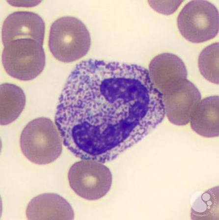
Dohle Bodies
Aggregates of ribosomal rER that appear in the peripheral cytoplasm of neutrophils
Blue-grey, rod-shaped, or oval inclusions
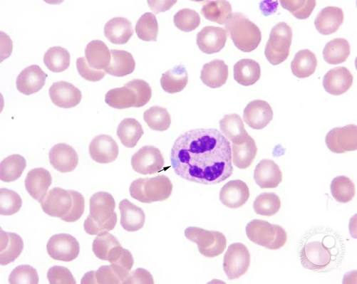
What are the three toxic changes seen in neutrophilia?
Toxic Granules
Dohle Bodies
Vacuoles
Causes of Neutropenia
Bone Marrow Failure
Chemotherapy
Drug Intoxication
Immune Destruction
Pseudoneutropenia
Caused by neutrophils clumping together
Results in falsely decreased white cell and neutrophil counts
Pelger-Huet Anomaly - Inheritance
Autosomal Dominant
Pelger-Huet Anomaly
The neutrophils function normally, but don’t segment beyond two lobes (hyposegmentation)
Nucleus may also appear round (mononuclear) with more mature chromatin
Alder-Reilly Anomaly
Large purple granules in the cytoplasm of all white cells
Alder = Abundance of granules

What disorders are associated with Alder-Reilly syndrome?
Decreased mucopolysaccharide production
(Hunter’s syndrome, Hurler’s syndrome)
How does Alder-Reilly Syndrome differ from toxic granulation?
All white cells are affected in Alder-Reilly syndrome
Chediak-Higashi Anomaly - Inheritance
Autosomal Recessive
Chediak-Higashi Anomaly
Fusion between primary and secondary granules in all white cells
How is function impacted in Chediak-Higashi anomaly?
Granules do not function properly
Reduced chemotaxis
Reduced bactericidal capabilities
What CBC findings are associated with Chediak-Higashi anomaly?
Neutropenia and Thrombocytopenia
May-Hegglin Anomaly
Giant (cigar-shaped) platelets and randomly distributed Dohle-like bodies in all white cells
May = Mega Platelets
What CBC finding is associated with May-Hegglin anomaly?
Thrombocytopenia
Chronic Granulomatosus Disease
Defect in the respiratory burst
Patients can phagocytize bacteria, but cannot form the superoxides needed to kill the bacteria
Bacteria survive; patients often have frequent infections
What test is used to confirm chronic granulomatosus disease?
Nitroblue Tetrazolium Dye Test
Neutrophils are mixed with nitroblue tetrazolium dye and bacteria
If they can take up the bacteria and initiate oxygen intake, the solution turns blue
In CGD patients, the solution will stay yellow
Myeloperoxidase Deficiency
Cells are deficient in myeloperoxidase
Neutrophils will show up on the PBS and appear morphologically normal
They will not show up on the automated CBC scatterplot
What disorders are associated with pseudo Pelger-Huet cells?
Myelodysplastic Disorders
Myeloproliferative Neoplasms
How can Pelger-Huet and Pseudo Pelger-Huet cells be distinguished?
In Pelger-Huet anomaly, all neutrophils will be hyposegmented
There will be a mix of “normal” and hyposegmented neutrophils in pseudo Pelger-Huet cases
Where are the findings in Alder-Reilly anomaly most prominent?
Cells of the bone marrow
Causes of Eosinophilia
Parasitic Infections
Hypersensitivity Reactions
Allergies
Malignancies
Hypereosinophilic Syndrome
An eosinophil count >1.5 × 10⁹/L with tissue infiltration and no apparent cause
Associated with Charcot-Leyden crystals & heart damage
How can HES and eosinophilic leukemia be distinguished?
HES will generally involve mature eosinophils
EL will exhibit increased eosinophilic myelocytes and myeloblasts
Charcot-Leyden Crystals
Formed from eosinophil breakdown products (eosinophil granules + cytoplasm)
They are found in tissues or fluids where eosinophils migrate and then break down
Causes of Basophilia
Hypersensitivity Reactions
Chronic Myeloproliferative Disorders
Causes of Monocytosis
Malignancies
Inflammation
Immune Disorders
Chronic Infection
What malignancies are associated with monocytosis?
Acute monocytic and myelomonocytic leukemias
Chronic myelomonocytic leukemia
Gaucher Disease - Cause
Glucocerebrosidase deficiency
Macrophages can’t digest the stroma of ingested cells, and glucocerebrosidase accumulates
Gaucher Cells
20-80 microns
Small, eccentric nucleus
Wrinkled/striated “tissue paper” cytoplasm filled with lipid

Gaucher Disease - Inheritance
Autosomal Recessive
Gaucher Disease - Clinical Findings
Hepatosplenomegaly
Anemia
Leukopenia
Thrombocytopenia (& bleeding)
Elevated Serum Acid Phosphatase
Niemann Pick Disease
Missing/deficient sphingomyelinase
Niemann-Pick Cells
Lymphoid tissue and bone marrow
20-100 microns
Eccentric nucleus
Globular, foamy cytoplasm filled with lipid droplets

Niemann-Pick Disease - Inheritance Pattern
Autosomal Recessive
Associated with Ashkenazi Jewish populations
Niemann-Pick Disease - Clinical Characteristics
Leukopenia
Thrombocytopenia
Poor physical development
Fatal by age 3