Anatomy Exam 3
 Call Kai
 Learn
 Practice Test
 Spaced Repetition
 Match
 Flashcards
 Knowt Play
1/187

There's no tags or description
Looks like no tags are added yet.
188 Terms
What are the general characteristics of muscle cells
specialized for contraction, aggregated into bundles, elongated, arranged in parallel bundles, and contain actin and myosin
What are the specialized structures of muscle cells?
Sarcoplasm: cytoplasm (mostly contains the proteins myosin and actin)
Sarcolemma: cell membrane (surrounds the muscle cell and carries electrical signals)
Sarcoplasmic reticulum: modified ER (stores and releases calcium needed for contraction)
What are the three types of Muscle Tissue?
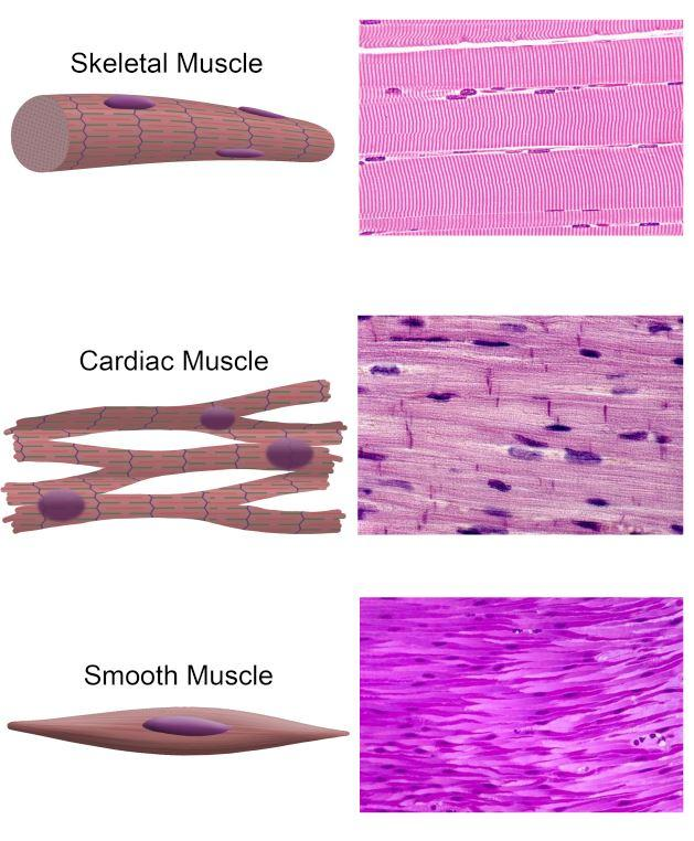
Skeletal muscle, Cardiac muscle, and Smooth muscle
What types of muscle tissues are striated (marked with a series of parallel lines, bands, stripes, or grooves) v non-striated
Striated: skeletal and Cardiac muscle
non-straited: Smooth muscle
How do skeletal muscle fibers develop?
Myoblasts are immature muscle cells that fuse together to form a long, multinucleated cell called myotubes
Myotubes differentiate (mature) into skeletal muscle fibers
Satellite cells remain alongside muscle fibers to help with muscle repair and limited regeneration

What are the characteristics of skeletal muscle and how does it attach to bone
Parallel, striated, multinucleated fibers with peripheral nuclei and rich blood supply
Can NOT divide so limited regeneration (get from satellite cell)
Attaches to bone through tendons made of collagenous fibers
What are the three connective tissue layers of skeletal muscle?
Endomysium: surrounds each muscle cell, containing capillaries
Perimysium: Surrounds bundles of muscle cells, contains larger blood vessels (fascicles)
Epimysium: surrounds entire muscle

What are myofibrils made of and what do they do?
Myofilaments: thick (myosin) and thin (actin)
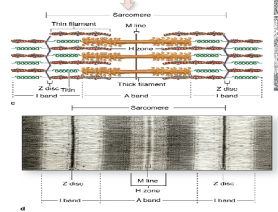
myofibrils contain the bands that create the striated (striped) appearance of skeletal and cardiac muscle (made of many repeating sacromeres)
What is a sacromere?
Functional unit of muscle, segment of myofibrils between two z-lines
contains the myosin filaments (A-band)

What are the A-band and I-band, and what do actin filaments do
A-band: contains thick myosin filaments and remains constant (does not change in size)
I-band: region between adjacent A-bands, contains only actin filaments and shortens as actin filaments slide
Actin filaments attach to Z-line and extend toward center of the sarcomere through I-band and partly into A-band
Repeating sarcomeres with alternating A-bands and I-bands is what produces the striated appearance
What are T-tubules
Inward extensions of the muscle fiber's plasma membrane (sarcolemma) that carry action potentials deep into the muscle fiber, triggering muscle contraction.
What is a triad?
One T-tubule and two terminal cisternae (enlarged sacs of the sarcoplasmic reticulum (SR) that store calcium (Ca²⁺) needed for muscle contraction)
What is a synapse
Junction where a neuron communicates with another cell, such as a muscle or gland
(neuromuscular junction is the location where they communicate and it has the presynaptic, synaptic, and postsynaptic parts)
What happens at the neuromuscular junction
The presynpatic terminal of a motor neuron releases acetylcholine (ACh) into the synaptic cleft, which binds to receptors on the sarcolemma, intiating a muscle action potential along sarcolemma and into T-tubules, Terminal cisternae release calcium, Actin and myosin interact, sarcomeres shorten
How does the action potential spread through the muscle
Travels down the sarcolemma and down the T-tubules
What causes muscle contraction
shortening of sarcomeres
What is a motor unit?
single motor neuron axon and all muscle fibers it innervates
What are Type 1 muscle fibers
Slow-twitch, red, oxidative, fatigue-resistant
What are Type 2a muscle fibers
Fast-twitch, intermediate, oxidative-glycolytic, fatigue-resistant
What are type 2b muscle fibers?
Fast-twitch, white, glycolytic, fatigue-prone.
How do the soleus and extensor digitorum longus (EDL) differ?
Soleus: slow-twitch, oxidative, fatigue-resistant. EDL: fast-twitch, glycolytic, fatigable
skeletal muscle organization
Whole muscle → fascicle → muscle fiber → myofibril → sarcomere → myofilaments
What are the characteristics of cardiac muscle
Striated, mononucleated (one central nucleus), smaller branching cells that interconnect, and no satellite cells (meaning cant repair).
What are intercalated discs?
Connections between cardiac muscle cells containing desmosomes and gap junctions
What is the function of gap junctions in cardiac muscle?
Allow excitation to spread between cardiac cells for coordinated contraction.
How does cardiac muscle contraction begin?
Spontaneously through pacemaker cells; signals travel through conducting fibers, including Purkinje fibers.
How does cardiac muscle's blood supply compare to skeletal muscle?
Cardiac muscle has more capillaries because it is highly sensitive to lack of oxygen
What are the characteristics of smooth muscle?
Non-striated, involuntary, spontaneously active, maintains tone, and can regenerate.
What does the nucleus of smooth muscle look like?
Ellipsoid (elongated); may appear corkscrew-shaped during contraction.
How are smooth muscle myofilaments organized?
Arranged in multiple planes and anchored to the plasma membrane.

How do smooth muscle cells contract
Actin and myosin interact and slide past each other, causing the cell to shorten and thicken. Unlike skeletal and cardiac muscle, smooth muscle does not contain sarcomeres
Where is smooth muscle found?
Walls of hollow organs, blood vessels, digestive tract, uterus, respiratory and urogenital ducts.
What are myoepithelial cells?
Specialized contractile cells associated with exocrine glands, such as salivary and mammary glands. (basically smooth muscle cells are found in a variety of other organs)
What makes up the musculoskeletal system
Bones, muscles, joints, tendons, cartilage, ligaments, and connective tissue.
What are the functions of the musculoskeletal system?
Support/structure
movement
protection of organs
mineral storage (calcium and phosphorus)
blood cell production (bone marrow)
shock absorption and reduces friction during movement
What is the difference between a muscle's origin and insertion?
Origin: typically the more stationary attachment. Insertion: typically the more movable attachment.

How do skeletal muscles produce movement?
Sarcomeres shorten → causing the muscle to contract.
Muscles pull (never push) → pulling on bones through tendons.
Bones move at joints → producing movement through a lever system
In order to produce a movement, a muscle must be attached to two bones and cross at least one joint
What is an agonist vs. antagonist?
Agonist (prime mover): muscle primarily responsible for a movement. (ex: (biceps) contracts and shortens to bend the elbow)
Antagonist: opposes the agonist's movement. (ex: (triceps) relaxes and lengthens to allow the movement)
What is fascia?
Dense irregular connective tissue that covers, separates, or holds together muscles, organs, blood vessels, and nerves.
What is muscle tone?
Slight, continuous contraction of muscles even at rest.
What is an isometric contraction?
Muscle tension increases (sarcomeres shorten bc of increased tension), but overall muscle length does not change.
What is an isotonic contraction?
Muscle changes length (shortens or lengthens) while moving a load.
concentric: tension development while shortening
What is the difference between shunt and spurt muscles?
Shunt: pull parallel to the bone to stabilize a joint. Spurt: pull obliquely to the bone to produce effective movement.
Which muscle contraction is associated with the most injuries?
Isotonic eccentric contractions (Because the muscle lengthens while contracting, creating high tension that can damage or tear muscle fibers)
How are muscles named?
By shape, size, attachments, and functions
What is an aponeurosis?
A wide, flat tendon
What makes up the axial skeleton?
Skull, vertebral column, and thoracic cage
What makes up the appendicular skeleton?
Pectoral (shoulder) girdle, Upper limb, Pelvic girdle, Lower limb
What are the 3 main types of joints?
Fibrous: little/no movement
Cartilaginous: slight movement
Synovial: freely movable
What are fibrous joints (synarthroses)? and what does it have (2 things)
Bones joined by fibrous connective tissue with limited or no movement.
sutures: Fibrous joints connecting skull bones.
syndesmosis: Bones joined by dense fibrous connective tissue, such as interosseous ligaments
What are Cartilaginous joints? and what does it have (2 things)
joints where bones are connected by cartilage rather than a joint cavity (little to no movement)
synchondrosis: Cartilaginous joint connected by hyaline cartilage (flexible tissue), e.g., epiphyseal plate or costochondral joint
symphysis: Cartilaginous joint connected by fibrocartilage (ex: stable hard to move), e.g., pubic symphysis
What are synovial joints (diarthroses)
freely movable joints that have a fluid-filled joint cavity between the bones
What is synostosis?
Fusion of bones
What are the 2 types of synoviocytes in the synovial membrane?
Type A: macrophage-like (remove debris and waste from the joint) Type B: fibroblast-like (produces components of synovial fluid)
What are the 4 main structures of a synovial joint?
Hyaline articular cartilage covers bone ends; joint cavity contains synovial fluid; fibrous capsule stabilizes the joint; synovial membrane lines the capsule and helps produce synovial fluid.
dermatome
an area of skin innervated by a single spinal nerve
What is the brachial plexus?
A network of nerves from C5–T1 that supplies movement and sensation to the upper limb.
What are the five organizational levels of the brachial plexus?
Roots (5) → Trunks (3) → Divisions (3 anterior and 3 posterior) → Cords (3: lateral, medial, and posterior) → Branches (lot but 5 main ones)
What are the five major terminal branches?
Musculocutaneous, Axillary, Radial, Median, Ulnar (MARMU)

Movements of the Pectoral Girdle
depression and elevation
retraction and protraction

Three Main Muscles that move the Pectoral Girdle (Posterior)
Posterior:
Trapezius (one of the largest muscles on the back, elevates, retracts, depresses, and upwardly rotates scapula)
Levator scapulae (elevates the scapula (shrugs shoulders) and assists in downward rotation)
Rhomboids (Retract the scapula (pull shoulder blades together))

Anterior Muscles that move the Pectoral Girdle
Anterior:
Serratus anterior (Protracts the scapula (pulls shoulder blade forward), upwardly rotates it, and holds it against the rib cage)
Pectoralis minor (Protracts, depresses, and downwardly rotates the scapula. Innervated by the medial pectoral nerve)
Subclavius (Depresses and stabilizes the clavicle)

What muscles move the arm and originate from the axial skeleton?
Pectoralis major and latissimus dorsi
What muscles move the arm and originate from the scapula?
Deltoid, coracobrachialis, teres major, triceps brachii, and rotator cuff muscles
What are the actions and innervation of pectoralis major?
Actions: flexion, adduction, rotation of arm
Innervation: pectoral nerves

What are the actions and innervation of latissimus dorsi?
Actions: extension, adduction, rotation of arm
Innervation: thoracodorsal nerve

What are the actions and innervation of deltoid?
Actions: flex, extend, and abduct the arm
Innervation: axillary nerve
What are the actions and innervations of the rotator cuff (what is it composed of)
Composed of: infraspinatus, supraspinatus, subscapularis, and teres minor
surround shoulder joint: stabilize joint, abduct and rotate arm at shoulder
innervation: posterior branches of brachial plexus
all except the supraspinatus rotate the humerus (assists with abduction instead)
What do both the Coracobrachalis and Biceps brachii do and are innervated by what nerve? And what is the Triceps Brachii innervated by?
Flex
Musculocutaneous nerve
Triceps: radial

What is the glenohumeral joint and what helps stabilize it?
The shoulder joint between the head of the humerus and glenoid cavity of the scapula
shallow ball-and-socket joint so it has Rotator cuff muscles and their tendons to help stabilize it
What muscles are in the anterior compartment of the arm, what are their actions, and what nerve innervates it?
Biceps brachii (also supinates!!), brachialis, coracobrachialis
Actions: Flexion at shoulder/elbow, adduction at shoulder, and supination
Innervations: Musculocutaneous nerve
What muscle is n the posterior compartment of the arm, what are their actions, and what nerve innervates it?
Triceps brachii
Actions: extension at elbow and shoulder
Innervation: radial nerve
Which muscles flex the arm at the shoulder and Which muscles flex the forearm at the elbow?
flex arm at shoulder: Coracobrachialis and biceps brachii
flex forearm at elbow: Biceps brachii and brachialis
What are the two bones of the forearm and what connects them?
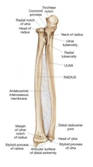
Radius (lateral) and ulna (medial) connected by Interosseous membrane, a type of fibrous joint called a syndesmosis
What are the actions of the anterior forearm compartment and what nerves innervate them?
Flexes wrist/digits and pronates forearm
Innervated primarily by Median nerve, but also Ulnar
Brachioradialis is the exception and is innervated by the Radial nerve
What are the actions of the posterior forearm compartment and what nerves innervate them?
Actions: extension of wrist and digits
Innervation: radial nerve
Carpel Tunnel Syndrome
a medical condition that happens when swelling or pressure squeezes the median nerve inside the rigid passageway made of bones and a tough ligament
What muscles supinate the forearm?
Biceps brachii (musculocutaneous nerve) and supinator (radial nerve)
What muscles pronate the forearm?
Pronator teres and pronator quadratus (median nerve)
What are the 3 groups of intrinsic hand muscles?
Thenar, hypothenar, and midpalmar
What are the functions and innervation of thenar muscles?
Flex, abduct, and oppose thumb. Median nerve
What are the functions and innervation of hypothenar muscles?
Flex, abduct, and oppose 5th digit. Ulnar nerve
What are the functions and innervation of midpalmar muscles
Fine movements of digits. Primarily ulnar nerve
What are the subdivisions and major joints of the lower extremity?
Subdivisions: gluteal region, thigh, leg, foot
Joints: hip, knee, ankle, foot
What 3 bones form the pelvic bone? And where does one of them articulate
Ilium, ischium, and pubis (fused together)
Where does the ilium articulate: With the sacrum; the two pubic bones meet at the pubic symphysis

Why are females pelvis wider than mens
Because it is adapted for childbirth and makes the angle more obtuse for giving birth
What are the major bones of the ankle?
Talus: articulates with tibia
Calcaneus: heel bone, transmits most weight to ground
What are the two arches of the foot and what forms them?
Metatarsals and ankle bones form arches, longitudinal and transverse

What are the main muscles and actions of the gluteal region?
Muscles: gluteus maximus, medius, minimus, tensor fasciae latae
Actions: primarily extension and abduction of thigh
Innervation: gluteal nerves
What is the primary action of gluteus maximus?
Extension of the thigh at the hip
What is the primary action of gluteus medius and minimus?
Abduction of the thigh at the hip
What are the main muscles and actions of the anterior thigh?
Actions: flex thigh at hips
Muscles: Iliopsoas (iliacus + psoas major), sartorius, and rectus femoris
Innervates: femoral nerve (lumbar plexus)
What are the three compartments of the thigh?
Anterior, medial (adductor), and posterior
What are the four muscles of the quadriceps femoris? and what are the actions and innervation of quadriceps femoris?
Rectus femoris, vastus lateralis, vastus medialis, vastus intermedius
Action: extends leg at knee
Innervation: femoral nerve
What are the muscles of the posterior thigh? and what are the actions and innervation of the posterior thigh muscles?
Biceps femoris, semitendinosus, semimembranosus
Actions: extend thigh at hip and flex leg at knee
Innervation: sciatic nerve (largest nerve of the body)
hamstrings!
What muscles are in the medial thigh compartment and what are the actions and innervation
Adductor magnus, longus, brevis, and gracilis
Action: adduction of thigh at hip
Innervation: obturator nerve
What are the two bones of the leg and their locations?
Tibia: medial, weight-bearing, articulates with femur and ankle. Fibula: lateral, mainly muscle attachment and ankle stabilization

What connects the tibia and fibula?
Interosseous membrane (fibrous connective tissue). Unlike the radius and ulna, they have very little movement relative to each other
What are the 3 articulations of the knee joint?
Two femorotibial articulations (lateral and medial) and one femoropatellar articulation
What are the two layers of the knee joint capsule?
External: fibrous layer
Internal: synovial membrane