Unit 2 Anatomy/Physiology Written
 Call Kai
 Learn
Learn Practice Test
 Spaced Repetition
 Match
Match Flashcards
 Knowt Play
1/36

There's no tags or description
Looks like no tags are added yet.
Name | Mastery | Learn | Test | Matching | Spaced | Call with Kai | Chat |
|---|
No analytics yet
Send a link to your students to track their progress
37 Terms
Functions of Bone and Skeletal System:
Provide structural framework
Support soft tissue
Points of attachment for skeletal muscles
Protect internal organs
Assist in movement (with muscles)
Mineral storage of calcium and phosphorous
Production of blood cells in red bone marrow
Triglyceride storage in yellow bone marrow
Diaphysis
Bone shaft (portion between the epiphysis)
Articular cartilage
Located where bones come together to form a joint
Reduces shock and friction
Medullary cavity
Located within the shaft (diaphysis) of the long bone, inter most part of the bone
Hollow cavity filled with yellow bone marrow in adults
Osteons
Circular shaped units that make up compact bone
Formed by concentric layers of matrix called lamellae and has a central canal with a nervous and blood supply.
Also known as the Haversian system
Trabeculae
Latticework
cancellous bones containing numerous bars or plates that forms partitions in organs
support/strengthens: follow the lines of stress in spongy bone
Epiphysis
Ends of long bones
Periosteum
dense connective tissue that forms the outer covering of a bone
Compact bone
Solid dense bone tissue, no air spaces
Strong support
In diaphysis of long bones
Central haversian canal: blood vessels and nerves
Concentric lamellae: rings of hard, calcified extracellular matrix
Osteocytes: bone cells built by osteoblasts
Spongy bone
Lightweight
Covered by a protective layer of compact bone (inside the layer)
Contains latticework called trabeculae
Supports red bone marrow
Does not contain osteons
Makes up epiphysis of long bone and other types of bone (short, flat and irregular shaped bones)
Red bone marrow
Specialized tissue that produces blood cells, found in medullary cavity in most long bones (in infants)
Development of cartilaginous model
Structure is covered by perichondrium, cells gather in the site where the bone will form and become chondroblasts
Growth of cartilage model
Chondroblasts are now called chondrocytes. Chrondocytes increase in size. Cartilage begins to clacify and die.
Formation of primary ossification center
Perichondrium is now called the periosteum because the bone is present. Small cavities become the primary ossification center. A nurient artery appears in the center.
Development of medullary cavity
Osteoclasts break down spongy bone and begin to form the medullary cavity. Spongy bone remains present in the epiphysis and is not replaced with a medullary cavity or compact bone. Wall of diaphysis is replaced with compact bone. Primary ossification center expands to ends of bone.
Formation of primary ossification center
Secondary ossification center forms in the epiphysis. Medullary cavity fills with red bone marrow.
Formation of articular cartilage and epiphyseal plate
Line of cartilage remains between the epiphysis and diaphysis until adulthood
Stress Fracture
Tiny cracks in the bone by repetitive overuse (smallest type)
Comminuted Fracture
Bone breaks into more than two pieces
Greenstick fracture
Incomplete break where a bone bends and cracks on one side instead of snapping completely into separate pieces
Buckle or Torus Fracture
Incomplete fracture in which the break doesn’t go all the way through the bone
They are compression fractures which means the break is caused by sudden pressure on the bone
Avulsion Fracture
It’s where a small piece of bone attached to the tendon or ligament gets pulled away form the main part of the bone
Growth Plate Fracture
Break in the developing layer of soft cartilage near the ends of a child/teenager’s long bones
Compression Fracture
Partial collapse/break in the spinal bone (vertebrae)
How many regions does the vertebrae have and how many are there within each region?
5 regions
Cervical vertebrae: 7 bones, neck
Thoracic vert: 12 bones, chest/ribs
Lumbar vert: 5 bones, lower back
Sacrum: made of fused vert
Coccyx: made of fused vert
Normal curves of vertebral column
Infant has 1 concave curve
Adult has 4 curves
Purpose
Increase strength
Help maintain balance
Absorb shock
Protects vertebrae from breaks
Ribs
12 pairs
In back connects to: Corresponding thoracic vertebrae
In front connect to:
Pairs #1-7 Sternum by costal cartilage = true ribs
Pairs #8-10 Indirectly to rib #7= false ribs
Pairs #11-12 Do not connect = floating ribs
Ear
Hemostasis
It constricts blood vessels and forms blood clot to stabilize the broken ends of the bone

Inflammatory phase
Immune system sends white blood cells, macrophages to fracture site in which its purpose is to clear the germs around the break to reduce infection

Reparative phase
Chondroblasts are dispersed, which are responsible for forming cartilage, and they form a soft callus made of fibrous tissue which acts as a temporary bridge between the broken ends

Development of Hard Callus
Soft callus gets replaced by hard callus made of bone tissue, specialized cells called osteoblasts which deposit minerals like calcium and phosphorus making the callus more rigid

Remodeling phase
The body enters this phase by sending osteoclasts to breakdown and absorb the extra tissues formed earlier
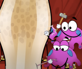
Steps of Fracture Repair
Hemostasis
Inflammatory Phase
Reparative Phase
Development of hard callus
Remodeling Phase
Vertebrae
1. Protects spinal cord, supports head
2. Attaches ribs, pelvic girdle, muscles of back
3. Intervertebral disc: cartilage between
How many regions does vertebrae have and how many are there?
5 regions
Cervical vertebrae: 7 bones, neck
Thoracic vert: 12 bones, chest/ribs
Lumbar vert: 5 bones, lower back
Sacrum: made of fused vert
Coccyx: made of fused vert
Normal curves of vertebral column:
Infant has 1 concave curve
Adult has 4 curves
Purpose
Increase strength
Help maintain balance
Absorb shock
Protects vertebrae from breaks