Biology Unit 3 Exam
 Call Kai
 Learn
 Practice Test
 Spaced Repetition
 Match
 Flashcards
 Knowt Play
1/42

There's no tags or description
Looks like no tags are added yet.
43 Terms
Mitosis
production of somatic cells (non-reproductive)
daughter cells identical to parent cell
Meiosis
production of gametes (eggs/sperm)
daughter cell = ½ genetic material as parent cell
involved in sexual reproduction & genetic inheritance
Cytokinesis
cytoplasm of cell divides into 2 distinct daughter cells
Mitosis + Cytokinesis Function
growth
wound repair
asexual reproduction
Chromosome
single, long, DNA double helix
encodes cell’s hereditary info
DNA wrapped around histone (protein) in highly organized manner
Chromatin
entire complex of DNA + protein that makes up chromosomes in eukaryotes
macro-structure formed by DNA wrapping around histones (DNA-protein complex)
Gene
region of DNA in chromosome that codes for particular protein or RNA
Chromosome Replication
before mitosis/meiosis, each chromosome is replicated
when cell division begins, chromosomes condense into compact structures that can be moved around cell efficiently
half of a replicated chromosome: chromatid
Replicated Chromosome Structure
2 sister chromatids joined along length by proteins called cohesions
centromere — attaches 2 sister chromatids at specialized region; hub for kinetochore assembly and spindle attachment
2 chromatids in a single replicated chromosome
Replicated/Replicated/Condensed Chromosome
Un-replicated chromosome — single, long DNA double helix wrapped around proteins
Replicated chromosome — consists of 2 copies same DNA double helix (2 sister chromatids/2 identical DNA double helices)
Condensed replicated chromosome — DNA condensed around histones (10,000x shorter than o/g length)
Cell Cycle Events
Interphase — growth and replication
DNA replication (S-phase)
M-Phase — partitioning
Mitosis
Cytokinesis
Overview Cell Cycle
M-phase
cells are in process of separating chromosomes
Interphase
chromosomes uncoil into long, thin structures
cell is either growing/preparing to divide/fulfilling specialized function in multicellular individual
cells spend most of their time in interphase
S-Phase: DNA replication
sandwiched between Gap Phases 1 (and G0) & 2
Overview Cell Cycle
Gap phases (G1 & G2) — organelles replicate and additional cytoplasm is made
cells grow and replicate organelles so it can divide into 2 cells that function normally
G1 - cell decides to begin replication and transition to S-phase
G2 - preparation for M-phase
Synthesis Phase — DNA replication
M phase — mitosis (separation of chromosomes & nuclear division) / cytokinesis (cytoplasmic division)
M Phase
Mitosis — division of replicated chromosomes (in nucleus) and formation of two daughter nuclei with identical chromosomes and genes
daughter cells identical to parent cell
Cytokinesis — division of cytoplasm into 2 daughter cells
5 Phases of Mitosis
Interphase — chromosomes are replicated & centrosomes are replicated
Prophase — chromosomes condense; spindle apparatus begins to form
Prometaphase — nuclear envelope breaks down; microtubules contact chromosomes at kinetochores
Metaphase — chromosomes complete migration to middle of cell
Anaphase — sister chromatids separate into daughter chromosomes, pulled to opposite poles of spindle apparatus
Telophase — nuclear envelope reforms and chromosomes decondense
Cytokinesis — cell division begins (actin-myosin ring causes plasma membrane to pinch in) & cell division completes with formation of 2 daughter cells
Prophase
chromosomes condense into compact structures
spindle apparatus forms
moves replicated chromosomes during early mitosis
pulls chromatids apart during late mitosis
in S phase, centrosome has replicated along with DNA; 2 centrosomes move to opposite sides of nucleus to form spindle apparatus
centrosomes (2 centrioles each — microtubules arranged in 9 sets of triplets)
radiate microtubules and move to opposite poles, generating mechanical pushing force
helped by motor proteins
Prometaphase
once chromosomes condensed, nuclear envelope disintegrates
microtubules attach to the chromosomes' kinetochores, and motor proteins push and pull the chromosomes toward the center of the cell
allows cytoplasmic microtubules to attach to chromosomes at structures called kinetochores
each sister chromosome has its own kinetochore
kinetochore assembled at centromere
each chromosome has 2 kinetochores attached to microtubules originating from opposite sides of spindle apparatus
chromosomes secured to plus ends of microtubule via kinetochore proteins
chromosomes begin to be pushed & pulled by microtubules and motor proteins until they reach the middle of the spindle
Metaphase
once all chromosomes have migrated to middle of spindle, metaphase begins
all chromosomes are fully aligned along the metaphase plate (the imaginary equator in the middle of the spindle), ready to be separated
spindle apparatus completely formed so that each sister chromatid is connected to microtubules at its kinetochore
astral (free) microtubules extend from MTOC and interact with plasma membrane proteins to help hold spindle poles in place
microtubules are continually grow and shrink by adding and losing α/β-tubulin dimers (tug of war/highly dynamic)
polymerize at (+) end — equator
depolymerize at (-) end — poles
keeping length steady
Anaphase
sister chromatids separate and travel to opposite poles
become independent chromosomes when centromere splits
doubles number of chromosomes in cell
centromere (consisting of DNA & specialized chromatin) splits & cohesin (holding sister chromatids together) is cleaved by enzyme; tension pulls chromatids apart
movement
daughter chromosomes move to opposite poles via attachment of kinetochore proteins to shrinking microtubules
two poles of spindle apparatus are pushed and pulled further apart
Chromosome Movement During Anaphase
tubulin subunits of microtubule are lost from (+) ends at the kinetochore as (+) end frays and disassembles
dynein & other kinetochore motor proteins attach to kinetochore’s fibrous crown
motor proteins “walk” towards - end of spindle fiber (microtubule)
chromosome is pulled to one end of mitotic spindle
by force generated when kinetochore microtubules depolymerize
Telophase
complete set of chromosomes arrive at each spindle pole
microtubules of mitotic spindle break down & disappear (disintegrate)
nuclear envelope reforms around each set of chromosomes = 2 new nuclei
chromosomes decondense and become less visible
Cytokinesis
cytoplasm divides → 2 daughter cells
each daughter cells has its own nucleus and complete set of organelles
ring of actin & myosin filaments contract inside cell membrane, causing it to pinch inward (cleavage furrow)
contraction — myosin motor proteins that bind to actin filaments use ATP to slide filaments past one another
ring shrinks & tightens
contracting ring pulls plasma membrane with it
starts in telophase and finishes shortly after
Control of Cell Cycle
regulation varies among cells in the same organism/different organisms/cell types
variations in length of G1 phase
rapidly dividing cells = very short G1
nondividing cells = permanent G1 (G0)
rate of cell division varies in response to changing environmental conditions
Variation in Cell Cycle Example: Liver Cells
Normal state (G0): In a healthy adult liver, hepatocytes exit G1 into a quiet G0 resting state, dividing only 1-2x a year to replace worn-out cells.
Environmental trigger (Injury/Loss): If the liver is damaged—toxins (alcohol), viral infection, or surgical removal of part of the liver—surrounding environment floods with growth factors
Re-entering G1: Those chemical signals act as a green light, pushing hepatocytes out of G0 and back into G1
Rapid division & return to G0: The cells pass through G1, S, G2, and M phases rapidly to regenerate the lost liver mass.
Can divide every 2 days!
Once the liver returns to its normal size, inhibitory signals tell the cells to exit the cycle and return to G0
Cell Cycle Regulation
2 main proteins
CDKs (cyclin-dependent kinase)
Cyclins
ex) MPF (Maturation Promoting Factor)
Cyclin-Cdk Complex Regulates Cell Cycle
Cyclins
protein
binds to Cdks
activates them only at right time in cell cycle
cyclin concentration fluctuates in dividing cells
KEY: when concentration is high, CDK activity is high & target proteins are phosphorylated
Cyclin-dependent Kinase
enzyme
phosphorylates (activates/inactivates) proteins important for different stages of cell cycle
catalyzes transfer of Pi from ATP → target protein
Summary
cyclin binds to & activates CDK to control progression through cell cycle
cyclin CDK complex phosphorylates target proteins that promote cell division

Example of Cyclin-Dependent Kinase: MPF
Maturation Promoting Factor
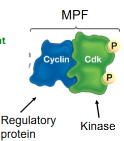
Cyclin-CDK complex (MPF only exists when Cyclin B is bound to Cdk1)
triggers cell’s passage past G2 checkpoint and into the M phase (G2 → M)
Present in the cytoplasm of M-phase cells///starts in the cytoplasm and moves into the nucleus just before cell division (M-phase)
present in all actively dividing eukaryotic cells → induces meiosis in all eukaryotes
non-dividing cells (mature adult neurons & heart muscle cells) lack MPF bc. they are not preparing to undergo mitosis
MPF Cycle
Cyclin B and Cdk 1 complexes form and accumulate (in cytoplasm)
MPF becomes active around M phase entry
MPF phosphorylates target proteins involved in chromosome condensation, spindle assembly, and nuclear envelope breakdown (i.e., nuclear lamins and pore subunits)
MPF is inactivated and degraded via cyclin B destruction during M phase progression/exit
What does MPF Phosphorylate?
MPF - CDK1 & cyclin B
dephosphorylation of CDK at Site 2 activates MPF → MPF is now active kinase that can phosphorylate many substrates
activating effect via phosphorylation
chromosomal proteins (initiate M phase/chromosome condensation)
nuclear lamina (initiate nuclear envelope breakdown)
microtubule-associated proteins (activates mitotic spindle)
inhibitory effect via phosphorylation
enzyme that degrades cyclin (cyclin concentration declines → MPF concentration decreases)
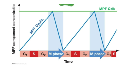
Fluctuating Concentrations of CDK & Cyclin
Cdk concentration remains constant throughout cell cycle while cyclin concentration peaks & decreases at different points in cell cycle
specific cyclin-CDK pairs are active during different cell cycle phases & cyclin concentrations rise and fall at specific stages
different cyclins are synthesized when needed and broken down once their phase ends
each cyclin's peak aligns with the phase where its CDK complex is needed (for example, cyclin B with CDK1 right before M phase; cyclin D with CDK4/6*** from G1 into S phase)
cyclin concentration is a primary driver, but CDK activity is also shaped by activating/inhibitory phosphorylation

CDK-Cyclin Complex
2 subunits: a) cyclin-dependent protein kinase, & b) cyclin protein
cyclin-dependent protein kinase
2 phosphorylation sites
Site 1: activated
Site 2: deactivated (inhibited)
both are phosphorylated simultaneously after cyclin binds to Cdk subunit, allowing the concentration of the dimer to increase without starting M phase early
(in G2 phase for MPF), PHOSPHATASE removes inhibitory phosphate
dephosphorylation + Pi present at site 1 → confirmational change in Cdk → kinase activity is ON
MPF created when cyclin binds to protein kinase
dephosphorylation of MPF at its inhibitory sites
carried out by phosphatase
triggered as the cell prepares to enter M phase once G2 checkpoints are satisfied and pro-mitotic signals rise
How is MPF turned OFF?
via negative feedback!
enzyme name: APC/C (Anaphase-Promoting Complex/Cyclosome)
an enzyme complex activated by events in mitosis deactivates MPF
remember, active MPF phosphorylates this enzyme (that degrades cyclin)
the enzyme complex adds UBIQUITINS to MPF cyclin subunit
ubiquitin = small regulatory protein/peptide tag that marks cyclin proteins for destruction by PROTEOSOME (large cylindrical/barrel-shaped protein complex)
proteosome breaks cyclin into small peptides, leaving CDK intact
in response to MPF activity, therefore, concentration of cyclin rapidly declines
builds slowly back up during interphase
Cell Cycle Regulation → There’s More!
changes in cyclin concentration & Cdk actively drive ordered events of cell cycle
many other protein complexes, however, regulate the cell cycle at cell cycle checkpoints
hold cells in particular stage or stimulate passage to next stage
What are Cell Cycle Checkpoints?
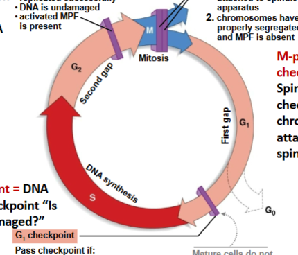
protein brakes that stop cell cycle if something is wrong & cell division need not proceed
3 critically regulated points in a cell cycle
at each checkpoint, interactions among regulatory molecules determine whether cell proceeds w/ division or not
G1 Checkpoint
Will cell divide or exit cell cycle & enter G0? Is DNA damaged?
Passes checkpoint if:
cell size is adequate
nutrients are sufficient
social signals are present (signaling molecules from other cells)
DNA is undamaged
if DNA is physically damaged, p53 protein (tumor suppressor) either
activates genes that stop cell cycle until damage is repaired
causes cell’s programmed death (apoptosis)
defective tumor suppressor = uncontrolled cell division
G2 Checkpoint
Will cell split into daughter cells? Is all DNA replicated?
Passes checkpoint if:
chromosomes replicated successfully
DNA is undamaged
activated MPF (triggers G2 → M)
first phosphatase must remove inhibitory Pi on Cdk subunit
M Phase Checkpoint
2 checkpoints:
Metaphase → anaphase
Passes if chromosomes attached to spindle apparatus & lined up at mitotic plate
KEY: defects at this checkpoint → chromosomes don’t separate correctly & daughter cells have too many/few chromosomes
Mitotic Exit
Passes if
chromosomes have properly segregated
MPF is absent (enzyme degrades cyclins, turning off Cdk1 activity)
Cancer
caused by cells that divide in uncontrolled fashion, invade other tissues, and spread to other sites in body
cancer cells use nutrients and space needed by unaffected cells & disrupt function of unaffected tissues
tumor formation: cells in multicellular organisms divide in uncontrolled fashion
benign — noninvasive
malignant — invasive; spreads via blood/lymph; initiates secondary tumors
mestasis: cancer cells detach from original tumor and invade other tissues
200+ cancer types
not a single illness — complex family of diseases that affect many organs
varies in time of onset, growth rate, cause
2 Cancer-Causing Defects in Cell Cycle
Activation: Activated proteins required for cell growth when the shouldn’t be active
ex) Ras
Inhibition: Tumor suppressor genes prevented from shutting down the cycle
ex) p53 — cells with damaged DNA keep dividing
Ras overview
small monomer G-protein
small monomeric GTPase
one subunit
acts as a molecular switch downstream of growth factor receptors (like receptor tyrosine kinases, or RTKs) to activate the MAPK signaling cascade
KEY: if Ras is “stuck on” (permanently active/cannot be inactivated), it continuously sends signals that trigger mitosis and cell division
normally, Ras turns itself off by hydrolyzing GTP to GDP
If a mutation prevents GTP hydrolysis, Ras stays permanently active ("stuck on"), continuously driving signals for cell growth, mitosis, and division → cancer
MAPK Pathway Review
Signaling molecule (such as a growth factor/mitogen) binds to a Receptor Tyrosine Kinase (RTK) on the plasma membrane, causing two receptor monomers to dimerize and cross-phosphorylate each other.
Adaptor proteins bind to the activated RTK and stimulate Ras to swap GDP for GTP, turning Ras "on."
Active Ras triggers a sequential chain of phosphorylation events that amplify the signal:
Active Ras recruits and activates Raf (a MAP kinase).
Activated Raf phosphorylates and activates MEK (a MAP kinase).
Activated MEK phosphorylates and activates ERK (the final MAP kinase).
Activated MAPK3 moves into the nucleus and phosphorylates transcription factors, turning on genes that drive mitosis, cell division, and survival.
G1 Checkpoint & Cancer
cancer = loss of cell cycle control
many types of cancer involve defects at G1 checkpoint (commitment to DNA replication)
passage through G1 depends on available nutrients & cell size
key mechanism of G1 checkpoint: social control
cells divide in response from signals to other cells (→ cells divide only when their growth benefits the whole organism)
social control is based on growth factors
polypeptides/small proteins that bind to specific cell surface receptors
stimulate cell division in other cells