Week 3: Neuropathic/Diabetic foot ulcers
 Call Kai
 Learn
 Practice Test
 Spaced Repetition
 Match
 Flashcards
 Knowt Play
1/88

There's no tags or description
Looks like no tags are added yet.
89 Terms
1. Diabetes mellitus
2. Neuropathy
3. Vascular disease
4. Impaired ROM
5. Foot deformities
6. Mechanical stress
7. Prior ulcer or amputation
Risk factors for neuropathic ulcers:
1. D______ mellitus
2. N________
3. V_______ disease
4. Impaired R__
5. F____ deformities
6. M________ stress
7. P_____ ulcer or amputation
True
True or false: Diabetic foot is the most common type of neuropathic ulcer.
All - Gotchu
Hyperglycemia impairs which pauses of healing?
A. Proliferation
B. Inflammation
C. Remodeling
D. Hemostasis
1. Increase bacteria proliferation in high glucose environment
2. Decrease production and migration of cells required for healing
3. Decrease epithelial migration
4. Decrease blood flow
Hyperglycemia impairs healing by:
1. (Increase/decrease) bacteria proliferation in high glucose environment
2. (Increase/decrease) production and migration of cells required for healing
3. (Increase/decrease) epithelial migration
4. (Increase/decrease) blood flow
Symmetrical
Diabetic Neuropathy is usually symmetrical or asymmetrical?
Distal - feet and hands
Diabetic Neuropathy affects distal or proximal nerves first?
1. Age
2. Disease duration (~10 yrs)
3. Poor glucose control
Diabetic Neuropathy severe increases with:
1. A___
2. Disease d_____ (~10 yrs)
3. Poor g_____ control
Sensory
_________: type of diabetic neuropathy; poor awareness of trauma to the feet; occurs gradually; presents w/ paresthesia (burning, tingling, aching) that are painful and debilitating w/ false sense of protective sensation
A. Sensory
B. Autonomic
C. Motor

Motor
_________: type of diabetic neuropathy; paralysis of foot intrinsic w/ increased plantar forces, hallux valgus, claw toe, hammer toe
A. Sensory
B. Autonomic
C. Motor

Autonomic
_________: type of diabetic neuropathy; altered sweating, callus formation, blood flow (AV shunting --> less perfusion at skin level, vasodilation --> increase blood to bone, leaches calcium --> osteopenia/charcot arthropathy)
A. Sensory
B. Autonomic
C. Motor

Inflammatory & remodeling
Peripheral arterial disease slows which healing phases? Select all:
A. Hemostasis
B. Inflammatory
C. Proliferative
D. Remodeling
1. Decreased blood flow causing poor oxygen and nutrient delivery
2. Increased risk of infection due to limited delivery of immune cells and antibiotic to wound sites
Peripheral artery disease causes:
1. (Increased/decreased) blood flow causing poor oxygen and nutrient delivery.
2. (Increased/decreased) risk of infection due to limited delivery of immune cells and antibiotic to wound sites.
1. Leads to formation of products that stiffen collagen in tendons, ligaments, and joint capsules
2. Reduce ankle and 1st MTP mobility altering gait mechanics
3. Leads to abnormally high plantar pressures, especially under the forefoot and toes
4. Loss of protective sensation also causes abnormal gait
Hyperglycemia impact on ROM:
1. Leads to formation of products that stiffen c_____ in tendons, ligaments, and joint capsules
2. Reduce a____ and 1st M__ mobility altering gait mechanics
3. Leads to abnormally high plantar pressures, especially under the forefoot and toes
4. Loss of p______ sensation also causes abnormal gait
Motor
______ neuropathy causes flexor/extensor muscle imbalances of the toes and foot .... overtime this can cause foot deformities such as hammertoes, claw toes, prominent metatarsal heads, pes cavus or planus, chariot foot.
It may prevent patients from noticing tight shoes, foreign objects, or minor injuries, leading to repetitive trauma and skin breakdown
How does loss of protective sensation (LOPS) increase the risk of skin breakdown?
A. It increases the foot's ability to absorb shock
B. It allows patients to detect pressure from tight shoes earlier
C. It may prevent patients from noticing tight shoes, foreign objects, or minor injuries, leading to repetitive trauma and skin breakdown
D. It increases joint mobility and distributes pressure more evenly
It reduces the foot's ability to absorb shock and adapt to surface, concentrating stress in specific areas
How does limited joint mobility contribute to skin breakdown?
A. It improves the foot's ability to adapt to different surfaces
B. It reduces the foot's ability to absorb shock and adapt to surfaces, concentrating stress in specific areas
C. It eliminates repetitive stress during walking
D. It decreases plantar pressure throughout the foot
1. Shear = parallel
2. Pressure = Perpendicular
3. Force = Pressure/Area & Body weight
Match the following:
1. Shear
2. Pressure
3. Force
- Parallel
- Perpendicular
- Pressure/Area
- Body weight
False - Are
True or false: Diabetic foot ulcers and amputations are not highly recurrent.
Clinical exam
_________ _____:
1. Laboratory values
2. Inspection of skin and toenails
3. Foot deformity
4. Vascular
5. Motor/ROM (MMT, flexibility, gait/balance)
6. Sensory testing (Monofilament)
7. Lower extremity reflexes
8. Leg length discrepancy
9. Check other foot (b/w toes + both shoes)
Apply it perpendicular to the skin with enough force to bend the filament
What is the correct technique when using a 10-g monofilament to test sensation?
A. Apply it parallel to the skin until the patient reports pain
B. Apply it perpendicular to the skin with enough force to bend the filament
C. Slide it repeatedly across the skin
D. Apply it directly over calluses and ulcers
Ulcers, calluses, scars, or necrotic tissue
Where should you avoid testing with a 10-g monofilament?
A. First and third toes
B. First and third metatarsal heads
C. Ulcers, calluses, scars, or necrotic tissue
D. Plantar surface of the foot
First and third toes, progressing to the first and third metatarsal heads
Which areas are likely to first show loss of sensation with peripheral neuropathy?
A. Heel and fifth metatarsal
B. First and third toes, progressing to the first and third metatarsal heads
C. Medial and lateral malleoli
D. Dorsum of the foot and ankle
1.5 seconds
Approximately how long should the approach, skin contact, and departure of the monofilament take?
A. 0.5 seconds
B. 1.5 seconds
C. 5 seconds
D. 10 seconds
Bunion
Match the deformity: A prominent bump at the medial side of the 1st MTP joint, commonly associated with hallux valgus.
A. Hallux valgus
B. Bunion
C. Bunionette
D. Hammer toe
E. Claw toe
F. Mallet toe
G. Pes equinus
H. Pes cavus
I. Pes planus
J. Hallux limitus
H. Hallux rigidus
I. Charcot arthropathy
J. Plantar flexed 1st ray

Hammer toe
Match the deformity: Primarily affects the PIP joint, causing it to flex/curl downward.
A. Hallux valgus
B. Bunion
C. Bunionette
D. Hammer toe
E. Claw toe
F. Mallet toe
G. Pes equinus
H. Pes cavus
I. Pes planus
J. Hallux limitus
H. Hallux rigidus
I. Charcot arthropathy
J. Plantar flexed 1st ray

Hallux limitus
Match the deformity: Limited motion, especially extension/dorsiflexion, of the great toe at the 1st MTP joint PROM
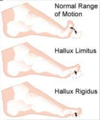
Claw toe
Match the deformity: MTP extension with PIP and DIP flexion, giving the toe a claw-like appearance.
A. Hallux valgus
B. Bunion
C. Bunionette
D. Hammer toe
E. Claw toe
F. Mallet toe
G. Pes equinus
H. Pes cavus
I. Pes planus
J. Hallux limitus
H. Hallux rigidus
I. Charcot arthropathy
J. Plantar flexed 1st ray

Bunionette
Match the deformity: A prominent bump at the lateral side of the 5th MTP joint near the little toe.
A. Hallux valgus
B. Bunion
C. Bunionette
D. Hammer toe
E. Claw toe
F. Mallet toe
G. Pes equinus
H. Pes cavus
I. Pes planus
J. Hallux limitus
H. Hallux rigidus
I. Charcot arthropathy
J. Plantar flexed 1st ray

Mallet toe
Match the deformity: Primarily affects the DIP joint, causing the tip of the toe to curl downward.
A. Hallux valgus
B. Bunion
C. Bunionette
D. Hammer toe
E. Claw toe
F. Mallet toe
G. Pes equinus
H. Pes cavus
I. Pes planus
J. Hallux limitus
H. Hallux rigidus
I. Charcot arthropathy
J. Plantar flexed 1st ray

Charcot arthropathy
Match the deformity: Progressive collapse and destruction of the bones and joints of the foot due to neuropathy, potentially producing a rocker-bottom deformity. Plantar midfoot ulcers.
A. Hallux valgus
B. Bunion
C. Bunionette
D. Hammer toe
E. Claw toe
F. Mallet toe
G. Pes equinus
H. Pes cavus
I. Pes planus
J. Hallux limitus
H. Hallux rigidus
I. Charcot arthropathy
J. Plantar flexed 1st ray

Pes cavus
Match the deformity: An abnormally high medial longitudinal arch of the foot. Load is concentrated at heels and MET heads.
A. Hallux valgus
B. Bunion
C. Bunionette
D. Hammer toe
E. Claw toe
F. Mallet toe
G. Pes equinus
H. Pes cavus
I. Pes planus
J. Hallux limitus
H. Hallux rigidus
I. Charcot arthropathy
J. Plantar flexed 1st ray

Pes equinus
Match the deformity: The ankle is maintained in excessive plantarflexion, limiting the ability to dorsiflex or get the heel to the ground. Forefoot load.
A. Hallux valgus
B. Bunion
C. Bunionette
D. Hammer toe
E. Claw toe
F. Mallet toe
G. Pes equinus
H. Pes cavus
I. Pes planus
J. Hallux limitus
H. Hallux rigidus
I. Charcot arthropathy
J. Plantar flexed 1st ray

Hallux Valgus
Match the deformity: Great toe deviates laterally >15 deg.
A. Hallux valgus
B. Bunion
C. Bunionette
D. Hammer toe
E. Claw toe
F. Mallet toe
G. Pes equinus
H. Pes cavus
I. Pes planus
J. Hallux limitus
H. Hallux rigidus
I. Charcot arthropathy
J. Plantar flexed 1st ray

Pes planus
Match the deformity: A decreased or flattened medial longitudinal arch, commonly called flatfoot. Medial midfoot load.
A. Hallux valgus
B. Bunion
C. Bunionette
D. Hammer toe
E. Claw toe
F. Mallet toe
G. Pes equinus
H. Pes cavus
I. Pes planus
J. Hallux limitus
H. Hallux rigidus
I. Charcot arthropathy
J. Plantar flexed 1st ray

Hallux rigidus
Match the deformity: Severe restriction or essentially no motion of the great toe at the 1st MTP joint, often due to arthritis. Plantar hallux IP ulcers.
A. Hallux valgus
B. Bunion
C. Bunionette
D. Hammer toe
E. Claw toe
F. Mallet toe
G. Pes equinus
H. Pes cavus
I. Pes planus
J. Hallux limitus
H. Hallux rigidus
I. Charcot arthropathy
J. Plantar flexed 1st ray

Plantar flexed 1st ray
Match the deformity: The 1st metatarsal sits lower (more plantarflexed) than the other metatarsals, causing increased pressure under the 1st metatarsal head during weight bearing.
A. Hallux valgus
B. Bunion
C. Bunionette
D. Hammer toe
E. Claw toe
F. Mallet toe
G. Pes equinus
H. Pes cavus
I. Pes planus
J. Hallux limitus
H. Hallux rigidus
I. Charcot arthropathy
J. Plantar flexed 1st ray

1. Protective sensation is lost due to peripheral neuropathy
2. Repeated microtrauma goes unnoticed
3. Excessive inflammation develops
4. Bone resorption and ligament laxity occurs
5. Joint collapse and deformity develops
Put in order how Charcot arthropathy develops:
- Protective sensation is lost due to peripheral neuropathy
- Joint collapse and deformity develops
- Bone resorption and ligament laxity occurs
- Repeated microtrauma goes unnoticed
- Excessive inflammation develops
Diabetes mellitus
Which of the following is the MOST common risk factor associated with Charcot arthropathy?
A. Obesity
B. Chronic kidney disease
C. Diabetes mellitus
D. Previous foot trauma
1. Diabetes mellitus
2. Peripheral neuropathy
3. Chronic kidney disease
4. Previous foot trauma
5. Obesity
6. History of foot ulcer
What are common risk factors for Charcot arthropathy:
1. D_____ mellitus
2. Peripheral n_______
3. Chronic k_____ disease
4. Previous f____ trauma
5. O________
6. History of foot u_______
Midfoot (Lisfranc joint)
What is the MOST common location for Charcot arthropathy?
A. Forefoot
B. Hindfoot
C. Midfoot (Lisfranc joint)
D. Ankle
1. Swelling: Sig. unilateral edema
2. Temperature: Foot >2C warmer than opposite side
3. Redness: Diffuse erythema
4. Pain: Often minimal
5. Pulses: Usually intact
6. Sensation: Peripheral neuropathy present
Clinical presentation of acute Charcot:
1. Swelling: Sig. (unilateral/bilateral) edema
2. Temperature: Foot >2C (warmer/colder) than opposite side
3. Redness: Diffuse erythema
4. Pain: Often (significant/minimal)
5. Pulse: Usually (intact/diminished)
6. Sensation: Peripheral neuropathy (present/absent)

Warm swollen foot in patient with diabetes, no obvious wounds, minimal pain, and normal or mildly elevated inflammatory markers
Which of the following combinations represents red flags for acute Charcot arthropathy?
A. Cold foot, severe pain, open wound, and elevated inflammatory markers
B. Pale foot, absent pulses, severe pain, and delayed capillary refill
C. Warm foot, purulent drainage, severe pain, and highly elevated inflammatory markers
D. Warm swollen foot in a patient with diabetes, no obvious wound, minimal pain, and normal or mildly elevated inflammatory markers
Charcot Arthropathy
Differential Diagnosis: Warm, swollen, minimal pain, neuropathy.
A. Charcot Arthropathy
B. Cellulitis
C. Osteomyelitis
D. Deep vein thrombosis
E. Gout
Gout
Differential Diagnosis: Sudden severe pain, usually very tender
A. Charcot Arthropathy
B. Cellulitis
C. Osteomyelitis
D. Deep vein thrombosis
E. Gout
Deep vein thrombosis
Differential Diagnosis: Calf pain/swelling, vascular findings
A. Charcot Arthropathy
B. Cellulitis
C. Osteomyelitis
D. Deep vein thrombosis
E. Gout
Cellulitis
Differential Diagnosis: Fever, elevated WBC, skin infection
A. Charcot Arthropathy
B. Cellulitis
C. Osteomyelitis
D. Deep vein thrombosis
E. Gout
X-rays may appear normal, while MRI may show bone marrow edema and microfractures
Which of the following correctly describes imaging findings associated with early stage Charcot arthropathy?
A. X-rays may appear normal, while MRI may show bone marrow edema and microfractures
B. Include bone fragmentation, joint subluxation, and rocker-bottom deformity
Include bone fragmentation, joint subluxation, and rocker-bottom deformity
Which of the following correctly describes imaging findings associated with later stage Charcot arthropathy?
A. X-rays may appear normal, while MRI may show bone marrow edema and microfractures
B. Include bone fragmentation, joint subluxation, and rocker-bottom deformity
Prevent further joint destruction
What is the primary goal of management during the acute phase of Charcot arthropathy?
A. Prevent further joint destruction
B. Increase weight bearing
C. Restore full ankle ROM
D. Strengthen the intrinsic foot muscles
Immediate immobilization
Which of the following is considered the standard of care for acute Charcot arthropathy?
A. Immediate weight bearing as tolerated
B. Immediate immobilization with strict offloading
C. Aggressive ROM exercises
D. Surgical fixation for all patients
Total contact cast
Which device is considered the gold standard for immobilization of acute Charcot arthropathy?
A. Ankle brace
B. Compression stocking
C. Total contact cast
D. Night splint
Knee scooter, walker, wheelchair, or crutches
Which of the following may be used to assist with strict offloading in acute Charcot arthropathy?
A. Cane only
B. Shoe insert only
C. Compression wrap
D. Knee scooter, walker, wheelchair, or crutches
Endocrinology/podiatry follow-up, glycemic control, and monitoring skin integrity
Which of the following is an important component of medical management for Charcot arthropathy?
A. Endocrinology/podiatry follow-up, glycemic control, and monitoring skin integrity
B. Encourage barefoot walking
C. Avoid monitoring the skin while immobilized
D. Discontinue diabetes management during offloading
Severe instability, recurrent ulceration, or major deformity
Surgical management of Charcot arthropathy is generally reserved for which situation?
A. Mild swelling without deformity
B. Severe instability, recurrent ulceration, or major deformity
C. All patients immediately after diagnosis
D. Patients with normal foot alignment
Do
Do or Do NOT acute stage PT management for Charcot Arthropathy: Recognize early signs (warm, swollen, erythematous foot with neuropathy)
Do NOT
Do or Do NOT acute stage PT management for Charcot Arthropathy: Continue gait training without medical clearance
Do NOT
Do or Do NOT acute stage PT management for Charcot Arthropathy: Encourage weight bearing on an acutely inflamed Charcot foot.
Do
Do or Do NOT acute stage PT management for Charcot Arthropathy: Immediately refer the patient to a physician, podiatrist, or foot and ankle specialist.
Do
Do or Do NOT acute stage PT management for Charcot Arthropathy: Reinforce strict offloading using the prescribed device. (e.g., total contact cast or CAM boot)
Do NOT
Do or Do NOT acute stage PT management for Charcot Arthropathy: Treat the condition as a simple ankle sprain or minor MSK injury.
Do NOT
Do or Do NOT acute stage PT management for Charcot Arthropathy: Delay referral when Charcot arthropathy is suspected, as early intervention is critical to prevention deformity.
Do
Do or Do NOT acute stage PT management for Charcot Arthropathy: Educate patients on limb protection, daily foot inspection, and the importance of adhering to offloading recommendations.
1. Protective footwear education
2. Balance training
3. Strengthening
4. Gait training
5. Fall prevention
6. Skin inspection education
7. Pressure redistribution
Chronic Stage PT focus for Charcot Arthropathy includes:
1. P______ footwear education
2. B______ training
3. S_______
4. G___ training
5. F___ prevention
6. S___ inspection education
7. P_________ redistribution
Diabetic foot ulcers
What is the Wagner Grading Scale primarily used to classify?
A. Pressure injuries
B. Venous leg ulcers
C. Diabetic foot ulcers
D. Arterial insufficiency
Grade 0
Wagner Grading: No ulcer in a high-risk foot.
A. Grade 0
B. Grade 1
C. Grade 2
D. Grade 4
E. Grade 5
Grade 1
Wagner Grading: Superficial ulcer involving the full skin thickness but not underlying tissue.
A. Grade 0
B. Grade 1
C. Grade 2
D. Grade 4
E. Grade 5

Grade 2
Wagner Grading: Deep ulcer, penetrating down to ligaments and muscle, but no bone involvement or abscess formation.
A. Grade 0
B. Grade 1
C. Grade 2
D. Grade 4
E. Grade 5

Grade 3
Wagner Grading: Deep ulcer with cellulitis or abscess formation, often with osteomyelitis.
A. Grade 0
B. Grade 1
C. Grade 2
D. Grade 4
E. Grade 5

Grade 4
Wagner Grading: Localized gangrene.
A. Grade 0
B. Grade 1
C. Grade 2
D. Grade 4
E. Grade 5

Grade 5
Wagner Grading: Extensive gangrene involving the whole foot.
A. Grade 0
B. Grade 1
C. Grade 2
D. Grade 4
E. Grade 5

Increase
To decrease pressure, (decrease/increase) area....

Offloading for pressure relief
What is the MOST important treatment to address the underlying cause of neuropathic ulcers and promote healing?
A. Wound cleansing
B. Antibiotics
C. Offloading for pressure relief
D. Moist wound dressings
Custom diabetic shoe
Match the following:
A. Non-custom diabetic shoe/therapeutic shoe/extra depth shoe
B. Custom diabetic shoe
C. Custom diabetic insoles/orthotics
D. Peg assist insole
E. Post op shoe/surgical shoe
F. Ortho wedge shoe
G. Total contact cast
H. Diabetic healing walker
I. Wound healing sandals

Ortho wedge shoe
Match the following:
A. Non-custom diabetic shoe/therapeutic shoe/extra depth shoe
B. Custom diabetic shoe
C. Custom diabetic insoles/orthotics
D. Peg assist insole
E. Post op shoe/surgical shoe
F. Ortho wedge shoe
G. Total contact cast
H. Diabetic healing walker
I. Wound healing sandals

Post op shoe/ surgical shoe
Match the following:
A. Non-custom diabetic shoe/therapeutic shoe/extra depth shoe
B. Custom diabetic shoe
C. Custom diabetic insoles/orthotics
D. Peg assist insole
E. Post op shoe/surgical shoe
F. Ortho wedge shoe
G. Total contact cast
H. Diabetic healing walker
I. Wound healing sandals

Non-custom diabetic shoe/therapeutic shoe/extra depth shoe
Match the following:
A. Non-custom diabetic shoe/therapeutic shoe/extra depth shoe
B. Custom diabetic shoe
C. Custom diabetic insoles/orthotics
D. Peg assist insole
E. Post op shoe/surgical shoe
F. Ortho wedge shoe
G. Total contact cast
H. Diabetic healing walker
I. Wound healing sandals

Wound healing sandal
Match the following:
A. Non-custom diabetic shoe/therapeutic shoe/extra depth shoe
B. Custom diabetic shoe
C. Custom diabetic insoles/orthotics
D. Peg assist insole
E. Post op shoe/surgical shoe
F. Ortho wedge shoe
G. Total contact cast
H. Diabetic healing walker
I. Wound healing sandals

Total contact cast
Match the following:
A. Non-custom diabetic shoe/therapeutic shoe/extra depth shoe
B. Custom diabetic shoe
C. Custom diabetic insoles/orthotics
D. Peg assist insole
E. Post op shoe/surgical shoe
F. Ortho wedge shoe
G. Total contact cast
H. Diabetic healing walker
I. Wound healing sandals

Diabetic healing walker
Match the following:
A. Non-custom diabetic shoe/therapeutic shoe/extra depth shoe
B. Custom diabetic shoe
C. Custom diabetic insoles/orthotics
D. Peg assist insole
E. Post op shoe/surgical shoe
F. Ortho wedge shoe
G. Total contact cast
H. Diabetic healing walker
I. Wound healing sandals

Peg assist insole
Match the following:
A. Non-custom diabetic shoe/therapeutic shoe/extra depth shoe
B. Custom diabetic shoe
C. Custom diabetic insoles/orthotics
D. Peg assist insole
E. Post op shoe/surgical shoe
F. Ortho wedge shoe
G. Total contact cast
H. Diabetic healing walker
I. Wound healing sandals

Custom diabetic insoles/orthotics
Match the following:
A. Non-custom diabetic shoe/therapeutic shoe/extra depth shoe
B. Custom diabetic shoe
C. Custom diabetic insoles/orthotics
D. Peg assist insole
E. Post op shoe/surgical shoe
F. Ortho wedge shoe
G. Total contact cast
H. Diabetic healing walker
I. Wound healing sandals

Extra depth and width, protective materials, firm heel counter, non-binding uppers, and shock absorption
Which of the following are key features of diabetic/therapeutic shoes?
A. Pressure relief, immobilization, A. Pressure relief, immobilization, accommodation of dressings, prevention of further injury, and custom fit
B. Custom fit, rigid structure, and seamless interior
C. Extra depth and width, protective materials, firm heel counter, non-binding uppers, and shock absorption
D. Rigid sole, adjustable straps, rockered bottom sole, padded interior
Pressure relief, immobilization, accommodation of dressing, prevention of further injury, and custom fit
Which of the following are key features of a post-op/surgical shoe?
A. Pressure relief, immobilization, accommodation of dressings, prevention of further injury, and custom fit
B. Custom fit, rigid structure, and seamless interior
C. Extra depth and width, protective materials, firm heel counter, non-binding uppers, and shock absorption
D. Rigid sole, adjustable straps, rockered bottom sole, padded interior
Total contact cast (TCC)
What is the gold standard of care for offloading diabetic foot ulcers?
A. Compression stockings
B. Total contact cast (TCC)
C. Standard walking shoe
D. Ankle-foot orthosis (AFO)
Custom fit, rigid structure, and seamless interior
Which of the following is a feature of a total contact cast (TCC)?
A. Loose fit with a flexible structure
B. Custom fit, rigid structure, and seamless interior
C. Removable design with minimal foot contact
D. Soft structure with an open interior
True
True or false: Total contact casts requires frequent reapplication.
Diabetic foot ulcers, Charcot foot, post-surgical recovery, and foot fractures in patients with diabetes
Which of the following conditions can be treated using a total contact cast?
A. Diabetic foot ulcers, Charcot foot, post-surgical recovery, and foot fractures in patients with diabetes
B. Only diabetic foot ulcers
C. Venous ulcers and lymphedema only
D. Acute arterial insufficiency only
Rigid sole, adjustable straps, rockered bottom sole, padded interior
Which of the following correctly describes the features and purposes of a DH Walker?
A. Pressure relief, immobilization, accommodation of dressings, prevention of further injury, and custom fit
B. Custom fit, rigid structure, and seamless interior
C. Extra depth and width, protective materials, firm heel counter, non-binding uppers, and shock absorption
D. Rigid sole, adjustable straps, rockered bottom sole, padded interior
Diabetic foot ulcers, post-surgical healing, and Charcot foot
Which of the following conditions are commonly treated with a DH Walker?
A. Arterial insufficiency, DVT, and lymphedema
B. Venous ulcers, arterial ulcers, and cellulitis
C. Plantar fasciitis, ankle instability, and Achilles tendinopathy only
D. Diabetic foot ulcers, post-surgical healing, and Charcot foot