A&P Exam 1
 Call Kai
 Learn
 Practice Test
 Spaced Repetition
 Match
 Flashcards
 Knowt Play
1/183

There's no tags or description
Looks like no tags are added yet.
184 Terms
Nucleus — what is it and what does it do?
The nucleus is the cell's largest organelle and acts as its control center. It holds the DNA instructions used to build nearly all of the cell's proteins, and it adjusts which proteins are produced, and in what amounts, in response to signals the cell receives.
How many nuclei can a cell have? (uninucleate, multinucleate, anucleate)
Most cells are uninucleate, with just one nucleus. A few cell types are multinucleate and contain many nuclei — examples include B lymphocytes, skeletal muscle cells, and some liver cells — while mature red blood cells are anucleate and have no nucleus at all.
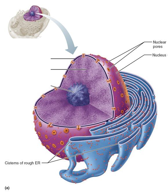
What are the three main structures that make up the nucleus?
The nuclear envelope, the membrane barrier around the nucleus; the nucleolus, where ribosomal RNA is made; and chromatin, the cell's DNA.
Nuclear envelope
The double-membrane barrier that wraps around the nucleus and separates its contents from the rest of the cell. It encloses the nucleoplasm, the fluid interior of the nucleus.
How is the outer membrane of the nuclear envelope connected to the rough endoplasmic reticulum?
The envelope's outer membrane is directly continuous with the rough ER, and just like the rough ER, its surface is studded with ribosomes.
Nuclear lamina
A network of proteins lining the inner surface of the nuclear envelope. It helps the nucleus hold its shape and acts as a scaffold that helps organize the DNA.
Nucleoplasm
The jellylike fluid that fills the inside of the nucleus. The other nuclear components, such as chromatin and the nucleolus, are suspended in it.
Nuclear pores and the nuclear pore complex
Nuclear pores are channels through the nuclear envelope that allow substances to move into and out of the nucleus. Each pore is guarded by a nuclear pore complex, which regulates the passage of specific large molecules.

Nucleolus
A dark-staining, spherical body found inside the nucleus, usually one or two per nucleus. It is the site where ribosomal RNA (rRNA) is synthesized and where ribosome subunits are assembled.
Chromatin
The cell's DNA in its usual, loosely arranged form inside the nucleus, organized as long strands rather than compact rods. When a cell gets ready to divide, chromatin condenses and coils up into chromosomes.
What are the four primary tissue types, and what is each one's main job?
Epithelial tissue forms coverings, linings, and glands. Connective tissue protects and supports the body. Muscle tissue produces movement. Nervous tissue carries messages so different parts of the body can communicate.
What are the key characteristics of epithelial tissue?
It is densely packed with cells, it is polar (a free apical surface and an attached basal surface), it has no blood vessels of its own (avascular), it has a nerve supply (innervated), it regenerates quickly, and it sits anchored on a basement membrane.
Apical surface vs. basal surface of an epithelium — what is the difference?
The apical surface is the free top surface, exposed to the body's exterior or to the inside of a hollow organ. The basal surface is the bottom surface, attached to the basement membrane that anchors the epithelium to the tissue below.
Epithelial tissue contains no blood vessels. What is this property called, and how does the tissue get its nutrients?
The property is called being avascular. Oxygen and nutrients reach the epithelial cells by diffusion from blood vessels in the connective tissue underneath the basement membrane.

Cilia vs. microvilli — what does each structure do?
Both project from the apical surface. Cilia are longer, hair-like structures that beat in waves to sweep materials such as mucus across the surface. Microvilli are much smaller folds that increase surface area so the cell can absorb more. The diagram makes the size difference obvious — panel A shows cilia as long, sparse, hair-like projections on the apical surface, while panel B shows microvilli as much shorter, denser, brush-like folds on the same surface.
How are epithelial tissues named?
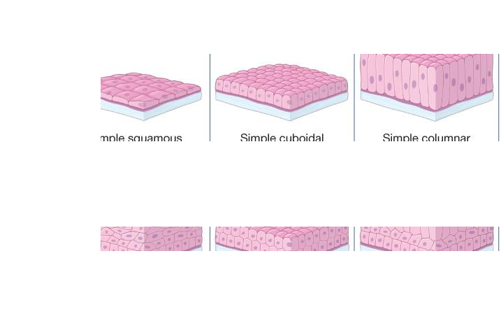
By two features combined: the shape of the cells at the apical surface (squamous = flat, cuboidal = cube-shaped, columnar = tall and column-like) and the number of cell layers (simple = one layer, stratified = two or more layers). The figure is a 2x3 grid of the naming system: rows are layer count (simple = one layer, stratified = multiple layers) and columns are apical cell shape (squamous = flat, cuboidal = cube-shaped, columnar = tall), producing simple squamous, simple cuboidal, simple columnar, stratified squamous, stratified cuboidal, and stratified columnar epithelium.
Simple vs. stratified vs. pseudostratified epithelium — define each.
Simple means a single layer of cells. Stratified means two or more stacked layers. Pseudostratified is a single layer whose cells vary in height, making it look layered even though every cell still touches the basement membrane.

What is a goblet cell?
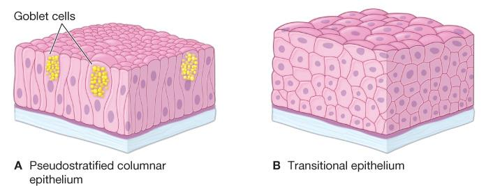
A single-celled (unicellular) gland scattered among the cells of simple and pseudostratified epithelia. It produces and secretes mucus, which protects and lubricates the surface being lined.

Simple squamous epithelium — structure, locations, and function.
One layer of thin, flat cells. It lines the air sacs (alveoli) of the lungs and the inside of the heart, blood vessels, and lymphatic vessels. Because it is so thin, substances cross it easily by diffusion and filtration, and it can secrete lubricating substances such as surfactant.
Simple cuboidal epithelium — locations and function.
A single layer of cube-shaped cells. It lines kidney tubules and the ducts and secretory parts of small glands, where its jobs are secretion and absorption of substances such as sodium, potassium, and glucose.
Simple columnar epithelium — locations and function.
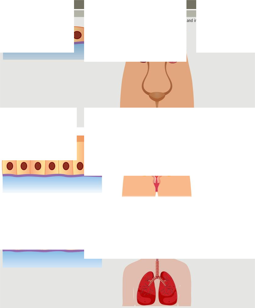
A single layer of tall, column-shaped cells. It lines much of the digestive tract and parts of the reproductive tract, where it absorbs nutrients and secretes materials such as mucus and enzymes.
Pseudostratified columnar epithelium — why the name, where is it found, and what does it do?
It is named "pseudo" (false) stratified because the nuclei sit at different heights, faking a layered look, yet it is one layer and all cells contact the basement membrane. It lines the trachea and most of the upper respiratory tract, where goblet cells secrete mucus and cilia sweep it away. Panel A shows pseudostratified columnar epithelium with yellow, mucus-filled goblet cells labeled among the columnar cells — the nuclei at staggered heights give the false layered look. Panel B, for contrast, shows transitional epithelium: genuinely many layers thick, with rounded, dome-like surface cells (see also card 15).

Stratified squamous epithelium — locations and function.
Many layers of cells that are flat at the surface. It lines the esophagus, mouth, and vagina — surfaces exposed to friction — and its main job is protection against abrasion.

Stratified cuboidal and stratified columnar epithelium — where are they found and what do they do?
Both are uncommon and both secrete and protect. Stratified cuboidal occurs in sweat glands, salivary glands, and mammary glands. Stratified columnar occurs in the male urethra and in the ducts of some glands. This is Table 5.1 (continued). It also shows stratified squamous epithelium (top row): lines the esophagus, mouth, and vagina and protects against abrasion. Stratified cuboidal: sweat glands, salivary glands, and mammary glands — secretes and protects. Stratified columnar: the male urethra and ducts of some glands — secretes and protects.

Transitional epithelium — where is it found and what makes it special?
It is a stratified epithelium lining the bladder, ureters, and urethra. Its surface cells can change shape and flatten out as the organ fills, letting urinary organs expand and stretch without tearing.
In a stratified epithelium, which layer's cell shape gives the tissue its name, and where do replacement cells come from?
The tissue is named for the shape of the cells in the apical (top) layer. Replacement cells come from stem cells dividing in the basal (bottom) layer; new cells push upward to replace the surface cells that are shed.
Endocrine glands vs. exocrine glands — how do they differ?
Endocrine glands are ductless and release hormones directly into the blood; examples are the pituitary gland, adrenal glands, and thymus. Exocrine glands release their products through ducts onto a surface or into a cavity; examples include sweat and salivary glands, which produce sweat, saliva, mucus, and milk.
What is connective tissue made of, and how does it differ from epithelial tissue?
Connective tissue is mostly extracellular matrix — ground substance plus protein fibers — with relatively few cells that are widely spaced and rarely touching. Unlike epithelium, it is generally well supplied with blood vessels (vascularized).
What are the two components of the extracellular matrix (ECM)?
Ground substance, the material filling the spaces between the cells and fibers, and protein fibers embedded in it, such as thick collagen fibers and thin, branched elastin fibers. Cells such as fibroblasts build and maintain the matrix.
What are the three categories of connective tissue, and what belongs in each?
(1) Connective tissue proper: areolar, adipose, reticular, dense regular, and dense irregular. (2) Supportive connective tissue: hyaline cartilage, fibrocartilage, elastic cartilage, and compact and spongy bone. (3) Fluid connective tissue: blood and lymph.
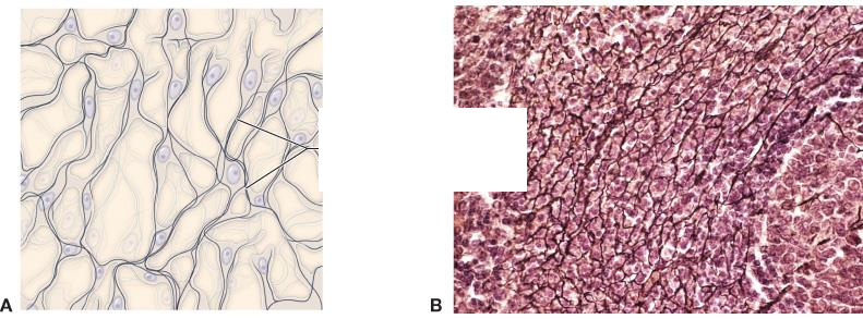
Reticular connective tissue — structure and location.
It is a delicate, sponge-like meshwork of fine reticular fibers. It forms the internal framework (stroma) that holds up the working cells of lymphatic tissue, the spleen, and the liver.
Dense regular vs. dense irregular connective tissue — how do they differ?
In dense regular tissue the collagen bundles all run parallel in one direction, so it resists pulling along that line; it makes up tendons and ligaments. In dense irregular tissue the bundles are interwoven in many directions, so it resists tension from all sides; it is found in the skin (dermis).
Hyaline cartilage — properties and locations.
It is the most abundant cartilage and has the smoothest, glassiest matrix. It covers the ends of bones at joints and forms the costal cartilages connecting ribs to the sternum, giving smooth movement and flexible support.
Fibrocartilage — properties and locations.
It is the strongest cartilage, packed with collagen fibers so it can absorb heavy compression and shock. It forms the intervertebral discs between the vertebrae and the meniscus pads in the knee.
Elastic cartilage — properties and locations.
It is the most flexible cartilage because its matrix is full of elastic fibers, so it can bend and spring back into shape. It forms the external ear (and the epiglottis).
Why does a torn meniscus in the knee heal so poorly?
Cartilage is largely avascular, meaning it has little or no direct blood supply. Without adequate blood flow to deliver nutrients and repair cells, cartilage heals very slowly or not at all, so a torn meniscus often requires surgery.

Compact bone vs. spongy bone — how do they differ?
Bone is the most rigid connective tissue and it protects and supports internal organs. Compact bone is dense and solid and provides most of the bone's strength; spongy bone is an open lattice of bony struts, and the spaces within it hold red bone marrow.

Why are blood and lymph classified as connective tissues, and how do they differ from each other?
Both are fluid connective tissues: cells suspended in a fluid matrix that transports molecules and cells around the body. Blood is cellular, containing erythrocytes (red blood cells), leukocytes (white blood cells), and platelets. Lymph is mostly fluid with very few cells (nearly acellular).

What are the four types of tissue membranes?
Mucous membranes, serous membranes, the cutaneous membrane (the skin), and synovial membranes. The figure maps all four membranes on the body: serous membranes wrap organs such as the heart (visceral pericardium on the organ, parietal pericardium outside it); mucous membranes line tracts and are coated with mucus; synovial membranes line joint cavities such as the knee and contain synovial fluid; and the cutaneous membrane is the skin, layered epidermis over dermis over hypodermis above muscle.
Mucous membranes vs. serous membranes — how do they differ?
Mucous membranes line body cavities that open to the outside world — the digestive, respiratory, urinary, and reproductive tracts — and usually contain mucus-secreting goblet cells. Serous membranes line closed internal cavities and cover organs (pericardium around the heart, pleura around the lungs, peritoneum in the abdomen) and secrete a fluid that reduces friction as organs move.
Cutaneous membrane — what is it, and what does it do?
The cutaneous membrane is the skin, made of stratified squamous epithelium over connective tissue, with keratin adding toughness. It protects the body from drying out (desiccation) and from invasion by pathogens.
Synovial membranes — where are they found and what do they do?
They line the inside of freely movable joints such as the knee, hip, and elbow. Their cells secrete synovial fluid, which lubricates the joint, cuts friction between the moving bones, and nourishes the cartilage inside the joint.

Identify this tissue
The four primary tissue types, each shown as a micrograph beside the body system it serves: nervous tissue (brain, spinal cord, nerves), muscle tissue (cardiac, smooth, skeletal), epithelial tissue (skin, GI tract lining), and connective tissue (bone, fat, tendon).
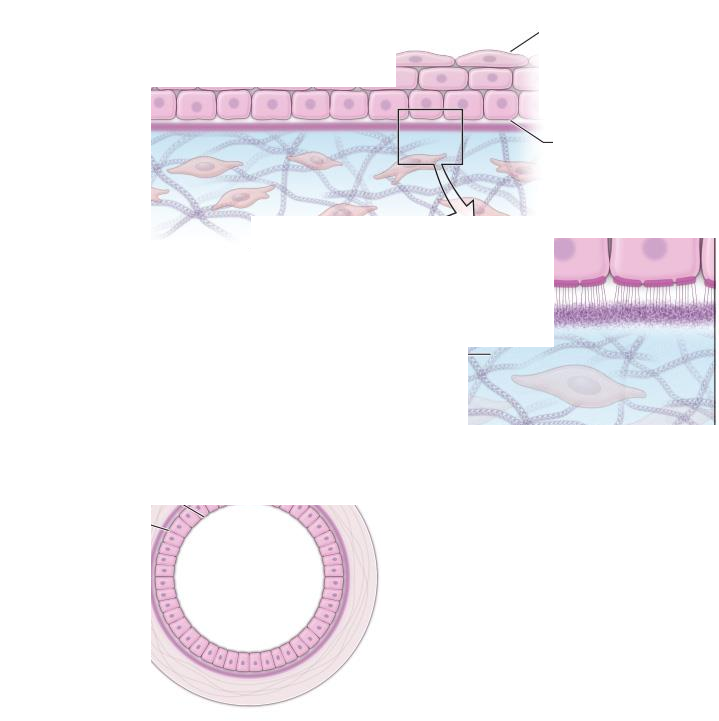
Identify the structures in this diagram of an epithelium
Epithelium anatomy: the apical side is the free surface, the basal side faces down, and the basement membrane (made of the lamina lucida and lamina densa) anchors the epithelium to the underlying connective tissue. Epithelia are highly cellular, polar, avascular, and innervated.

Identify this tissue
Simple squamous epithelium — a single layer of flat, scale-like cells resting on a basement membrane. It lines the air sacs of the lungs and the heart, blood vessels, and lymphatic vessels, where its thinness allows rapid diffusion and filtration.

Identify this tissue (the three panels show three related types)
Top: simple cuboidal epithelium — one layer of cube-shaped cells; lines kidney tubules and gland ducts; secretes and absorbs. Middle: simple columnar epithelium — one layer of tall cells; lines the digestive tract; absorbs and secretes mucus and enzymes. Bottom: pseudostratified columnar epithelium — one layer that looks layered, with cilia; lines the trachea; secretes and moves mucus.

Identify this tissue
Stratified squamous epithelium — many cell layers thick. Stem cells in the basal layer keep dividing, and the cells flatten as they are pushed toward the apical surface, which is why the tissue is named for its flat surface cells. It protects surfaces such as the esophagus, mouth, and vagina against abrasion.

Identify this tissue
Transitional epithelium — a stratified epithelium lining the bladder, ureters, and urethra. Its rounded surface cells can flatten when the organ stretches, allowing the urinary organs to expand as they fill.

Identify this tissue (note the pale, mucus-filled cells)
Goblet cells — single-celled glands wedged between columnar epithelial cells, packed with mucus secretory vesicles (diagram, left) and visible as pale, clear cells in the micrograph of pseudostratified columnar epithelium (right). They secrete the mucus that protects and lubricates body linings.

Identify the components in this diagram of connective tissue
Connective tissue anatomy: fibroblast cells scattered through extracellular matrix composed of ground substance plus fibers — thick, loosely woven collagen fibers and thin, branched elastin fibers. The blood vessels in the image are the giveaway that connective tissue is vascularized, unlike epithelium.

Identify this tissue
Reticular connective tissue — a delicate, branching network of thin reticular fibers (diagram, left; micrograph, right) forming a sponge-like framework that supports the cells of lymphatic tissue, the spleen, and the liver.

Identify this tissue
Dense irregular connective tissue — thick bundles of collagen fibers interwoven in many different directions, with fibroblast nuclei scattered between them. Because the fibers run every which way, it resists tension from all directions; it is found in the dermis of the skin.

Identify this tissue
Dense regular connective tissue — collagen fiber bundles all arranged in parallel rows in a single direction, with fibroblast nuclei squeezed between the bundles. This alignment resists strong pulling along one direction, as in tendons and ligaments.

Identify this tissue (panels A–C show its three types)
Cartilage, a supportive connective tissue whose cells (chondrocytes) sit in small cavities called lacunae within a firm matrix. A: elastic cartilage — most flexible, forms the external ear. B: fibrocartilage — strongest, forms intervertebral discs and the knee meniscus. C: hyaline cartilage — smoothest and most abundant, found at joints and in the ribs.

Identify this tissue (note the two regions)
Bone tissue, the most rigid connective tissue. Compact bone is the dense, solid region that provides strength; spongy bone is the inner lattice of bony struts whose open spaces contain red bone marrow.

Identify this tissue
Blood, a fluid connective tissue. Panel A shows its formed elements: very numerous small red erythrocytes (red blood cells) and a few larger purple-stained leukocytes (white blood cells), all suspended in fluid plasma. Panel B shows the artery and vein it travels through.
What are the main jobs of the plasma (cell) membrane?
It forms the cell's outer boundary, keeping the inside of the cell separate from the fluid around it, and it controls which substances are allowed to enter or leave.
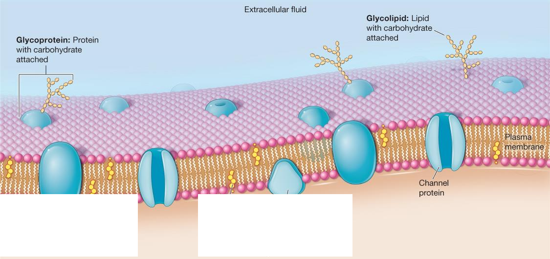
What is the fluid-mosaic model?
The model that describes the cell membrane as a flexible, constantly shifting layer: a phospholipid bilayer with proteins and other molecules embedded in it and able to drift within it, like tiles in a mosaic.
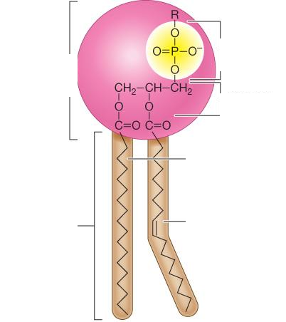
What is a phospholipid, and what does "amphipathic" mean?
A phospholipid is a membrane-building molecule with a phosphate-containing head that is attracted to water (hydrophilic) and two fatty acid tails that avoid water (hydrophobic). Having both a water-loving and a water-fearing part makes it amphipathic. Diagram labels: a single phospholipid — hydrophilic head (phosphate group + glycerol) and two hydrophobic fatty-acid tails, one saturated and one unsaturated.
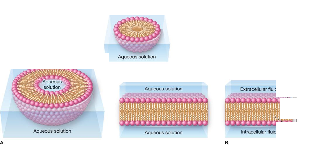
How are phospholipids arranged in the membrane, and why?
They form two layers (a bilayer) with the hydrophilic heads pointing outward toward the watery fluids on both sides of the membrane and the hydrophobic tails tucked together in the middle, away from water. Diagram info: in water, phospholipids self-assemble — a single layer forms a sphere with heads facing the water and tails hidden inside, while a bilayer can close into a hollow sphere or spread as a flat sheet. In the membrane sheet, hydrophilic heads face the extracellular and intracellular fluids, with the hydrophobic tails forming the bilayer's middle.
What does it mean that the membrane is selectively permeable, and what crosses it freely?
It means the membrane lets some substances through while blocking others. Only small, nonpolar molecules can slip directly through the lipid bilayer without help.

What is the difference between a transmembrane protein and a peripheral protein?
A transmembrane protein passes all the way through the membrane from one side to the other. A peripheral protein sits on just one surface of the membrane (inside or outside) without spanning it. Diagram labels: a transmembrane/channel protein spanning the bilayer, a peripheral protein on the cytosol side, glycoprotein, glycolipid, and cholesterol, with extracellular fluid above and cytosol below.
What jobs do membrane proteins do?
They work as channels that let specific substances cross, receptors that receive signals, enzymes that speed up reactions at the membrane, and markers that let cells recognize each other (including recognizing "self").
What are glycoproteins and the glycocalyx?
Glycoproteins are membrane proteins with carbohydrate chains attached; they help with cell recognition. The glycocalyx is the fuzzy coating formed by many of these carbohydrate chains on the cell's outer surface, which can also protect the cell and help it attach to other cells.

What is the difference between intracellular fluid (cytosol) and extracellular fluid?
Intracellular fluid is the watery fluid inside the cell; the portion surrounding the organelles is called the cytosol. Extracellular fluid is the fluid outside the cell.
What is passive transport?
Movement of substances across a membrane that costs the cell no energy. It works only when a concentration gradient exists, so substances move from where they are more concentrated to where they are less concentrated.

What is simple diffusion?
A form of passive transport in which molecules spread from an area of higher concentration to an area of lower concentration by passing directly through the membrane, with no help from proteins, until their concentrations even out (equilibrium). Diagram: three-panel sequence — small uncharged molecules pass directly through the lipid bilayer from extracellular fluid to cytosol until evenly distributed.

What is facilitated diffusion, and which molecules need it?
Passive movement down a concentration gradient that requires help from a transmembrane (channel or carrier) protein. Polar and charged (ionic) molecules need it because they cannot pass through the lipid bilayer on their own. Diagram: two panels — molecules cross through a channel protein (A) or through a carrier protein that changes shape to release its cargo (B).

What is osmosis?
The movement of water across a selectively permeable membrane toward the side with the higher solute concentration (equivalently, from lower solute toward higher solute concentration) until concentrations balance. Diagram: beakers divided by a semipermeable membrane; the water level rises on the side with more solute as water moves toward the higher solute concentration.

What happens to a cell placed in a hypotonic solution?
The surrounding fluid has less solute than the cell's interior, so water moves into the cell. The cell swells and may burst. Diagram: red blood cells shown as a normal disc in isotonic solution, swollen/spherical in hypotonic solution, and shriveled (crenated) in hypertonic solution, with arrows for net water movement and micrographs under each panel.

What happens to a cell placed in a hypertonic solution?
The surrounding fluid has more solute than the cell's interior, so water leaves the cell. The cell shrinks (crenates).

What happens to a cell placed in an isotonic solution?
The fluid around the cell has the same solute concentration as the cell's interior, so water moves in and out at equal rates. The cell keeps its normal shape and functions normally.

What do isosmotic, hyperosmotic, and hypoosmotic mean?
These terms compare the total solute concentration (osmolarity) of two solutions: isosmotic means equal, hyperosmotic means one has more solute than the other, and hypoosmotic means one has less solute than the other. Diagram info: beakers compare solute particle counts — a hypoosmotic solution contains a lower concentration of solutes than the solution it is compared with, a hyperosmotic solution contains a higher concentration, and two isosmotic solutions contain the same total solute concentration.
What is active transport?
Energy-requiring movement of substances across the membrane against their concentration gradient — from an area of lower concentration to an area of higher concentration.
What is primary active transport?
Active transport powered directly by ATP: the energy released from ATP drives a membrane pump to move substances uphill against their gradient.
What is secondary active transport?
Active transport that does not burn ATP directly. Instead it harnesses the energy stored in an existing electrochemical gradient (for example, sodium rushing back into the cell) to drag another substance, such as glucose, along with it.
What is the difference between a symporter and an antiporter?
A symporter carries two substances across the membrane in the same direction. An antiporter carries two substances in opposite directions — one in while the other goes out.

How does the sodium-potassium (Na+/K+) pump work?
It is a primary active transport pump that uses ATP to push 3 sodium ions out of the cell and bring 2 potassium ions into the cell, both against their concentration gradients. Diagram: four-step pump cycle — Na⁺ binds from the cytosol, ATP is split to ADP, Na⁺ is released outside, K⁺ binds outside and is released inside; Na⁺ and K⁺ concentration-gradient bars shown alongside.
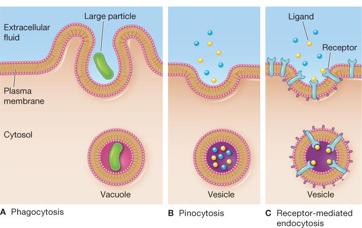
What is endocytosis?
An energy-using process in which the cell membrane folds around material outside the cell and pinches off, trapping the material inside a vesicle so it enters the cell. Diagram: three panels — phagocytosis engulfing a large particle into a vacuole, pinocytosis taking dissolved molecules into a small vesicle, and receptor-mediated endocytosis with ligands bound to membrane receptors.

How do phagocytosis and pinocytosis differ?
Phagocytosis ("cell eating") extends the membrane around large particles or whole cells to engulf them. Pinocytosis ("cell drinking") uses small membrane indentations to take in tiny droplets of fluid with dissolved substances.

What is receptor-mediated endocytosis?
A selective form of endocytosis in which a specific molecule (a ligand) must first bind to a matching receptor on the membrane; only then does the membrane pinch off and bring that cargo into the cell.

What is exocytosis?
The process a cell uses to export material: a vesicle inside the cell fuses with the plasma membrane and releases its contents outside. Cells secrete hormones and digestive enzymes this way. Diagram info: a vesicle in the cytosol moves to the plasma membrane, fuses with it, and opens so its contents spill out into the extracellular fluid.

How do cytoplasm, cytosol, and organelles relate to each other?
The cytoplasm is everything inside the cell membrane except the nucleus — the fluid plus the structures suspended in it. The cytosol is just the gel-like fluid portion, and the organelles are the membrane-bound structures within it that each perform a specific job. Diagram labels: fully labeled model human cell — nucleus, nucleolus, chromatin, rough and smooth ER, Golgi apparatus and vesicle, mitochondria, lysosome, peroxisome, ribosomes, cytoskeletal filaments, centrosome, cytoplasm, plasma membrane.

How do the rough ER and smooth ER differ in structure and function?
The rough ER is studded with ribosomes and is involved in making proteins. The smooth ER has no ribosomes and is involved in making lipids. Both are networks of channels connected to the nuclear membrane used for synthesis, transport, and storage. Diagram info: the flattened rough ER, studded with ribosomes, wraps around the nucleus and is continuous with the smooth ER, which appears as a network of tubules with no ribosomes.

What does the Golgi apparatus do?
It receives products made by the rough ER at its cis face, then sorts, modifies, and packages them, releasing the finished products from its trans face for transport where they are needed. Diagram info: the figure traces the pathway — products leave the rough ER in a transport vesicle, enter the Golgi apparatus at its cis face, exit at the trans face inside a secretory vesicle, and are released where the vesicle fuses with the plasma membrane; a lysosome is also shown.
How do lysosomes and peroxisomes differ?
Both are small membrane-bound sacs of enzymes. Lysosomes contain digestive enzymes that break down wastes and worn-out parts inside the cell. Peroxisomes contain enzymes that produce hydrogen peroxide and are used for detoxification and lipid metabolism.

What is the mitochondrion, and what is special about its inner membrane?
The mitochondrion is the site of aerobic respiration, where nutrients are broken down to produce most of the cell's ATP. It has two membranes, and the inner one is folded into ridges called cristae; mitochondria are especially numerous in energy-hungry cells like muscle and nerve cells. Diagram labels: the outer membrane, inner membrane, intermembrane space, and the folded cristae.

What is the cytoskeleton, and what are its three filament types?
The cytoskeleton is an internal network of protein filaments that supports the cell's shape, organizes the cytoplasm, moves materials inside the cell, and helps pull the cell apart during division. Its filaments are microtubules (made of tubulin), intermediate filaments (made of keratin), and microfilaments (made of actin). Diagram info: each panel pairs a fluorescence micrograph with the filament's structure — A: microtubules are hollow tubes built of tubulin subunits; B: microfilaments are paired strands of actin subunits; C: intermediate filaments are rope-like bundles of fibrous keratin subunits coiled together.
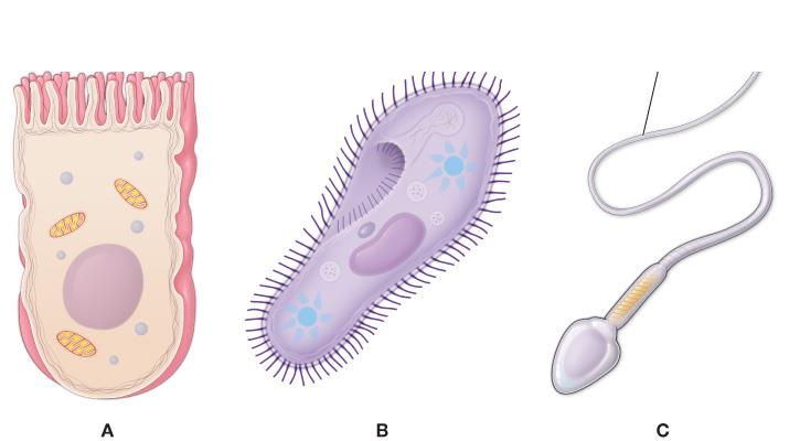
How do microvilli, cilia, and flagella differ?
Microvilli are tiny folds of the membrane that increase a cell's surface area for absorption. Cilia are short projections that beat to move substances across the cell's surface (or move the cell). Flagella are long, whip-like appendages used to propel the cell itself. Diagram info: panel A shows many short microvilli covering the surface of an epithelial cell; panel B shows a Paramecium covered in short cilia; panel C shows a sperm cell with a single long flagellum.

How are vesicles transported along microtubules, and which motor proteins move them?
Microtubules act as tracks for intracellular transport: the motor protein kinesin carries vesicles toward the plus (+) end of a microtubule, while dynein carries vesicles in the opposite direction. In the figure, vesicles near the rough ER and Golgi apparatus are hauled along microtubules by these motors.
Matter, mass, and weight — how do they differ?
Matter is anything that takes up space and has mass. Mass is the amount of matter in an object and stays the same anywhere; weight depends on gravity, so the same object weighs less on the Moon but has identical mass.

Element vs. atom vs. molecule vs. compound
An element is a pure substance made of just one kind of atom. An atom is the smallest unit of an element that still keeps that element's properties. A molecule is two or more atoms bonded together; a compound is two or more different elements joined by chemical bonds. The periodic table arranges all elements in order of increasing atomic number; each box gives the atomic number, symbol, name, and atomic weight, and the color key shows which elements are solids, liquids, gases, or artificially prepared at room temperature.

What are the three subatomic particles and their charges?
Protons carry a positive charge, neutrons have no charge, and electrons carry a negative charge. Protons and neutrons sit in the nucleus, while electrons move around it in shells.

Atomic number
The count of protons in an atom's nucleus. It identifies which element the atom is, and in a neutral atom it also equals the number of electrons. In an element's periodic-table box, the atomic number sits at the top (helium = 2, its proton count), the symbol and name are in the middle, and the atomic weight (4.002602 for helium) is at the bottom.
Atomic weight
Approximately the total of an atom's protons and neutrons; electrons contribute only a negligible amount of mass.
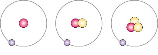
Isotopes
Forms of the same element with identical proton counts but different numbers of neutrons, giving them different atomic weights. Hydrogen has three: protium (0 neutrons), deuterium (1), and tritium (2). Unstable isotopes are radioactive and shed particles.

Ion, cation, and anion
An ion is an atom that gained or lost electrons and therefore carries an electrical charge. Losing electrons produces a positive cation; gaining electrons produces a negative anion. Potassium (19 protons, 19 electrons) becomes the cation K⁺ by giving up its single outer-shell electron (19 protons, 18 electrons); fluorine (9 protons, 9 electrons) becomes the anion F⁻ by gaining one electron to nearly fill its outer shell (9 protons, 10 electrons).

Electron shells and the valence shell
Electrons are arranged in shells around the nucleus — the first shell holds at most 2 electrons, the second up to 8. The outermost (valence) shell controls reactivity: atoms share, donate, or accept electrons in order to fill it.
What are the three main types of chemical bonds in the body?
Ionic bonds (attraction between oppositely charged ions), covalent bonds (electrons shared between atoms), and hydrogen bonds (weak attractions between polar molecules). Whether an atom bonds depends on how many electrons are in its valence shell.

Ionic bonds
Bonds created when one atom gives up electrons to another, producing oppositely charged ions that attract each other. Example: sodium donates an electron to chlorine, yielding Na+ and Cl- — table salt (NaCl).