Exam 5
 Call Kai
 Learn
 Practice Test
 Spaced Repetition
 Match
 Flashcards
 Knowt Play
1/237

There's no tags or description
Looks like no tags are added yet.
238 Terms
Nervous system is
master controlling and communicating system of body
Nervous System functions (3)
Sensory Input- Information gathered by sensory receptors about internal and external changes
Integration- Processing and interpretation of sensory input
Motor Output- activation of effectors produces a response

Nervous system is divided into two parts
Central Nervous System-Brain and spinal cord of dorsal body cavity
Integration and control center
Interprets sensory input and dictates motor output
Peripheral Nervous System-
The portion of nervous system outside CNS
Consists mainly of nerves that extend from brain and spinal cord–
Spinal nerves to and from spinal cord
Cranial nerves to and from brain

Walls of gastrointestinal tract also contain neurons called the
enteric nervous system
Peripheral nervous system (PNS) has two functional divisions
Sensory (afferent) division
Somatic sensory fibers: convey impulses from skin, skeletal muscles, and joints toCNS
Visceral sensory fibers: convey impulses from visceral organs toCNS
Motor (efferent) Division
Transmits impulses fromCNS to effectors–Muscles and glands
Two divisions–Somatic nervous system–Autonomic nervous system

Somatic nervous system, or voluntary nervous system function
Somatic motor nerve fibers conduct impulses from CNS to skeletal muscle
Conscious control of skeletal muscles

Autonomic nervous system (ANS), or involuntary nervous system functions
Consists of visceral motor nerve fibers
Regulates smooth muscle, cardiac muscle, and glands
Two functional subdivisions work in opposition of each other ▪
Sympathetic division- mobilize body systems during activity
Parasympathetic division- conserves energy, promotes house-keeping functions during rest

Neuroglia (glial cells)
small cells that surround and wrap delicate neurons
Neurons (nerve cells)
excitable cells that transmit electrical signals
Four main neuroglia support CNS neurons
Astrocytes
Microglial cells
Ependymal cells
Oligodendrocytes
Astrocytes
Most abundant, versatile, and highly branched of glial cells
Cling to neurons, synaptic endings, and capillaries
Functions:
Support and brace neurons
Play role in exchanges between capillaries and
neurons
Guide migration of young neurons
Control chemical environment around neurons
Clean up leaked potassium ions
Recapture and recycle neurotransmitters
Influence neuronal functioning

Microglial
Small, ovoid cells with thorny processes that touch and monitor neurons
Migrate toward injured neurons
Can transform to phagocytize microorganisms and neuronal debris

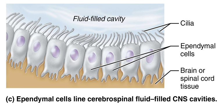
Ependymal cells
Range in shape from squamous to columnar
May be ciliated
Cilia beat to circulate CSF
Line the central cavities of the brain and spinal column
Form permeable barrier between cerebrospinal fluid (CSF) in cavities and tissue fluid bathing CNS cells
Oligodendrocytes
Branched cells with fewer processes than astrocytes
Processes wrap CNS nerve fibers forming insulating myelin sheath

Two major neuroglia seen in PNS
Satellite cells–
Surround neuron cell bodies in PNS
Function similar to astrocytes of CNS
Schwanncells, or neurolemmocytes
Surround all peripheral nerve fibers and form myelin sheaths around the thicker nerve fibers
Similar function as oligodendrocytes–
Vital to regeneration of damaged peripheral nerve fibers

Neurons, or nerve cells, are
structural units of nervous system
Large, highly specialized cells that conduct impulses
Special characteristics
Extreme longevity (lasts a person’s lifetime)
Amitotic (lose their ability to divide) with few exceptions
High metabolic rate and require continuous supply of oxygen and glucose
All have a cell body and one or more slender processes

Cell body, soma, contains
spherical nucleus with nucleolus, surrounded by cytoplasm
Most neuron cell bodies are located in CNS
Nuclei: clusters of neuron cell bodies in CNS
Ganglia: clusters of neuron cell bodies in PNS
Neuron Processes
Processes extend from cell body of all neurons
CNS contains both neuron cell bodies and their processes
PNS contains chiefly of neuron processes (most cell bodies in CNS)
Two types of processes–Dendrites–Axon
Dendrites
Motor neurons can contain hundreds of these short, tapering, diffusely branched processes
Contain same organelles as in cell body
Receptive(input) regionsof neuron
Convey incoming messages toward cell body as graded potentials(short-distance signals)
In many brain areas, finer dendrites are highly specialized to collect information
Contain dendritic spines, appendages with bulbous or spiky ends

Axon Structure
Each neuron has only one axon
Axon length varies
Some axons branch
Bundles of axons in CNS are called
tracts
Bundles of axons in PNS are called
nerves
Axon Functional Characteristics
Axon is the conducting region of neuron
Axon terminals are the secretory region of neuron
Each neuron receives and sends signals with different neurons simultaneously
Axons rely on cell bodies for membrane components and mRNA
Axons quickly decay if cut or damaged
Axonal Transport
Axons have efficient internal transport mechanisms
Molecules and organelles are moved along axons by motor proteins and cytoskeletal elements
Movement occurs in both directions
Myelin sheath function
Protect and electrically insulate axon
Increase speed of nerve impulse transmission
segmented sheath surrounds most long or large-diameter axons
Myelinated axons
do not contain sheath
Nonmyelinated axons
Conduct impulses more slowly
Myelination in the PNS
Formed by Schwann cells
Wrap around axon in jelly roll fashion
One cell forms one segment of myelin sheath

Myelin sheath gaps, or nodes of Ranvier
Gaps between adjacent Schwann cells
Sites where axon collaterals can emerge

Neuron Structural Classifications
Three types grouped by number of processes extending from cell body
Multipolar neurons: many processes (1 axon and many dendrites)–99% of neurons; major neuron type in CNS
Bipolar neurons: two processes (1 axon and 1 fused dendrite)
Rare neuron type; found in retina, ear, and olfactory mucosa
Unipolar neurons: one process (T-shaped process exits cell body and divides into 2 branches of axon)
Peripheral process: branch associated with sensory receptor
Central process: branch enters CNS
Also called pseudounipolar neurons
Found mainly in PNS ganglia where function as sensory neurons

Neuron Functional Classification
Three types of neurons grouped by direction nerve impulse travels relative to CNS
Sensory (afferent) neurons
Transmit impulses from sensory receptors toward CNS
Almost all are unipolar neurons
Cell bodies located in ganglia in PNS
Motor (efferent) neurons
Carry impulses from CNS to effectors
Multipolar neurons
Most cell bodies located in CNS (except some autonomic neurons)
Interneurons, or association neurons
Lie between motor and sensory neurons in neural pathways–
Shuttle signals through CNS pathways, most are entirely within CNS–99% of body’s neurons are interneurons

a measure of potential energy generated by separated charge
Voltage
Measured between two points in volts (V)or millivolts (mV) ▪
Called potentialdifferenceor potential
Charge difference across plasma membrane results in potential
Greater charge difference between points = higher voltage
flow of electrical charge (ions) between two points
Current
Can be used to do work
Flow is dependent on voltage and resistance
Resistance
hindrance to charge flow
Insulator
substance with high electrical resistance
Conductor
substance with low electrical resistance
Ohms Law
gives relationship of voltage, current, resistance Current(I) voltage(V)/resistance (R)
Role of membrane ion channels
Plasma membranes have large proteins that acts as ion channels
Two main types of ion channels
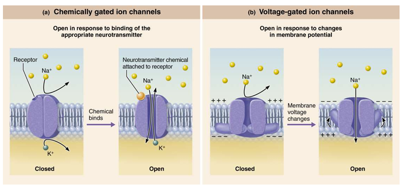
Leakage
Gated Channels
Leakage Channels
which are always open
Gated channels
in which part of the protein changes shape to open/close the channel–
Chemically gated channels or ligand gated channels
Open only with binding of a specific chemical (ex: neurotransmitter)
Voltage-gated channels
Open and close in response to changes in membrane potential–
Mechanically gated channels
Open and close in response to physical deformation of receptors, as in sensory receptors
Direction into or out of the cells is determined by the
Electrochemical gradient
ions move from area of higher concentration to area of lower concentration
Concentration Gradient
ions move toward area of opposite electrical change
Electrical gradient
Membrane Potential

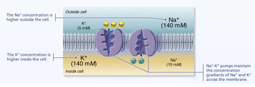
Differences in ionic composition
Cell cytosol has lower concentration of Na+ than ECF
Balanced chiefly by chloride ions Cl-
Cell cytosol has higher concentration of K+ than ECF
Balanced by negatively charged proteins
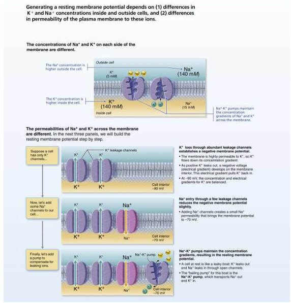
K+ plays most important role in membrane potential
Generating a resting membrane potential depends on
(1) differences in K and Na concentrations inside and outside cells,
(2) differences in permeability of the plasma membrane to these ions
stabilizes resting membrane potential
Sodium-potassium pump (Na KATPase)
Three Na are pumped out of cell while two K are pumped back in
Membrane potential changes when:
Concentrations of ions across membrane change
Membrane permeability to ions changes
Changes in membrane potential produce two types of signals
Graded potentials -Incoming signals operating over short distances
Action potentials -Long-distance signals of axons
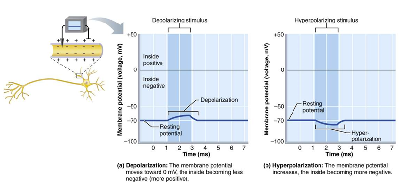
decrease in membrane potential (moves toward zero and above)
Depolarization
Inside of membrane becomes less negative than resting membrane potential Probability of producing impulse increases
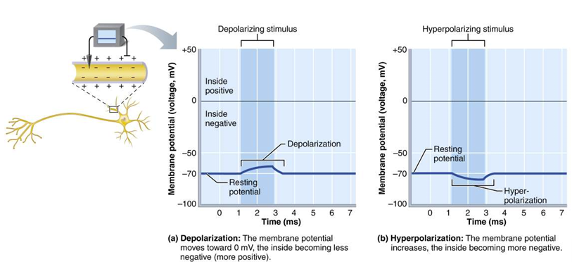
increase in membrane potential (away from zero)
Hyperpolarization
Inside of membrane becomes more negative than resting membrane potential ▪Probability of producing impulse decreases
short-lived, localized changes in membrane potential
Graded potentials
The stronger the stimulus, the more voltage changes and the farther current flows
Graded potentials are trigged by
stimulus that opens gated ion channels
graded potentials in receptors of sensory neurons
Receptor potential (generator potential):
neuron graded potential
Postsynaptic potential
special type of graded potential that occurs in muscle cell, instead of postsynaptic neuron, triggering action potential for muscle contraction
End-plate potential:
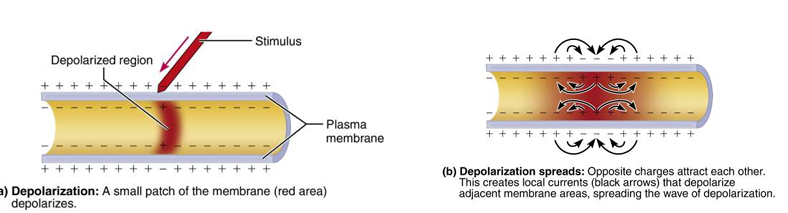
Once gated ion channel opens,
depolarization spreads from one area of membrane to next
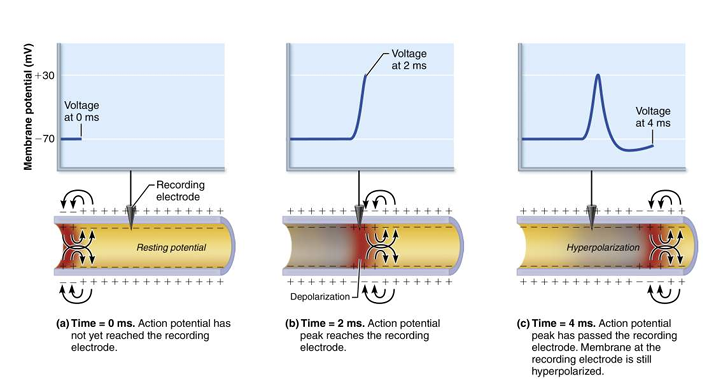
a brief reversal of membrane potential with a change in voltage of ~100 mV
action potential
Action potentials only occur in
excitable membranes–Neurons and muscle cells
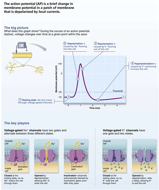
Steps in Generating an Action Potential - First Step
Resting state: All voltage-gated Na andK channels are closed
Only leakage channels for Na andK are open
Maintains the resting membrane potential
Each Na channel has two voltage-sensitive gates
Activation gates: closed at rest; open with depolarization, allowing Na to enter cell
Inactivation gates: open at rest; block channel once it is open to prevent more Na from entering cell ▪
Each K channel has one voltage-sensitive gate
Closed at rest
Opens slowly with depolarization

Steps in Generating an Action Potential - Second Step
Depolarization: voltage-gated Na channels open ▪
Depolarizing local currents open voltage-gated Na channels, and Na rushes into cell ▪
Na activation and inactivation gates open ▪
Na influx causes more depolarization, which opens more Na channels
As a result, ICF becomesless negative ▪
At threshold ( 55 to 50 mV), positive feedback causes opening of all Na channels–
Results in large action potential spike –
Membrane polarity jumps to +30 mV

Steps in Generating an Action Potential - Third Step
Repolarization: Na channels are inactivating, and voltage-gated K channels open ▪
Na channel inactivation gates close–
Membrane permeability to Na declines to resting state–
AP spike stops rising ▪
Voltage-gated K channels open–
K exits cell down its electrochemical gradient ▪
Repolarization: membrane returns to resting membrane potential

Steps in Generating an Action Potential - Fourth Step
Hyperpolarization: Some K channels remain open, and Na channels reset ▪
Some K channels remain open, allowing excessive K efflux–
Inside of membrane becomes more negative than in resting state ▪
This causes hyperpolarization of the membrane (slight dip below resting voltage) ▪
Na channels also begin to reset

Not all depolarization events produce APs
For an axon to “fire,” depolarization must reach threshold voltage to trigger AP
AP threshold
Membrane is depolarized by 15 to 20 mV
Na permeability increases–
Na influx exceeds K efflux
The positive feedback cycle begins
AP either happens completely, or does not happen at all
All-or-none phenomenon
allows AP to be transmitted from origin down entire axon length toward terminals
Propagation
time in which neuron cannot trigger another AP
Refractory period:
Absolute refractory period
Time from opening of Na channels until resetting of the channels
Ensures that each AP is an all-or-none event–
Enforces one-way transmission of APs
Relative refractory period
Follows absolute refractory period ▪
Most Na channels returned to their resting state, some K channels still open ▪
Repolarization is occurring–
Threshold for AP generation is elevated–Only exceptionally strong stimulus can stimulate an AP
Rate of AP propagation depends on two factors:
Axon diameter
▪Larger-diameter fibers have less resistance to local current flow, so have faster impulse conduction
2. Degree of myelination
▪Two types of conduction depending on presence or absence of myelin
APs can propagate in two ways:
Nonmyelinated axons: slower conduction, called continuous conduction, that occurs in nonmyelinated axons
Myelinated axons: faster conduction, called saltatory conduction, occurs only in myelinated axons and is about 30times faster
Axons (nerve fibers) are classified according to diameter, degree of myelination, and speed of conduction
A fiber, B fiber, C fiber
A Fiber
Largest diameter, heavily myelinated axons
Transmit at 150 m/s (over 300 mph)
B Fiber
Intermediate diameter, lightly myelinated axons
Transmit at 15 m/s ( 30 mph)
C Fiber
Smallest diameter, nonmyelinated axons ▪ Transmit at 1 m/s ( 2 mph) or less
junctions that mediate information transfer
From one neuron to another neuron
Or from one neuron to an effector cell
Synapses

neuron conducting impulses toward synapse (sends information)
Presynaptic neuron:
neuron transmitting electrical signal away from synapse (receives information)–In PNS may be a neuron, muscle cell, or gland cell
Postsynaptic neuron:
Types of Synaptic Connections
Axodendritic synapses: between axon terminals of one neuron and dendrites of others
Axosomatic synpases: between axon terminals of one neuron and soma (cell body) of others
Two main types of synapses
Electrical synapses
Chemical synapses
Electrical Synapse
Neurons are electrically coupled
Joined by gap junctions that connect cytoplasm of adjacent neurons–Communication is very rapid and may be unidirectional or bidirectional–
Found in some brain regions responsible for eye movements or hippocampus in areas involved in emotions and memory–
Most abundant in embryonic nervous tissue
Specialized for release and reception of chemical messengers
Neurotransmitters
Chemical Synapse composed of
Axon terminal of presynaptic neuron: contains synaptic vesicles filled with neurotransmitter
Receptor region on postsynaptic neuron’s membrane: receives neurotransmitter
Usually on dendrite or cell body
Two parts separated by fluid-filled synaptic cleft
Transmission across synaptic cleft
Synaptic cleft prevents nerve impulses from directly passing from one neuron to next
Chemical event (as opposed to an electrical one)
Depends on release, diffusion, and receptor binding of neurotransmitters
Ensures unidirectional communication between neurons
Chemical Synapse Information Transfer Steps
AP arrives at axon terminal of presynaptic neuron
Voltage-gated 2 Ca channelsopen, and 2 Ca enters axon terminal
Ca entry causes synaptic vesicles to release neurotransmitter by exocytosis
Neurotransmitter diffuses across the synaptic cleft and binds to specific receptors on the postsynaptic membrane
Binding of neurotransmitter opens ion channels, creating graded potentials
Neurotransmitter effects are terminated

Acetylcholine(ACh)
Released at neuromuscular junctions
Synthesized from acetic acid and choline by enzyme choline acetyltransferase
Degraded by enzyme acetylcholinesterase (AChE)
Catecholamines - Biogenic Amines
Dopamine, norepinephrine (NE), and epinephrine: made from the amino acid tyrosine
Indolamines - Biogenic Amines
▪Serotonin: made from the amino acid tryptophan
Histamine: made from the amino acid histidine
Amino Acids
Amino acids make up all proteins: therefore, it is difficult to prove which are neurotransmitters
Amino acids that are proven neurotransmitters ▪
Glutamate ▪Aspartate ▪Glycine ▪Gamma-( )aminobutyric acid (GABA
Peptides
Strings of amino acids that have diverse functions ▪
Substance P–Mediator of pain signals
▪Endorphins (beta endorphin, dynorphin, and enkephalins)–Act as natural opiates; reduce pain perception ▪
Gut-brain peptides–Somatostatin and cholecystokinin (CCK) play role in regulating digestion
Purines
Monomers of nucleic acids that have an effect in both CNS and PNS ▪
Adenosine triphosphate (ATP), the energy molecule, is now considered a neurotransmitter ▪
Adenosine, a part of ATP, is a potent inhibitor in brain
Caffeine blocks adenosine receptors ▪
Can induce 2 Ca influx in astrocytes
Gases and Lipids
Gasotransmitters ▪
Nitric oxide (NO), carbon monoxide (CO), hydrogen sulfide ( ) 2 HS gases
Central nervous system (CNS) consists of
brain and spinal cord
Adult brains have four regions:
Cerebral Hemispheres
Diencephalon
Brain stem
midbrain
pons
medulla oblongata
Cerebellum

short, nonmyelinated neurons and cell bodies
gray matter
myelinated and nonmyelinated axons
white matter
Basic pattern found in CNS:
central cavity surrounded by gray matter, with white matter external to gray matter