Ch 10 Sensory Physiology
 Call Kai
 Learn
Learn Practice Test
 Spaced Repetition
 Match
Match Flashcards
 Knowt Play
1/95

There's no tags or description
Looks like no tags are added yet.
Name | Mastery | Learn | Test | Matching | Spaced | Call with Kai | Chat |
|---|
No analytics yet
Send a link to your students to track their progress
96 Terms
What is the "common language" of the nervous system?
Action potentials. They carry info to the brain, and the brain sends responses back out through motor pathways.
What is the function of a sensory receptor?
It acquires the initial stimulus from the environment and converts it into APs that travel to the CNS.
What is modality?
The form of info a receptor is made for (e.g., light for vision, sound for hearing, pressure for touch).
What is transduction?
When a sensory receptor changes its modality (light, sound, pressure) into action potentials the nervous system can understand.
Trace sensory info from stimulus to response.
Modality → sensory receptor → transduction into APs → neuron to neuron → CNS integrates → response sent via motor pathways
Why do different modalities feel like different sensations?
Because each modality stimulates a different CNS pathway, so the brain processes them as different sensations.
What are the two structural forms a sensory receptor can take?
Simple dendritic endings of neurons, or specialized endings of neurons or non-neuronal cells (e.g., Pacinian and Meissner's corpuscles, which he said not to memorize).
How are sensory receptors grouped?
By the type of stimulus (modality) they transduce.
What do chemoreceptors sense? Give examples.
Chemical stimuli, such as calcium and CO₂ levels in the blood.
What do photoreceptors and thermoreceptors respond to?
Photoreceptors transduce light into APs. Thermoreceptors respond to temperature changes.
What do mechanoreceptors respond to?
Mechanical deformation of their cell membrane, such as pressure and touch.
What do nociceptors respond to?
Intense stimuli, which they signal as pain.
What do proprioceptors do? Give his example.
They signal the position of body parts. Reaching for a coffee cup without looking works because you know where your hand and fingers are.
What are cutaneous receptors, and what do they respond to?
Receptors near an epithelial surface (skin), also called general sense receptors. They respond to touch, pressure, temperature, or pain.
What are special sense receptors? Give examples.
Receptors that are part of a sensory organ, such as those for hearing, sight, and equilibrium.
What are the two key differences between special sense and cutaneous (general) receptors?
Special sense receptors are separate cells from the neuron and are found in only one specific region.
Cutaneous receptors are usually on a sensory neuron's dendrite and are found all over the body.
Are pain receptors special or general? What about smell receptors?
Pain is general (cutaneous) because pain can be felt anywhere in the body. Smell is special because those receptors are found only in the nose.
What is adaptation?
When a receptor stops responding to a stimulus even though the stimulus is still being applied. All receptors eventually adapt; they differ only in how fast.
What are phasic receptors? Give examples.
Fast-adapting receptors. They fire a burst of APs when the stimulus is applied, then quickly reduce their firing rate even though the stimulus continues. Examples: smell and touch.

Describe the quarter experiment and what it shows.
Place a quarter on your forearm with your eyes closed. You feel it at first, but within seconds you can't, even though it's still pressing. Touch receptors are phasic and adapt quickly.
What are tonic receptors? Give an example.
Slow-adapting receptors that keep firing at a constant rate as long as the stimulus is applied. They do adapt eventually, but it takes a very long time. Example: pain.

Can a receptor respond to a stimulus other than its modality?
Yes. Any receptor can be forced to respond to a strong enough stimulus (e.g., enough pressure on a vision receptor). That doesn't mean the receptor is made for that stimulus
What does the law of specific nerve energies state?
Stimulating a sensory fiber evokes only the sensation of its modality. A receptor's modality is the one that activates it with the least stimulus intensity.
What is the adequate stimulus?
The receptor's normal stimulus, the one that requires the least energy to activate it. For a vision receptor, a small amount of light activates it, so light is its adequate stimulus
What is the adequate stimulus for mechanoreceptors, pain receptors, chemoreceptors, and photoreceptors?
Mechanoreceptors respond to mechanical force, pain receptors to tissue damage, chemoreceptors to dissolved chemicals, and photoreceptors to light.

What is a generator potential?
A depolarizing graded potential in a sensory receptor, triggered by its adequate stimulus (modality).
It is the sensory receptor's equivalent of an EPSP.
How does a generator potential differ from an EPSP?
Both open ion channels that let Na⁺ in and depolarize the cell. In an EPSP, a neurotransmitter binding to a receptor opens the channels. In a generator potential, the modality itself opens them.
Trace how a sound stimulus becomes an AP in a hearing receptor.
Sound (modality) hits the receptor → ion channels open → Na⁺ enters → depolarization (generator potential) → if it reaches threshold, an AP fires
What determines whether a generator potential produces an AP?
Stimulus strength. A stronger stimulus makes a bigger generator potential, but if it stays below threshold (1–3 on the graph), no AP fires. Only when it reaches threshold (4) does an AP fire (5).

How does the brain know how strong a stimulus is?
By the frequency of APs reaching it. AP amplitude is always the same (+30 mV), so stimulus strength is coded by frequency, not size.
Trace how stimulus strength is coded.
Stronger stimulus → larger generator potential (amplitude is proportional to stimulus strength) → further above threshold → higher-frequency APs → brain reads it as a stronger stimulus
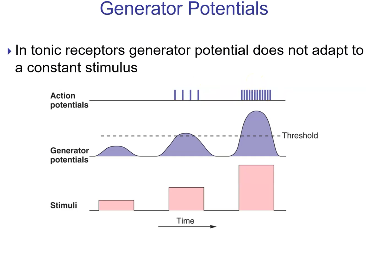
How do tonic receptors' generator potentials respond to a constant stimulus?
They don't adapt. The generator potential stays up as long as the stimulus is applied, so APs keep firing.
What is a receptive field?
The area of skin monitored by a single sensory neuron. Stimulating that area changes the neuron's firing rate.
How does receptive field size relate to receptor density and sensitivity?
Inversely. Smaller receptive fields mean more receptors and greater sensitivity (acuity). Larger fields mean fewer receptors and less sensitivity.
Compare receptive fields on the back/legs vs. the fingertips.
The back and legs have low receptor density, so their receptive fields are large and it's hard to pinpoint where you were touched. Fingertips have high receptor density and small receptive fields, so touch is very precise.
What is the two-point touch threshold?
The minimum distance at which 2 points of touch are perceived as separate. It measures tactile acuity. If both points fall in one neuron's receptive field, you feel only one point.

What are the 3 divisions of the ear, and where are their boundaries?
The outer (external) ear runs from the tympanic membrane outward.
The middle ear sits between the tympanic membrane and the cochlea and holds the ossicles.
The inner (internal) ear is the snail-like structure.
Which parts of the ear are involved in hearing vs. balance and equilibrium?
Hearing uses the outer, middle, and inner ear. Balance and equilibrium use only the inner ear.
What are the two types of sensory receptors in the ear, and what is the modality for hearing?
Receptors for hearing (modality is sound) and receptors for balance and equilibrium.
What does the vestibular apparatus provide, and what does it consist of?
A sense of equilibrium (orientation to gravity). It consists of the otolith organs (utricle and saccule) and the semicircular canals. Together with the cochlea, it forms the inner ear.
What do the utricle/saccule vs. the semicircular canals detect?
The utricle and saccule detect linear acceleration (forward, backward, up, down). The semicircular canals detect angular acceleration (rotation).
Why do the semicircular canals give us a 3D sense of movement?
They are oriented in 3 different planes.
Describe the structure of an equilibrium receptor.
Hair cells are the receptors. Each has 20–50 hairlike stereocilia, and one of these is the kinocilium (a true cilium). The stereocilia are mechanoreceptors, so physical bending stimulates them.

What happens when stereocilia bend toward vs. away from the kinocilium?
Toward the kinocilium, the hair cell is stimulated and AP frequency increases. Away from it, the hair cell is inhibited and AP frequency decreases. This applies to all receptors in the ear.
Trace what happens in the saccule when you tilt your head down.
Head tilts down → gravity pulls the otolithic membrane down → embedded stereocilia bend → hair cells fire APs → cerebellum → brain detects the head moving downward
Where are the hair cells of the semicircular canals embedded?
In the cupula of the crista ampullaris, a membrane surrounded by endolymph.
Trace how head rotation is detected by the semicircular canals.
Head rotates → endolymph moves → cupula bends → stereocilia bend (opposite the direction of angular acceleration) → hair cells fire APs → cerebellum interprets the rotation

What is the function of the stapedius muscle?
It's attached to the stapes and protects against loud noises by contracting to dampen large vibrations, which prevents nerve damage in the cochlea.
Trace sound vibrations from the outer ear to the cochlea.
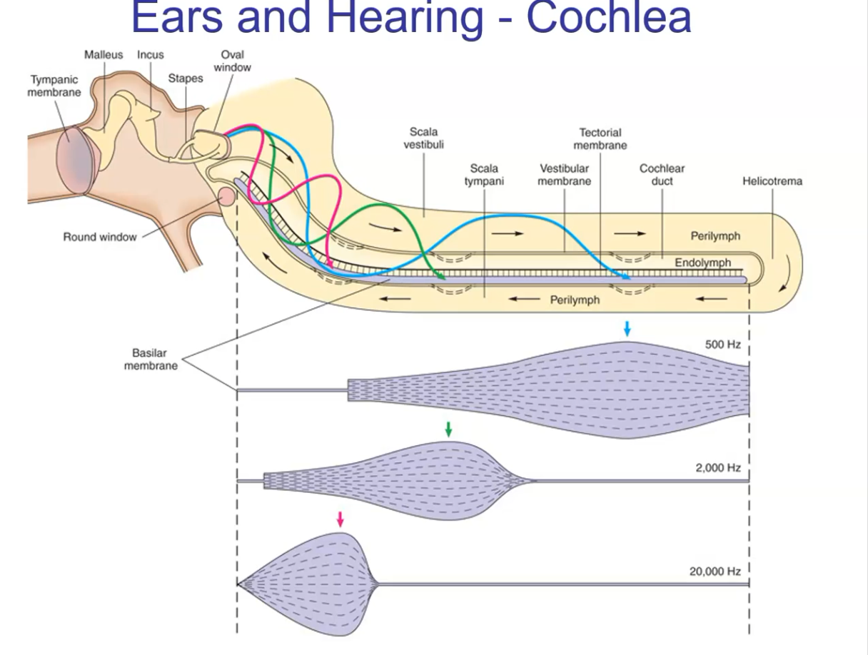
Sound waves → external auditory meatus → tympanic membrane vibrates → malleus → incus → stapes → oval window of the cochlea
What are the 3 chambers of the cochlea, from top to bottom?
Scala vestibuli (top), cochlear duct (middle), and scala tympani (bottom).

Where are the hearing receptors located?
In the organ of Corti (spiral organ), inside the cochlear duct.
What are the two membranes of the organ of Corti, and how is it different from other ear receptors?
The tectorial membrane holds the embedded stereocilia, and the basilar membrane sits below. Unlike other ear receptors, the basilar membrane is what moves, bending the stereocilia so the hair cells fire APs.
Trace sound from the oval window through the cochlea to the brain.
Stapes pounds the oval window → oval window vibrates → perilymph waves travel along the Scala vestibuli → waves cross into the cochlear duct (endolymph) → basilar membrane moves → stereocilia bend → hair cells fire APs → brain. Leftover energy → perilymph of the Scala tympani → round window vibrates → energy released into the middle ear (prevents extra receptor stimulation).

Which fluids fill each chamber of the cochlea?
Perilymph fills the scala vestibuli and scala tympani ("peri" means around). Endolymph fills the cochlear duct.
How does the ear determine pitch?
By which receptors along the cochlear duct are stimulated. The brain reads pitch from where along the cochlea the stimulation happens.
Where are low, mid, and high tones detected along the cochlear duct?
Low tones (bass) travel far, so receptors far from the oval window are stimulated. Mid tones (speech) are detected midway. High tones (opera) travel only a short distance, so receptors close to the oval window are stimulated.
What is tonotopic organization?
Each region of the cochlea sends signals to a matching area of the auditory cortex, and each area represents a different pitch.

How is sound volume encoded?
By AP frequency. Louder sound produces higher-frequency APs.
What determines pitch vs. volume?
Pitch is determined by which receptors fire (location). Volume is determined by how fast they fire (AP frequency).
What are 3 causes of hearing impairment?
The receptors don't respond, the nerve to the brain doesn't work, or the brain area that receives the signal doesn't work.
Trace light's path into the eye.
Cornea → anterior chamber → pupil → lens → vitreous → retina
What are the sclera and cornea?
The sclera is the white, outermost layer where the eye muscles attach. The cornea is its transparent front, which lets light in and does most of the light bending.
What holds the lens in place and changes its shape?
The ciliary bodies, through the suspensory ligaments.
What do the iris, pupil, and lens do?
The iris forms the pupil. The pupil controls how much light enters. The lens bends light so it lands focused on the retina.
Where are the photoreceptors located?
In the retina.
How does the iris constrict vs. dilate the pupil?
Constriction happens when the circular muscles contract (parasympathetic, in bright light). Dilation happens when the radial muscles contract (sympathetic, in dim light).

Why does the pupil constrict in bright light?
To keep from overstimulating the photoreceptors, which would cause temporary blindness.
Where does light that the retina doesn't absorb go?
It's absorbed by the dark choroid layer.
What is the fovea centralis?
The center of the macula lutea, where light produces the sharpest image.
What is the optic disc, and why is it the blind spot?
It's where retinal axons gather and exit as the optic nerve. It has no photoreceptors, so light landing there can't be detected.
What is accommodation?
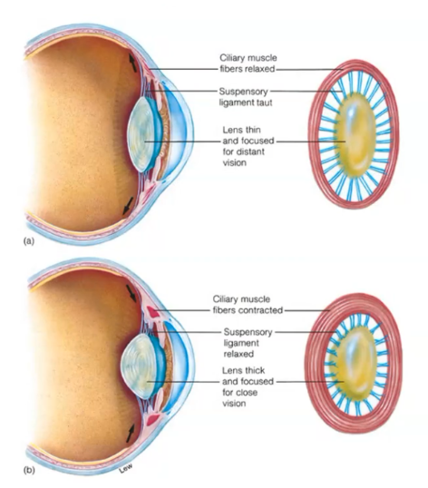
Changing the lens shape (via the ciliary bodies and suspensory ligaments) so light lands focused on the retina.
Trace accommodation for far (>20 ft) vs. near vision.
Far: ciliary muscles relax → suspensory ligaments taut → lens thin (least convex) → bends light less
Near: (as distance decreases): ciliary muscles contract → ligaments slack → lens recoils thick (more convex) → bends light more
Why does near vision get worse with age?
The lens loses elastic fibers and stiffens, so it can't recoil into a thick shape.
What is visual acuity, and what does it depend on?
Sharpness of vision. It depends on resolving power, the ability to tell apart 2 closely spaced dots.
What is myopia (nearsightedness)?
The image focuses in front of the retina because the eyeball is too long. Close objects are clear and far ones are blurry. It's corrected with a concave lens.

What is hyperopia (farsightedness)?
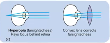
The image focuses behind the retina because the eyeball is too short. Far objects are clear and close ones are blurry. It's corrected with a convex lens.
What are emmetropia and astigmatism?
Emmetropia is normal vision, with light focused on the retina. Astigmatism is when parts of the light don't focus because the cornea or lens is unevenly shaped.
What does the retina consist of?
Neurons, pigmented epithelium, and photoreceptors (rods and cones). Its neural layers are an extension of the brain.
What must light pass through before reaching the rods and cones?
Several neural layers.
Trace the visual signal from photoreceptor to brain.
Rods/cones → bipolar cells → ganglion cells → ganglion axons form the optic nerve → primary visual cortex (occipital lobe)
What are the 2 segments of rods and cones, and what's in the outer segment?
The inner and outer segments. The outer segment holds stacks of photopigment discs; new discs are added at the base and removed at the tip.
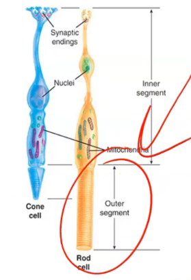
What do rods vs. cones detect?
Cones detect color ("C for color"). Rods detect black and white (grayscale).
Why are rods considered "night vision"?
Rods are very sensitive and activate in low light. Cones need bright light, so we can't see color in the dark.
What is rhodopsin, and what happens when light hits it?
The photopigment in rods. Light changes retinal from cis to trans, so rhodopsin dissociates into retinal and opsin. This is the bleaching reaction.

Can bleached rhodopsin respond to light again?
No. It must be replaced with unexposed pigment first.
How are photoreceptors different from other receptors?
Light inhibits them, and inhibiting them is what sends the signal to the brain. Other receptors signal when they're stimulated.
Trace what happens in a rod in the dark.
Na⁺ channels open (kept open by cGMP) → Na⁺ enters ("dark current") → rod depolarized → releases inhibitory NT → bipolar cell inhibited → no signal to ganglion cell → no signal to brain

Trace what happens in a rod in light.
Light → cGMP declines → Na⁺ channels close → rod hyperpolarizes → no inhibitory NT released (brakes removed) → bipolar cell releases excitatory NT → ganglion cell fires APs → brain

What keeps the rod's Na⁺ channels open in the dark?
cGMP bound to the channel.
Trace how light closes the rod's Na⁺ channels.
Light → 11-cis-retinal becomes all-trans-retinal (bleaching) → G-protein dissociates → α subunit activates phosphodiesterase → cGMP converted to GMP → Na⁺ channels close → dark current stops → rod hyperpolarizes

What is the dark current?
The depolarizing Na⁺ influx through open Na⁺ channels in rods and cones in the dark. Light stops it by closing those channels, which hyperpolarizes the cell.
Why do photoreceptors "work backwards"?
They're stimulated (depolarized) in the dark and inhibited (hyperpolarized) by light.
Why does the fovea centralis have the highest visual acuity?
The neural layers (ganglion and bipolar cells) are pushed to the sides, so light hits the photoreceptors directly. It also contains only cones, which give sharper images.

Where is the fovea located?
It's a pin-sized pit within the yellow macula lutea.
Compare cone vs. rod connections to ganglion cells.
In the fovea, each cone supplies 1 ganglion cell (no convergence), giving a small receptive field and high acuity. Many rods converge on 1 ganglion cell, giving a large receptive field, high sensitivity, and low acuity.
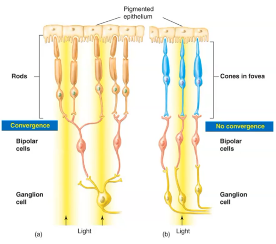
Why can't the brain pinpoint where light hits within a rod's receptive field?
All the rods in that field send signals through the same ganglion cell, so the brain can't tell which rod was activated.