A&P Lab Practical 3 and 4

0.0(0)
Studied by 0 people
call kaiCall Kai
learnLearn
examPractice Test
spaced repetitionSpaced Repetition
heart puzzleMatch
flashcardsFlashcards
GameKnowt Play
Card Sorting

1/96

encourage image

There's no tags or description

Looks like no tags are added yet.

97 Terms

1
New cards
<p class="question"></p><p>What type of glial cell is shown here?</p><p></p>


What type of glial cell is shown here?


Schwann cell
2
New cards
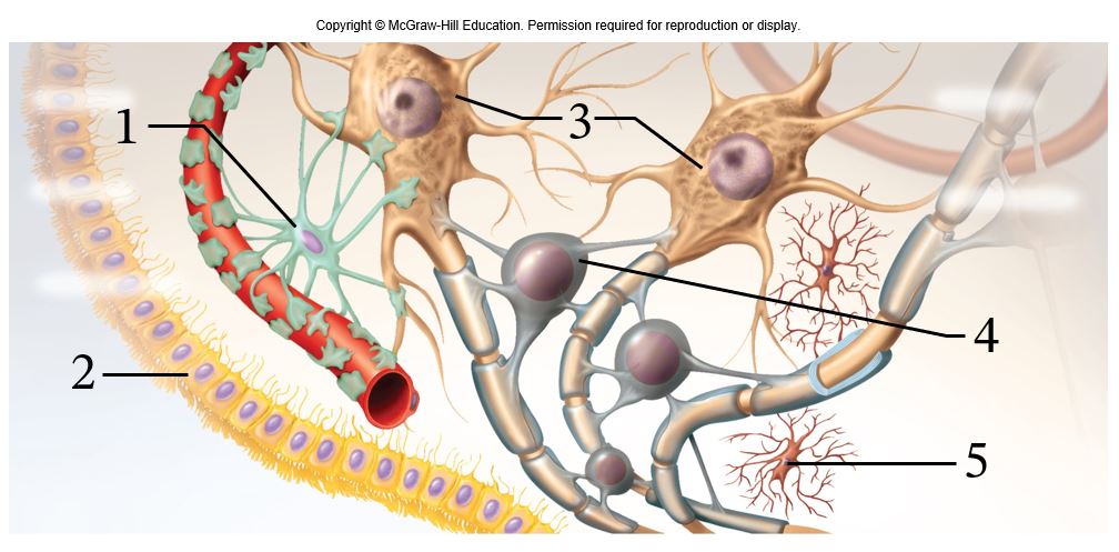
<p>What type of cell is labeled 1 in this figure?</p>

What type of cell is labeled 1 in this figure?

astrocyte

3
New cards
<p>What type of cell is labeled 2 in this figure?</p>

What type of cell is labeled 2 in this figure?

ependymal cell

4
New cards
<p>What type of cell is labeled 3 in this figure?</p>

What type of cell is labeled 3 in this figure?

neuron

5
New cards
<p>What type of cell is labeled 4 in this figure?</p>

What type of cell is labeled 4 in this figure?

oligodendrocyte

6
New cards
<p>What type of cell is labeled 5 in this figure?</p>

What type of cell is labeled 5 in this figure?

microglia

7
New cards
<p class="question"></p><p>Which option below correctly matches the types of neuron to the numeric labels on the figure?</p><p></p>


Which option below correctly matches the types of neuron to the numeric labels on the figure?


1-Afferent neuron, 2-Interneuron, 3-Efferent neuron

8
New cards
<p>Name the part of the neuron labeled 1.</p>

Name the part of the neuron labeled 1.

dendrites

9
New cards
<p>Name the part of the neuron labeled 2.</p>

Name the part of the neuron labeled 2.

cell body

10
New cards
<p>Name the part of the neuron labeled 3</p>

Name the part of the neuron labeled 3

nucleus

11
New cards
<p>Name the part of the neuron labeled 4</p>

Name the part of the neuron labeled 4

node of ranvier

12
New cards
<p>Name the part of the neuron labeled 5</p>

Name the part of the neuron labeled 5

internodes

13
New cards
<p>What is happening during the point on the graph labeled 1?</p>

What is happening during the point on the graph labeled 1?

The local current arrives at the axon hillock.

14
New cards
<p>Why is the membrane hyperpolarized at the point on the graph labeled 6?</p><p></p><p></p>

Why is the membrane hyperpolarized at the point on the graph labeled 6?


Voltage-gated potassium channels are open, so the membrane is more permeable to potassium than at rest.

15
New cards
<p>Why does the voltage peak at the point on the graph labeled 4?</p>

Why does the voltage peak at the point on the graph labeled 4?

Voltage-gated sodium channels innactivate.

16
New cards
<p>What is labeled 1 on this figure?</p>

What is labeled 1 on this figure?

Axon of presynaptic neuron

17
New cards
<p>What is labeled 2 on this figure?</p>

What is labeled 2 on this figure?

synaptic vesicle

18
New cards
<p>What is labeled 3 on this figure?</p>

What is labeled 3 on this figure?

Synaptic cleft

19
New cards
<p><span>The type of summation shown in this diagram is called </span><span style="line-height: inherit;">_________ summation.</span></p>

The type of summation shown in this diagram is called _________ summation.

temporal

20
New cards
<p><span>The type of summation shown in this diagram is called </span><span style="line-height: inherit;">_________ summation.</span></p>

The type of summation shown in this diagram is called _________ summation.

spatial

21
New cards
<p>What type of neural circuit is shown here?</p>

What type of neural circuit is shown here?

diverging

22
New cards
<p>What type of neural circuit is shown here?</p>

What type of neural circuit is shown here?

converging

23
New cards
<p>What type of neural circuit is shown here?</p>

What type of neural circuit is shown here?

reverberating

24
New cards
<p>What type of neural circuit is shown here?</p>

What type of neural circuit is shown here?

Parallel after-discharge

25
New cards

During the relative refractory period, the membrane is hyperpolarized and a larger incoming stimulus is required to trigger an action poential. How does this affect the response properties of a sensory neuron?

Higher stimulus intensity will increase the rate at which the sensory neuron fires action potentials.

26
New cards
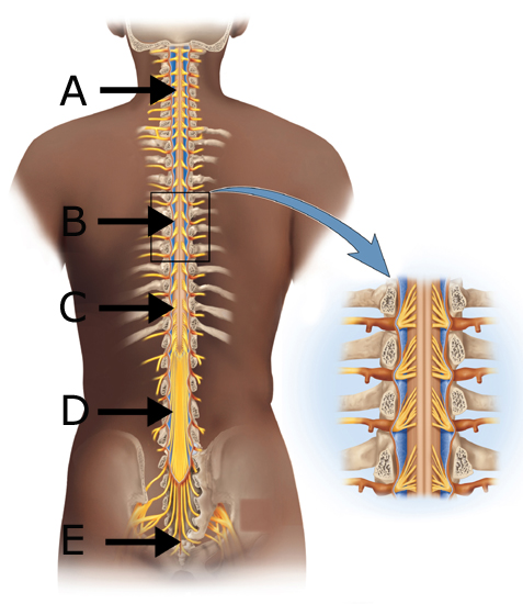
<p>Where do the nerves that innervate the lower limbs go in/out of the spinal cord?</p>

Where do the nerves that innervate the lower limbs go in/out of the spinal cord?

C

27
New cards
<p>What is the name of the structure at D?</p>

What is the name of the structure at D?

cauda equina
28
New cards
<p>What is the name of the structure at E?</p>

What is the name of the structure at E?

terminal filum

29
New cards
<p>In which space(s) is(are) CSF found?</p>

In which space(s) is(are) CSF found?

C and E

30
New cards
<p>Where is an epidural injection administered?</p>

Where is an epidural injection administered?

A

31
New cards
<p>Where is a spinal tap taken?</p>

Where is a spinal tap taken?

C

32
New cards
<p>Which connective tissue layer is the epineurium?</p>

Which connective tissue layer is the epineurium?

A

33
New cards
<p>Which connective tissue layer surrounds a fascicle?</p>

Which connective tissue layer surrounds a fascicle?

B

34
New cards
<p>Which connective tissue layer surrounds a fiber?</p>

Which connective tissue layer surrounds a fiber?

D

35
New cards
<p>The plexuses form from which of these structures?</p>

The plexuses form from which of these structures?

A

36
New cards
<p>Which structure carries only sensory information?</p>

Which structure carries only sensory information?

C

37
New cards
<p>Which structure(s) is(are) only found in the autonomic nervous system?</p>

Which structure(s) is(are) only found in the autonomic nervous system?

E

38
New cards
<p>Which reflex is pictured here?</p>

Which reflex is pictured here?

patellar tendon

39
New cards
<p>Which of the following accurately describes the reflex pictured here?</p>

Which of the following accurately describes the reflex pictured here?

polysynaptic

40
New cards
<p>Looking at the muscles at B and G, which are being stimulated to contract and which are being inhibited from contracting?</p>

Looking at the muscles at B and G, which are being stimulated to contract and which are being inhibited from contracting?

The muscles at B are being stimulated to contract;  the muscles at G are being inhibited from contracting.

41
New cards
<p class="question"></p><p>Which&nbsp;clinical test could be used to evaluate the function of this cranial nerve?</p><p></p>


Which clinical test could be used to evaluate the function of this cranial nerve?


Test the subject's ability to protrude their tongue against resistance.

42
New cards
<p class="question"></p><p>Which clinical test could be used to evaluate the function of this nerve?</p><p></p><p></p>


Which clinical test could be used to evaluate the function of this nerve?


Test the subject's ability to taste bitter and sour substances applied to the posterior one-third of their tongue.

43
New cards
<p class="question"></p><p>Which clinical test could be used to evaluate the function of this nerve?</p><p></p>


Which clinical test could be used to evaluate the function of this nerve?


Look for differences in the size and shape of the right and left pupils.

44
New cards
<p class="question"></p><p>Which clinical test could be used to evaluate the function of this cranial nerve?</p><p></p>


Which clinical test could be used to evaluate the function of this cranial nerve?


Test the subject's ability to make facial expressions such as smiling, frowning, and raising their eyebrows.

45
New cards
<p class="question"></p><p>Damage to this cranial nerve could result in which symptom?</p><p></p>


Damage to this cranial nerve could result in which symptom?


Impaired swallowing and gastrointestinal motility

46
New cards
<p>Name the embryonic brain vesicle labeled 1 in this diagram.</p>

Name the embryonic brain vesicle labeled 1 in this diagram.

Telencephalon

47
New cards
<p>Name the embryonic brain vesicle labeled 2 in this diagram.</p>

Name the embryonic brain vesicle labeled 2 in this diagram.

Diencephalon

48
New cards
<p>Name the embryonic brain vesicle labeled 3 in this diagram.</p>

Name the embryonic brain vesicle labeled 3 in this diagram.

Mesencephalon

49
New cards
<p>Name the embryonic brain vesicle labeled 4 in this diagram.</p>

Name the embryonic brain vesicle labeled 4 in this diagram.

Metencephalon

50
New cards
<p>Name the embryonic brain vesicle labeled 5 in this diagram.</p>

Name the embryonic brain vesicle labeled 5 in this diagram.

Myelencephalon

51
New cards
<p class="question"></p><p>This cranial nerve is shown entering the cranium through which of the following?</p><p></p>


This cranial nerve is shown entering the cranium through which of the following?


Internal accoustic meatus

52
New cards
<p class="question"></p><p>The masses of cerebral gray matter shown in this diagram play an important role in which of the following?</p><p></p>


The masses of cerebral gray matter shown in this diagram play an important role in which of the following?


motor control

53
New cards
<p class="question"></p><p>Based on this picture, what is the function of this cranial nerve?</p><p></p>


Based on this picture, what is the function of this cranial nerve?


lateral eye movement
54
New cards
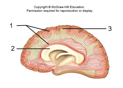
<p>What type of tract is labeled 1 in this diagram?</p>

What type of tract is labeled 1 in this diagram?

association tract

55
New cards
<p>What type of tract is labeled 2 in this diagram?</p>

What type of tract is labeled 2 in this diagram?

Commissural tract

56
New cards
<p>What type of tract is labeled 3 in this diagram?</p>

What type of tract is labeled 3 in this diagram?

Projection tract

57
New cards
<p>What is the function of the indicated brain region?</p>

What is the function of the indicated brain region?

Production of speech

58
New cards
<p>What is the function of the indicated brain region?</p>

What is the function of the indicated brain region?

Understanding written language

59
New cards
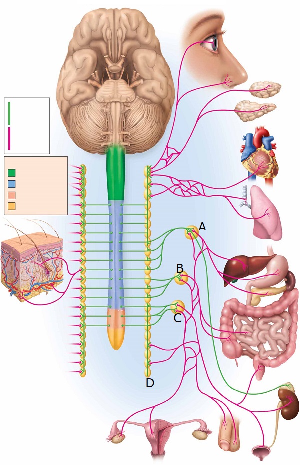
<p>Which ganglion is found at A?</p>

Which ganglion is found at A?

celiac

60
New cards
<p>Which ganglion is found at C?</p>

Which ganglion is found at C?

Inferior mesenteric

61
New cards
<p>Which ganglion is found at D?</p>

Which ganglion is found at D?

sympathetic chain

62
New cards
<p>Which ganglion is found at A?</p>

Which ganglion is found at A?

Pterygopalatine

63
New cards
<p>Which ganglion is found at B?</p>

Which ganglion is found at B?

Ciliary

64
New cards
<p>Which ganglion is found at E?</p>

Which ganglion is found at E?

celiac

65
New cards
<p>What type of sensory receptor is pictured here?</p>

What type of sensory receptor is pictured here?

Lamellar corpuscle

66
New cards
<p>What type of sensory receptor is pictured here?</p>

What type of sensory receptor is pictured here?

Muscle spindle

67
New cards
<p class="question"></p><p>What sensory phenomenon is illustrated here?</p><p></p>


What sensory phenomenon is illustrated here?


referred pain

68
New cards
<p class="question"></p><p>Name the cochlear chamber labeled 1.</p>


Name the cochlear chamber labeled 1.

Scala vestibuli

69
New cards
<p class="question"></p><p>Name the cochlear chamber labeled 2.</p>


Name the cochlear chamber labeled 2.

Cochlear duct

70
New cards
<p class="question"></p><p>Name the cochlear chamber labeled 3.</p>


Name the cochlear chamber labeled 3.

Scala tympani

71
New cards
<p class="question"></p><p>What type of fluid fills chambers 1 and 3?</p>


What type of fluid fills chambers 1 and 3?

Perilymph

72
New cards
<p class="question"></p><p>What type of fluid fills chamber 2?</p>


What type of fluid fills chamber 2?

Endolymph

73
New cards
<p class="question"></p><p>Name the structure labeled 1.</p>


Name the structure labeled 1.

cornea

74
New cards
<p class="question"></p><p>Name the structure labeled 2.</p>


Name the structure labeled 2.

Anterior chamber

75
New cards
<p class="question"></p><p>Name the structure labeled 3.</p>


Name the structure labeled 3.

posterior chamber

76
New cards
<p class="question"></p><p>Name the structure labeled 4.</p>


Name the structure labeled 4.

Vitreous chamber

77
New cards
<p>Which process is illustrated here?</p>

Which process is illustrated here?

transduction

78
New cards
<p><span>This picture illustrates the electrical excitation of hair cells. Vibrations of the cochlea cause the stereocilia to bend, pulling open K+ channels. K+ inflow through the open channels depolarizes the hair cell. Based on this, which of the following statements must be true?</span></p>

This picture illustrates the electrical excitation of hair cells. Vibrations of the cochlea cause the stereocilia to bend, pulling open K+ channels. K+ inflow through the open channels depolarizes the hair cell. Based on this, which of the following statements must be true?

There is a higher concentration of K+ in endolymph than in the intracellular solution of hair cells.

79
New cards
<p class="question"></p><p>Which cranial nerve innervates the eye muscle labeled 1 in this diagram?</p>


Which cranial nerve innervates the eye muscle labeled 1 in this diagram?

Trochlear nerve (IV)

80
New cards
<p class="question"></p><p>Which cranial nerve innervates the eye muscle labeled 2 in this diagram?</p>


Which cranial nerve innervates the eye muscle labeled 2 in this diagram?

Abducens nerve (VI)

81
New cards
<p class="question"></p><p>Which cranial nerve innervates the eye muscles labeled 3 in this diagram?</p>


Which cranial nerve innervates the eye muscles labeled 3 in this diagram?

Oculomotor nerve (III)

82
New cards
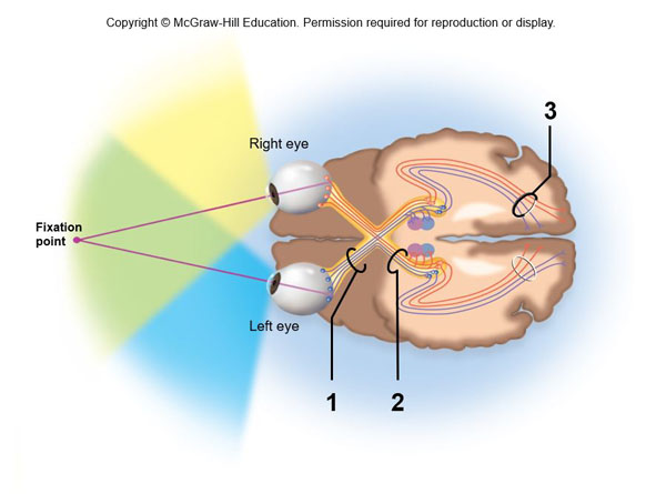
<p class="question"></p><p>What is labeled 1 in this diagram?</p>


What is labeled 1 in this diagram?

optic nerve

83
New cards
<p class="question"></p><p>What is labeled 2 in this diagram?</p>


What is labeled 2 in this diagram?

Optic tract

84
New cards
<p class="question"></p><p>What is labeled 3 in this diagram?</p>


What is labeled 3 in this diagram?

Optic radiation

85
New cards
<p class="question"></p><p>Which visual field deficit would result from damage at the point labeled 1?</p>


Which visual field deficit would result from damage at the point labeled 1?

Loss of sight in one eye

86
New cards
<p class="question"></p><p>Which visual field deficit would result from damage at the point labeled 2?</p>


Which visual field deficit would result from damage at the point labeled 2?

Loss of sight in the contralateral (left) visual field for both eyes
87
New cards
<p class="question"></p><p>Which visual field deficit would result from damage at the point labeled 3?</p>


Which visual field deficit would result from damage at the point labeled 3?

Loss of sight in the contralateral (left) visual field for both eyes

88
New cards
<p class="question"></p><p>Examine this figure, then determine which statement is true.</p><p></p>


Examine this figure, then determine which statement is true.


There is more convergence in the scotopic system, leading to high sensitivity but low resolution.

89
New cards

Which statement is true about scotopic vision as compared to photopic vision?

There is more convergence in the scotopic system, leading to high sensitivity but low resolution.

90
New cards
<p class="question"></p><p>Red lights are often used in photography dark rooms, in military bunkers, and other situations that require light to see by while also preserving night vision. Which of the following statements best explains why your eyes stay dark-adapted after exposure to red light?</p>


Red lights are often used in photography dark rooms, in military bunkers, and other situations that require light to see by while also preserving night vision. Which of the following statements best explains why your eyes stay dark-adapted after exposure to red light?

Since rods are not activated by long wavelengths, they are not subject to bleaching by red light.

91
New cards
<p>Use the absorption spectra graph to answer the following question.</p><p>&nbsp;</p><p>What percentages of maximum response in the three types of cones (S:M:L) will result in the perception of yellow light</p>

Use the absorption spectra graph to answer the following question.


What percentages of maximum response in the three types of cones (S:M:L) will result in the perception of yellow light

0:20:70

92
New cards
<p class="question"></p><p>Use the absorption spectra graph to answer the following question.</p><p>&nbsp;</p><p>Equal activation of M and L cones (but no activity in S cones) will create the perception of which color?</p>


Use the absorption spectra graph to answer the following question.


Equal activation of M and L cones (but no activity in S cones) will create the perception of which color?

green

93
New cards
<p></p>


orbicularis oris

94
New cards
<p> </p>

buccinator

95
New cards
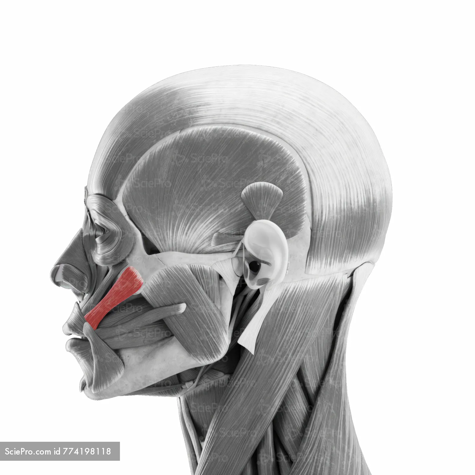
term image

zygomaticus major

96
New cards
term image
97
New cards
term image