Bone, Cartilage and Joints 1
 Call Kai
 Learn
 Practice Test
 Spaced Repetition
 Match
 Flashcards
 Knowt Play
1/49

Earn XP
Description and Tags
M.8R, W.2, L.1
50 Terms
List the 3 main types of CT
Embryonic CT
Adult CT
Special CT
List the subtypes of Adult CT
Loose CT
Dense CT
Dense Regular CT
Dense Irregular CT
Reticular CT
Elastic CT
List the 4 main types of Special CT and their subtypes
Adipose tissue
White adipose tissue
Brown adipose tissue
Cartilage
Hyaline Cartilage
Elastic Cartilage
Fibrocartilage
Bone
Compact Bone
Woven bone (in developing bone)
Cancellous bone
Bone Marrow

List the two main cells that make up the ECM of cartilage and their functions
ChondroBLASTS
cell that is responsible for producing ECM
ChrondroCYTES
maintenance of ground substances
Describe the ECM of cartilage
Made up of:
fibers
ground substance
Avascular
what do the fibers in the ECM of cartilage provide? what does the ground substance of the ECM of cartilage provide?
Fibers: Rigidity
Ground Substance: resiliency
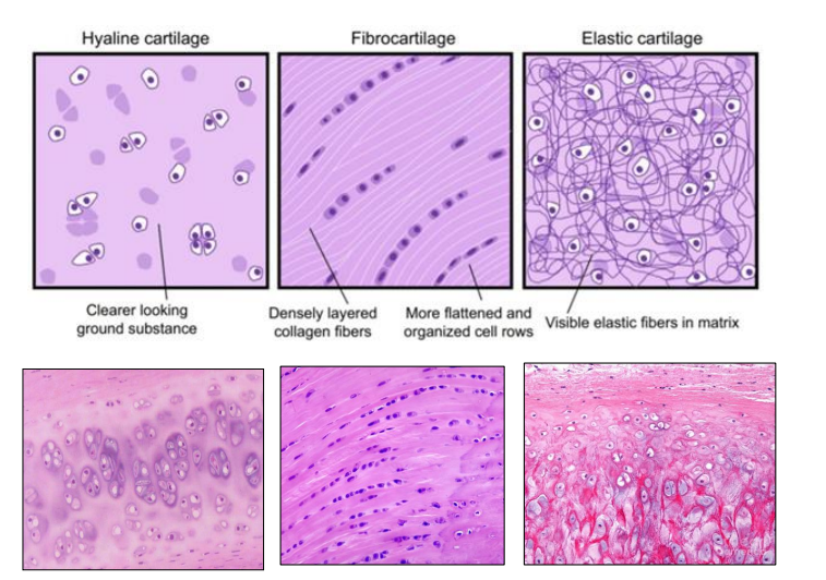
List the 3 types of cartilage and list a histological characteristic of each
Hyaline cartilage
clear looking ground substance
Fibrocartilage
densely layered collagen fibers
more flattened and organized cell rows
Elastic cartilage
visible elastic fibers in matrix
List the most common type of cartilage
Hyaline cartilage
What type of cartilage is present in embryo skeleton and what is it replaced by?
Hyaline cartilage
gradually replaced by bone
Define Hyaline cartilage (what it forms and what that enables, abundant in what, found in who, found in where, surrounded by what)
forms epiphyseal growth plate
enables longitudinal growth of long bones via endochondral ossification
abundant ground substance
rich in proteoglycans & fine Type II collagen fibrils
found in adults at
articular surfaces
costal cartilages
respiratory passages
nasal cartilages
portions of the larynx
surrounded by perichondrium

What does Hyaline Cartilage being surrounded by perichondrium enable?
Most hyaline cartilage is surrounded by perichondrium, except where?
Enables
appositional growth
Except
articular cartilage
growth plate cartilage
How does interstitial growth of hyaline cartilage occur?
through division of chondrocytes within cartilage matrix
Where is elastic cartilage found?
auricle (pinna) of the ear
epiglottis
auditory (Eustachian) tube
List the components of Elastic Cartilage ECM.
What do these components provide?
Components:
elastic fibers
type II collagen
provides greater flexibility and resilience

List the two ways how elastic cartilage grows and explain how these work
Appositional growth
addition at the surface via perichondrium
Interstitial growth
expansion from within by chondrocyte proliferation
List the regions fibrocartilage is found in
Regions subjected to tensile and compressive forces:
Intervertebral discs
Intra-articular discs (menisci)
Where ligaments and tendons attach to bones
Describe Fibrocartilage histologically (what is it made up of, what kind of connective tissue)
Type 1 collagen (dense bundles)
scattered chondrocytes in lacunae
minimal ground substance
lacks perichondrium
intermediate between dense regular CT and hyaline cartilage

What kind of growth does fibrocartilage undergo and why?
interstitial growth
bcus it lacks a perichondrium
Describe Appositional Growth (what kind of cartilage forms and where, types of cells involved and their function, where does this growth occur)
new cartilage forms at surface of existing cartilage from perichondrium
chondrogenic cells differentiate into chondroblasts and synthesize cartilage ECM
chondrocytes maintain and remodel surrounding ECM
occurs in elastic cartilage and most hyaline cartilage
Where does Appositional Growth not occur and why?
Articular cartilage
Growth plate cartilage
lack of perichondrium
Describe Interstitial Growth (where does growth occur, what cells are involved and their function, what forms and their function, and where does this growth occur)
growth occurs from within existing cartilage
chondrocytes proliferate; form isogenous groups which synthesize additional ECM
occurs in all types of cartilage
Describe Hyaline Cartilage (location, perichondrium?, describe the chondrocytes, describe ECM, type of collagen fibers)
Location
costal cartilages, nose, larynx, trachea, bronchi, epiphyseal plates, articular cartilage
most of fetal skeleton (until replaced by bones)
Perichondrium
Present
Chondrocytes
smaller compared to elastic cartilage
arranged in isogenous groups (2-8 cells)
ECM
Basophilic and homogenous
Collagen Fibers
Type II

Describe Elastic Cartilage (location, perichondrium?, describe the chondrocytes, describe ECM, type of collagen fibers)
Location
Auricle or pinna
Epiglottis
External auditory meatus
Perichondrium
Present
Chondrocytes
large
closely placed
isogenous groups (2-4 cells)
ECM
Abundance of elastic fibers
Collagen Fibers
Type II

Describe Fibrous Cartilage (location, perichondrium?, describe the chondrocytes, describe ECM, type of collagen fibers)
Location
Intervertebral discs
Pubic Symphysis
Articular discs in sternoclavicular and temporomandibular joints
Glenoidal mabrum and Acetabular labrum
Perichondrium
NOT present
Chondrocytes
small
arranged in rows between bundles of collagen fibers
ECM
Eosinophilic
abundance of collagen fibers
Collagen Fibers
Type I

Give an example of hyaline cartilage where only interstitial growth occurs
Articular cartilage and synovial joints

Define the Synovial Membrane / Synovium (what is it and what is it made up of)
specialized type of CT
made up of:
Type A Synoviocyte
Type B synoviocyte
Define Type A Synoviocyte (what are they and what is their function)
Macrophage-like cell
Responsible for:
maintenance and “clean up”

Define Type B Synoviocyte (what are they and what do they synthesize)
Fibroblast-like cell
Synthesizes:
hyaluronan and lubricin

Describe the sub-intima of the synovial membrane (what is it, what does it contain and the function of those structures)
deeper, supportive layer of the synovial membrane; lines synovial joint
contains blood vessels
provide nutrients to avascular intima
contribute to synovial fluid formation
Describe the composition of Synovial Fluid, what they are, and their functions
Hyaluronan
glycosaminoglycan
contributes to fluid’s viscosity + lubricating properties
Lubricin
glycoprotein
enhances lubrication
reduces friction in joint
Electrolytes and Proteins
derived from plasma ultrafiltration
List and describe the functions of Synovial Fluid
Lubrication
provided by hyaluronin and lubricin
reduces friction between articular cartilage during movement
bones can glide smoothly against eachother
Shock Absorption
helps distribute loads and absorb mechanical forces across joint
Nutrient Distribution
nutrients provided to avascular articular cartilage
removes waste products
maintains joint health
What is an Arthrocentesis and what is it used for?
Joint tap
cytology, microbial culture, assessment of joint disease

Which cell type is primarily responsible for the formation of the ECM in cartilage?
Which type of cartilage leads to the formation of isogenous groups?
Which type of cartilage is found at articulating surfaces of bones?
Chondroblasts
Interstitial
Hyaline cartilage
Define bone (what is bone, what kind of tissue, vascular or avascular, what cells maintain structure and function)
cells and collagen fibers embedded in hard mineralized ECM
dynamic tissue - continuously undergoes remodeling repair
highly vascular
structure and function maintained by osteoblasts, osteocytes, and osteoclasts

List the functions of bone
rigid and hard support
protects vital organs
houses bone marrow 9site of hematopoiesis)
reservoir (Calcium, phosphate, other ions)
transforms forces of skeletal muscle contraction into bodily movements

Label A-G of the long bone with the correct terminology

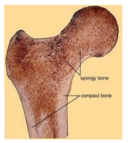
Describe Compact / Cortical Bone (type of bone, structural organization, orientation of collagen)
Lamellar bone
Structural Organization:
Haversian systems or osteons
specific orientation of collagen fibers

Describe Cancellous / Spongy / Trabecular Bone (type of bone, structural organization, function)
Lamellar Bone
Organization:
interconnected trabeculae
lacks Haversian systems
reduces weight of bone and supports marrow
Describe Woven Bone (type of bone, orientation of collagen, how fast is this bone deposited, when is this bone seen, and what is this bone replaced by)
Temporary / Immature bone
random orientation of collagen fibers
rapidly deposited bone during
development
fracture repair
some pathological conditions
replaced by lamellar bone
List the 3 bone cell types
Osteoblasts
Osteocytes
Osteoclasts
Describe Osteoblasts (where do they originate, function, what do they become, and why)
Originate from osteoprogenitor (osteogenic) cells associated with the periosteum and endosteum
Function
form Osteoid (organic part of ECM)
Become osteocytes
when entrapped in the mineralised matrix
Describe Osteocytes (where are they located, how they communicate, and function)
Located
within lacunae in calcified bone ECM.
Communicate
via cytoplasmic processes
extend through canaliculi
Function
maintain the bony matrix
Describe Osteoclasts (where are they found, describe the cells, list a histological characteristic, what are they derived from, and function)
Found
on endosteal and periosteal bone surfaces
Large, motile, multinucleated cells
Ruffled border
Derived:
from hematopoietic precursors
(of the monocyte macrophage linage)
Function
resorb mineralised bone matrix
during growth repair and remodelling
Label A-D in the photo

What do canaliculi contain and list their function
Cytoplasmic processes
connect neighboring osteocytes

What does the Lacunar-canalicular system allow?
communication between osteocytes + nutrient/waste exchange
Mechanosensation
osteocytes able to:
detect mechanical stress
regulate bone remodelling

List the components of Bone Matrix (ECM)
Osteoid (organic compound)
Fibers (collagen type I)
Glycoproteins
involved in mineralization and Ca2+ binding
Inorganic components
hydroxyapatite crystals
small amts of minerals (sodium, magnesium)
What is the function of the organic and inorganic components of bone matrix (ECM)
Organic Component
provides tensile strength
Inorganic Component
provides hardness + compressive strength
Which cell type in bone is responsible for forming new bone?
Osteoblasts
What is the functional role of the lacunar-canalicular system in compact bone?
communication and nutrient/waste exchange