Ch. 11 visual system
 Call Kai
 Learn
 Practice Test
 Spaced Repetition
 Match
 Flashcards
 Knowt Play
1/81

There's no tags or description
Looks like no tags are added yet.
82 Terms
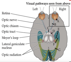
the visual system
- a greater portion of the brain is dedicated to vision versus any other sensory modality
- pathway of the visual system extends from retina --> lateral geniculate nucleus (LGN) --> primary visual cortex (PVC)
- lesions to the visual association cortex cause disorders of higher-order visual processing
eyes and retina
- light enters the eye through the lens to form image on the retina which is inverted or reversed
inverted (A)
- superior visual space/field projects onto inferior retina
- inferior visual space/field projects onto superior retina
reversed (B)
- right visual space/field projects to left side of retina
- left visual space/field projects to right side of retina
fovea
- central fixation point for each eye
- corresponds to central 1-2 degrees of visual space
- region of the retina with highest visual acuity
- light reaches the rods and cones directly
- the fovea is surrounded by the macula (also has high visual acuity and covers the central 5 degrees of visual space)
- what is represented on the fovea is projected to the occipital pole
fovea is represented by
about half of the fibers in the optic nerve and half of the fibers in the visual cortex
optic disk
- formed by axons leaving the retina as they enter the optic nerve
- there are no photoreceptors over the optic disc
- a small blind spot is formed here (located 15 degrees medial and slightly inferior to the central fixation point of each eye)
- no functional deficit when both eyes are used
- when one eye is used, our visual system seems to "fill in" the blind spot
retina: three layers
- photoreceptors
- bipolar cell layer
- ganglion cell layer
photoreceptors
- outermost layer of retina
- responds to light
- rods
- cones
rods
- for vision in low-level lighting conditions
- low resolution
- outnumber cones 20:1
cones
- high resolution
- highly represented in fovea where acuity is highest
- detect color
bipolar cell layer
- middle layer
- receive synapses from photoreceptors
ganglion cell layer
- innermost layer
- receives synapses from bipolar cells
- send their axons into the optic nerve
- fire action potentials: parasol cells and midget cells
parasol cells
respond to gross stimulus features and movement
midget cells
respond to fine visual detail and colors
optic nerve exits in
optic canal
optic chiasm
- optic nerves meet and there is partial crossing of the optic nerve fibers
- nasal (medial) retinal fibers crossover in chiasm
- nasal (medial) retinal fibers are responsible for temporal (lateral) hemifields (halves) of vision
- visual information from both eyes is contained in the optic chiasm
optic tracts
- posterior to optic chiasm
- end at the LGN
- contains visual information from ipsilateral (same) hemiretinas (half of retina) of each eye
- visual information from both eyes is now contained in each half of the brain
R hemiretinas of both eyes end up in
R optic tract (red)

L hemiretinas of both eyes end up in
L optic tract (blue)
ipsilateral hemiretinas represent
contralateral (opposite) visual fields
R hemiretinas represent what visual fields?
L visual fields
lesions of the retina or optic nerves produce
monocular visual field defects
monocular scotoma

monocular visual loss

lesions of the optic chiasm produce
bitemporal visual deficits
bitemporal hemianopia
homonymous visual field defects
- lesions posterior to chiasm (optic tracts, LGN, optic radiations, or visual cortex) will produce homonymous visual field defects
- visual field defect in the same region for both eyes
why will lesions posterior to the chiasm produce homonymous visual field defects?
information is from the same visual fields after the chiasm
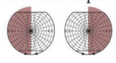
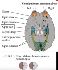
contralateral homonymous hemianopia (DGH)

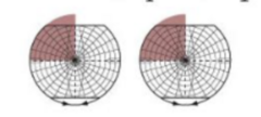
contralateral superior quadrantanopia (E,J)
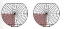
contralateral inferior quadrantopia (F,I)
lateral geniculate nucleus
- axons in the optic tracts wrap around the midbrain (laterally) to synapse at the LGN
- axons leaving the LGN then project back to the primary visual cortex
LGN has layers number 1-6 bottom to top
- layers 1-2: motion and spatial analysis
- layers 3-6: detailed form and color
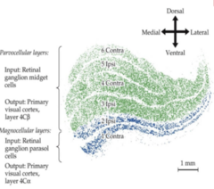
information from left and right eyes remains segregated while passing through the LGN
axons from left and right retinas synapse onto different layers of the LGN
right LGN = ________ hemiretinas of both eye = _____ visual field
right; left
extrageniculate pathways
a small number of fibers in the optic tract bypass the LGN to enter the superior colliculus and pretectal areas
pretectal area
- front portion of tectum
- involved in the pupillary light reflex
pretectal area and superior colliculus
involved in directing visual attention and eye movements toward visual stimuli (by projections to numerous brainstem areas, as well as lateral parietal cortex and frontal eye fields of prefrontal cortex via relays in the pulvinar)

optic radiations
- axons projecting from the LGN to the PVC
- axons sweep over and around the lateral ventricle
- lesions of optic radiations cause homonymous defects affecting the contralateral visual field
- these optic radiations are carrying information for what visual field?
- ipsilateral optic radiations carry info from contralateral visual fields
- lesions of ipsilateral radiations would cause a contralateral homonymous hemianopia visual field defect (G)
the optic radiations pass through
temporal and parietal lobes

inferior optic radiations
- fibers of inferior optic radiations (blue) arc slightly forward and into the temporal lobe forming Meyer's loop
- carry visual information from inferior retina (superior visual field)
- temporal lobe lesions cause contralateral homonymous superior quadrantanopia (pie in the sky) visual defect E

superior optic radiations
- fibers of superior optic radiations (green) pass through the parietal lobe (lower portion)
- carry visual information from superior retina (inferior visual field)
- lower parietal lobe lesions cause contralateral homonymous inferior quadrantanopia (pie on the floor) visual field defect F
primary visual cortex
- lies on the upper bank/gyrus and lower bank/gyrus of the calcarine fissure in the medial portion of the occipital lobe
- secondary visual cortex B18
- tertiary visual cortex B19
calcarine fissure
- cuneus (wedge): upper bank/gyrus
- lingula (little tongue): lower bank/gyrus
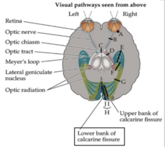
inferior optic radiations
- inferior optic radiations project to lower bank/gyrus of the calcarine fissure
- lower bank lesions (J) thus cause contralateral homonymous superior quadrantanopia
- lesion to right lower bank of PVC would cause what kind of visual defect? LEFT SUPERIOR quadrantanopia

superior optic radiations
- project to upper bank/gyrus of the calcarine fissure
- upper bank lesions (I) thus cause contralateral homonymous inferior quadrantanopia
- lesion to right upper bank of PVC would cause what visual field? LEFT INFERIOR quadrantanopia
primary visual cortex
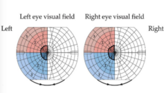
- PVC is retintopically organized
- fovea is represented at the occipital pole
- the fovea occupies about half of the PVC (the fovea contains the highest density of photoreceptors and has corresponding high visual acuity)
- fovea has a very disproportionate cortical representation despite the small retinal area
primary visual cortex: peripheral regions (2 and 3) of the visual fields are represented more
- anteriorly along the calcarine fissure
____ side of retinas = L visual fields = ____ side of PVC
right; right

analysis of motion and spatial relations
- dorsal/upper pathways project to pareito-occipital association cortex
- pathway answers the question of "where?" by analyzing motion and spatial relationships between objects as well as between the body and visual stimuli

analysis of form and color
- ventral/lower pathways project to occipitotemporal association cortex
- pathway answers the question of what? by analyzing form, with specific regions identifying colors, faces, letters, and other visual stimuli
assessment of visual disturbances: 2 major steps
- nature of the visual disturbance
- visual field
nature of the visual disturbance
- time course
- positive phenomena: simple and formed
- negative phenomena
visual field
description of the regions for each eye involved
simple phenomena
include lights, colors, or geometric shapes (arising from disturbances located anywhere from eye to cortex)
formed phenomena
include people, animals, or complex scenes (arising from inferior temporal-occipital visual association cortex)
negative phenomena
region of decreased vision or region of vision that a person cannot see (scotoma (region of decreased or no vision) or a homonymous defect)
assessment of visual disturbances
- distinction between a monocular or binocular visual disturbance is essential for localization
- visual disturbances are often described as being "in one eye", when in reality the left or right visual field is affected for both eyes
- blurred vision is hard to interpret without further description and examination (could be attributed to a variety of things from a cornea problem to a lesion in the visual cortex)
localization of visual field deficit
- visual field testing: tests for crude deficits in the visual filds
- test each quadrant while making sure patient's eyes stay centrally fixated
- test each eye using wiggling fingers and/or have pt count the number of fingers being held up
- fields recorded as if viewing own visual field
- blink to threat can be used on lethargic/uncooperative patients
visual field defects
- most important information for localization of problems in the visual pathways are: what is the position and shape of visual loss? does it affect one or both eyes?
- know the effects of lesions in the visual pathways from the retina to the PVC
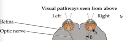
monocular scotoma
- lesion of the retina (A)
- location, size, and shape will vary depending on location and extent of the lesion
- causes: retinal infarcts, hemorrhage, degernation, and infection
- could also be from incomplete damage to optic nerve (trauma, or small, accidental cut of nerve during surgery in area of optic nerve)

monocular vision loss
- complete lesion of the optic nerve (B)
- fibers of the optic nerve contain information from entire retina of that eye
- causes: glaucoma, optic neuritis, neuropathy, elevated ICP, tumors, trauma
- a lesion that is severe enough to involve the entire retina would also produce a monocular vision loss
bitemporal hemianopia
- chiasm is made up of nasal (medial) retinal fibers which contain visual information for temporal (lateral) visual fields
- causes: pituitary adenoma, hypothalamus glioma, other tumors
- optic chiasm lies just above/in front of pituitary gland
- susceptible to pituitary tumors or other lesions in this area
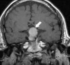
pituitary adenoma/tumor
many patients are not diagnosed until the tumor is large enough to cause visual symptoms
contralateral homonymous hemianopia
- lesions of optic tract (D)
- optic tract carries info for contralateral visual field
- right optic tract carries info for left visual field
- optic tracts: behind chiasm, where nasal (medial) retinal fibers for each eye responsible for temporal (lateral) hemifields have crossed to other side
- causes: tumors, infarct, demyelination
- lessons to the LGN also can cause contralateral homonymous hemianopia
contralateral homonymous hemianopia: lesions of entire optic radiations (G)
- entire optic radiations: superior (green, parietal) and inferior (blue, temporal/meyer's loop) optic radiations carry info for contralateral visual field
- right optic radiations carry info for left visual field
- causes: infarcts, tumors, demyelination, trauma, hemorrhage

contralateral homonymous hemianopia: lesions of entire PVC (H)
- optic radiations terminate on lower and upper banks of calcarine fissure
- causes: PCA infarcts, tumor, infection
contralateral inferior quadrantanopia (pie on the floor): lesions in the parietal lobe (F)
- superior optic radiations (green, parietal) carry info for inferior contralateral visual field
- right superior optic radiations carry info for left inferior visual field
- cause: MCA superior division infarcts

contralateral inferior quadrantanopia (pie on the floor): lesions to upper bank of calcarine fissure (I)
- superior optic radiations terminate on upper bank of calcarine fissure
- causes: PCA infarcts, hemorrhage, tumors, infections, trauma to occipital pole
contralateral superior quadrantanopia (pie in the sky): lesions in the temporal lobe (E)
- inferior optic radiations (blue, temporal/meyer's loop) carry info for superior contralateral visual field
- right inferior optic radiations carry info for left superior visual field
- cause: MCA inferior division infarcts

contralateral superior quadrantanopia (pie in the sky): lesions to lower bank of calcarine fissure (J)
- inferior optic radiations terminate on lower bank of calcarine fissure
- causes: PCA infarcts, hemorrhage, tumors, infections, trauma to occipital pole

homonymous hemianopia
- is the lesion anterior or posterior to the optic chiasm? POSTERIOR
- which side of the brain is the lesion on? RIGHT
- lesions to what areas could cause this visual defect? ENTIRE PVC, OPTIC RADIATIONS, LGN, OPTIC TRACT
infarct to inferior optic radiations: what type of visual field deficit would this patient have?
right superior quadrantanopia (pie in the sky)
localization of visual field defects
- fovea has a large representation (beginning in optic nerve and continuing to the PVC) given its actual size in the retina
- it would take a large lesion of the PVC to affect the entire fovea and macula
- thus, partial lesions of the visual pathways sometimes result in the central visual field being spared
- partial lesions would not usually affect entire fovea and macula representation in the PVC

macular sparing (macula is spared)
the visual field represented by the macula is intact

lesion to the occipital lobe
would involve the area represented by fovea and macula and would result in visual loss in the center of the visual field (central scotoma)
blood supply of the visual pathway
- optic tracts, optic chiasm, and optic nerves receive blood supply from numerous small branches of ACA and MCA
- clinically significant infarcts of these are rarely seen
- optic radiations passing through parietal lobes ( superior radiations): MCA superior division
- optic radiations passing through temporal lobe (meyer's loop) (inferior radiations): MCA inferior division
- primary visual cortex: posterior cerebral artery PCA
damage/infarct to the temporal (inferior/meyer's loop) optic radiations cause what type of visual field deficit?
contralateral superior quadrantanopia "pie in the sky"
damage/infarct to parietal (superior) optic radiations cause what type of visual field deficit?
contralateral inferior quadrantanopia "pie on the floor"
large infarcts involving the entire ipsilateral PVC will cause what visual field defect?
contralateral homonymous hemianopia

infarct of MCA inferior division: how would you explain this patient's deficits
- lower bank of the calcarine fissure: pie in the sky, contralateral superior quadrantanopia
- CANNOT SEE up right corner