msk- what to look out for
 Call Kai
 Learn
 Practice Test
 Spaced Repetition
 Match
 Flashcards
 Knowt Play
1/31

There's no tags or description
Looks like no tags are added yet.
32 Terms

•Usually result from a fall on the shoulder or outstretched hand.
•80% involve the middle third and are transverse.
•Typically, there is inferior displacement of the distal fragment.
•Overriding of the bone ends.
•Those involving the outer third are usually undisplaced due to the stabilisation of the ligament complex.
•The inferior surfaces of the clavicle and the acromion should be in a straight line.
•Any step in the alignment suggests a sub-luxation.
•More marked displacement suggests disruption of the coracoclavicular ligament.
•Frequently caused by a direct blow or occasionally by lifting a heavy weight.

Acromioclavicular joint dislocation
•The inferior surfaces of the clavicle and the acromion should be in a straight line.
•Any step in the alignment suggests a sub-luxation.
•More marked displacement suggests disruption of the coracoclavicular ligament.
•Frequently caused by a direct blow or occasionally by lifting a heavy weight.


Anterior dislocation of the glenohumeral joint
•Most common type of dislocation.
•Caused by excessive external rotation with the arm in abduction.
•The glenoid is empty.
•The humeral head lies antero-inferior to the glenoid.
•60% will have associated compression fracture of the humeral head (Hill-Sachs) causing hatchet deformity.
•Also associated with anterior and inferior glenoid rim fractures and greater tuberosity fractures.

Posterior dislocation of the glenohumeral joint
•Uncommon.
•Classically associated with epilepsy and electric shocks.
•Typically associated with antero-medial humeral head fractures.
•The humeral head appears like a light bulb. NB holding the arm in internal rotation produces the same appearance.
•Frequently missed on AP images, be sure to check that the joint space is visible and of normal width.

Scapular fracture
•Uncommon injuries.
•Usually caused by a direct crush injury.
•Fractures can occur to the body, the neck or the coracoid process.
•They are very easy to miss – especially on a single projection.
Glenoid fractures are associated with gleno-humeral dislocations
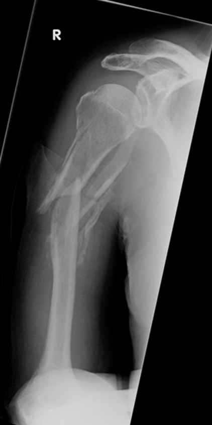
humeral fractures
•Greater and lesser tuberosity fractures can be associated with dislocations but can also exist in isolation.
•Shaft fractures can be oblique, transverse, spiral or comminuted.
•Surgical neck fractures are common, particularly in elderly women following a fall on the outstretched hand.

Supracondylar fracture- subtle breach in the posterior cortex, displaced anterior and posterior fat pad-sail sign
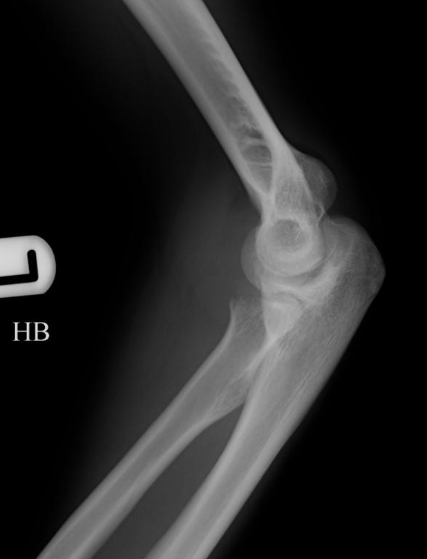
Radial head fracture
•Commonest adult elbow injury.
•The radial head is compressed against the capitellum.
•Clinically there is radial head tenderness and inability to pronate/supinate the forearm.
•Vertical fractures are the most common.
•Depressed fragments are common.
•Impacted fractures cause loss of the normal smooth radial neck contour.
•Radial neck fractures may not cause displaced fat pads because the fracture lies outside the joint capsule.

Radial neck fracture- loss of the normal smooth curve where the head and neck join
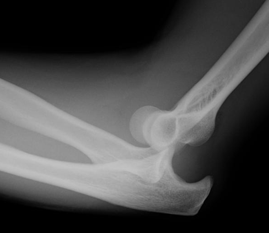
Dislocation of the elbow joint
Posterior is more common – caused by valgus angulation.
•Half are associated with fractures of the medial epicondyle, radial head/neck or coronoid process. These are often only seen post-reduction.
•Pulled elbow:- common between 2-4 years, occurs when the child is lifted by the hand, due to sub-luxation of the radial head out of the annular ligament. Diagnosis on soft tissue signs.

greenstick fracture- children
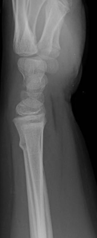
buckle/torus fracture

Galeazzi fracture- •Fracture of the radial shaft with dislocation of the head of the ulna.

•Monteggia Fracture:- Fracture of the proximal ulna with anterior dislocation of the radial head – use of radio-capitellar line.

•Most common in older women.
•Caused by a FOOSH.
•Dorsal displacement/angulation of the distal fragment.
•Roughly transverse fracture, which is often comminuted.
•Often has an accompanying fracture of the ulna styloid process.
•Can be impacted.

fractured waist of scaphoid

Perilunate dislocation
•The whole of the carpus except for the lunate is displaced posteriorly.
•The concavity of the lunate is empty.
•The radius and lunate remain in alignment.
•Often associated with scaphoid fractures.

Lunate dislocation
•The lunate dislocates anteriorly.
•The concavity of the lunate is empty on the lateral projection.
•The radius and capitate remain in a straight line.
•On a PA projection the lunate appears triangular instead of square.
•Much easier to see on the lateral.

Triquetral fracture
•Avulsion of the posterior surface by the radio-carpal ligament.
•Best seen on the lateral projection, with related soft tissue swelling.
•Also known as the pooping duck sign
There are no accessory ossicles in this area so we know its triquetral fracture
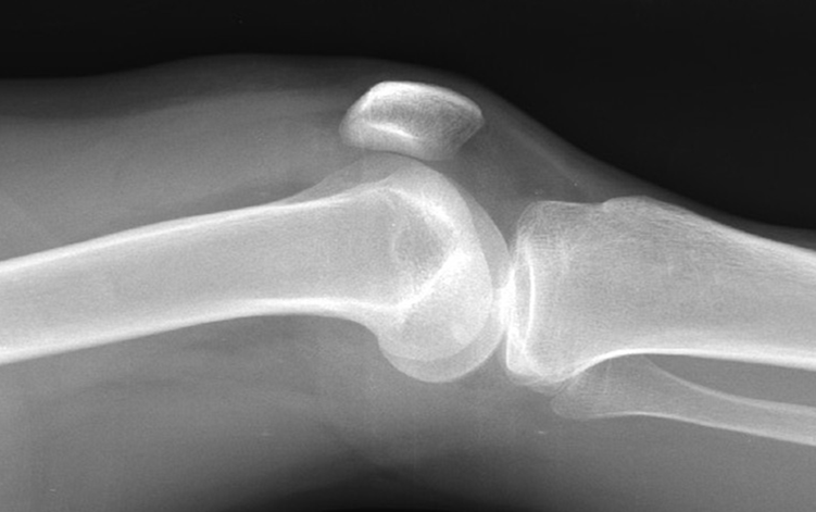
Knee joint effusion in supra patella bursa
•Increased amount of fluid in the supra-patella bursa.
•Can be due to inflammation, infection or trauma.
•Can be the only sign of a soft tissue injury.
•Supra-patella bursa should measure less than 1cm in AP diameter.

Lipohaemarthrosis - fat / fluid level
•Specific type of joint effusion.
•Occurs when there is an intra-articular fracture.
•Bone marrow fat leaks into the joint.
•Fat is less dense than blood, so it floats on top of it in the effusion – fat/fluid level.
•Only visible on HBL images.

TIbial stress fracture
ill defined oblique lucency indicates a fracture on AP within tibial diaphysis with subtle periosteal reaction on lateral
Common in female athletes.
Abnormal bone.
Sudden increase in activity.
Inadequate footwear.

Medial malleolar injuries
•Occur because of a tension force via the deltoid ligament or impaction of the talus.
•Fractures are transverse or oblique.
•Check the width of the joint space.
•Collateral ligament tension causes a fracture of the tip of the malleolus.

Lateral malleolar fractures
•Inversion:- Lateral collateral ligament sprain/rupture or a transverse fracture.
•External Rotation:- Oblique or spiral fracture.
•Abduction of the talus:- Oblique fracture.
•Collateral ligament tension:- Avulsion fracture of the tip of the malleolus.

Trimalleolar fracture
AP- medial ligaments disrupted- joint space widened, oblique fracture of distal fibula which involved distal tibio-fibular joint, lateral subluxation of the talus
Lateral- Comminuted fracture of the posterior malleolus

Maisonneuve fracture
medial side of the ankle mortice is widened
oblique fracture of the middle third of the fibula- would not be seen on ankle projections
•Caused by an eversion injury.
•Either the deltoid ligament is torn or there is a fracture of the medial malleolus.
•There may only be medial joint space widening.
•The tibio-fibular syndesmosis is disrupted, with a fracture of the proximal fibula.
•This happens because the tibia and fibula behave as a closed ring.

Osteochondral fracture of talar dome
•These are the result of inversion or eversion injuries.
•They can affect the medial or lateral aspects of the talar dome.
•The injury primarily affects the cartilage, but sometimes there is a small piece of bone involved as well.
•The fracture is painless, because the joint surface is insensitive.
•These injuries are frequently missed, (because they are very difficult to see), and the patient is treated for the accompanying ligament sprain.
•The result can degenerative joint disease many years later.

Tarsometatarsal Joint Alignment on the Oblique Projection
The medial margin of the base of the third metatarsal should be in line with the medial margin of the lateral cuneiform.

Tarsometatarsal Joint Alignment on the DP Projection
The medial margin of the base of the second metatarsal should be in line with the medial margin of the medial cuneiform.


Lisfranc injuries
Misalignment of the 2nd TMT joint with lateral displacement of the metatarsals. Misalignment of the 3rd TMT joint.
•Tarso-metatarsal dislocations – often caused by rotational injuries.
•Very easy to overlook if alignment is not checked.
•If a bony fragment is detached from the base of any of the four medial metatarsals, then a dislocation should be suspected.
•If there is a fracture of the second metatarsal distal to its base, the base is held by the ligaments, but the distal fragment and the remaining metatarsals dislocate.

Base of 5th metatarsal fracture
•Associated with inversion injuries.
•Caused by avulsion of the insertion of the peroneus brevis tendon.
•Should be visible on a lateral ankle projection, separate foot projections are not always necessary.
•In young patients there is an unfused apophysis at the base of the fifth MT, this lies longitudinal to the MT: fractures are usually transverse.

this image- stress fracture of nech of 2nd metatarsal
Metatarsal and Phalangeal Fractures and Dislocations.
•Metatarsal fractures are often caused by crush injuries or twist injuries.
•Can have any type of long bone fracture.
•Stress fractures are associated with prolonged standing/walking. These can be very hard to see initially, and some may never be visible on plain images.
•Phalangeal fractures and dislocations are common, but generally of little clinical importance. Be sure to check that the joint spaces are visible and congruous.
•Joint space incongruity after a dislocation is reduced suggests soft tissue entrapment in the joint.