P3 POSI PROJECTIONS (lower extremities)
 Call Kai
 Learn
Learn Practice Test
 Spaced Repetition
 Match
Match Flashcards
 Knowt Play
1/149

There's no tags or description
Looks like no tags are added yet.
Name | Mastery | Learn | Test | Matching | Spaced | Call with Kai | Chat |
|---|
No analytics yet
Send a link to your students to track their progress
150 Terms
PP: Supine/Seated; knee flexed; 15o foam wedge under foot
RP: 3rd MTP joint
CR: ┴ (15o foam wedge) or 15o posteriorly
SS: Phalanges & distal portion of metatarsals
AP/ AP AXIAL PROJECTION (TOES)
PP:
RP:
CR:
SS:
IP JOINTS ARE PARALLEL TO CR ANGLE
PP: Prone; dorsal aspect against IR
RP: 3rd MTP joint
CR: ┴
SS: MTP & IP joint spaces are well visualized
PA PROJECTION (TOES)
IP JOINTS ARE ___ TO CR ANGLE
PP:
RP:
CR:
SS:
PP: Supine/seated; knee flexed; plantar surface 30-45o from IR (lower leg & foot rotated medially )
RP: 3rd MTP joint
CR: ┴
SS: 2nd-5th MTP joint spaces; 1st-3rd toes
AP OBLIQUE PROJECTION (MEDIAL ROT.) (TOES)
PP: w angle
RP:
CR:
SS:
PP: Supine/seated; knee flexed; plantar surface 30-45o from IR (lower leg & foot rotated laterally)
RP: 3rd MTP joint
CR: ┴
SS: 4rd-5th toes; MTP joints overlapped
AP OBLIQUE PROJECTION (LATERAL ROT.) (TOES)
PP: w angle
RP:
CR:
SS:
PP: Lateral recumbent (affected side down); affected limb partially extended; ball of foot 30o to horizontal
RP: 3rd MTP joint
CR: ┴
SS: 2nd-5th MTP joint spaces; 1st-3rd toes
PA OBLIQUE PROJECTION (MEDIAL ROT.) (TOES)
PP: w what side down// ball of foot ____o to horizontal
RP:
CR:
SS:
PP: Lateral recumbent; toe in true lateral
RP: IP joint (1st toe); proximal IP joint (2nd -4th toes)
CR: ┴
SS: Phalanges in profile; open IP joints spaces
Lateromedial Projection: on the unaffected side- great toe and 2nd toe
Mediolateral Projection: on the affected side-3rd, 4th, and 5th toe
Lateral Projection (TOES)
PP:
RP: _____ (1st toe); ______ (2nd -4th toes)
CR:
SS:
Lateromedial Projection: on the _____ side- _____toe &_____ toe
Mediolateral Projection: on the _____ side- __, __, __ toe

WHAT ARE THE 5 PROJECTIONS OF THE TOE (8 in total if u breakdown)

what are the projection/ methods of the sesamoids
15-20
PP: Prone; dorsiflex great toe; ankle elevated; ball of foot ┴ to IR RP: 1st MTP joint
CR: ┴
SS: MT head & sesamoids in profile
LEWIS METHOD (tangential proj.) (sesamoids)
PLANTAR SURFACE OF FOOT ____ ANGLE FROM VERTICAL.
_____ perpendicular to IR
PP:
RP:
CR:
SS:
PP: Seated (more comfortable); plantar surface 75o to IR; toe flexed & hold w/ strip gauze bandage; medial border is vertical and plantar surface of foot ┴ to IR
RP: 1st MTP head
CR: ┴
SS: MT head & sesamoids in profile
HOLY METHOD (TANGENTIAL PROJ) (SESAMOIDS)
PP: ______; plantar surface ____ to IR; toe flexed & hold w/ ______; medial border is vertical
RP:
CR:
SS:
PP: Lateral recumbent (unaffected side down); knees flexed; limb partially extended; foot in lateral position; 1st MTP joint ┴ to IR
RP: Prominence of 1st MTP joint
CR: 40o toward the heel
SS: Sesamoids projection axiolaterally with slight overlap
CAUSTON METHOD (TANGENTIAL PROJ.) ( SESAMOID)
PP: _______(____side down); _____ ┴ to IR
RP:
CR:
SS:
PP: Supine; knee flexed; plantar surface against IR RP: 3rd MTP base
CR: ┴ or 10o posteriorly
SS: Metatarsals & Tarsals (┴);
TMT joint (10o)
AP/ AP AXIAL PROJECTION (FOOT)
PP:
RP:
CR:
SS:
AP PROJ. OF FOOT

PP: Supine; knee flexed; leg rotated medially; plantar surface of foot 30o to IR
RP: 3rd MTP base
CR: ┴
SS: • Cuboid in profile
• Sinus tarsi (well demonstrated)
• Interspaces b/n:
• cuboid & calcaneus;
• cuboid & 4th & 5th MT
• Talus & navicular bone
AP OBLIQUE PROJECTION (MEDIAL ROT.) (FOOT)
PP: w angle
RP:
CR:
SS: •
• Interspaces b/n:
PP: Supine; knee flexed; leg rotated laterally; plantar surface of foot 30o to IR
RP: 3rd MT base
CR: ┴
SS: • Navicular in profile •
Interspaces b/n:
• 1st & 2nd Metatarsal
• Medial & intermediate cuneiforms
• Separates 1st-2nd Metatarsal bases
• No superimposition of the medial and intermediate cuneiform
AP OBLIQUE (LATERAL ROT.) (FOOT)
PP: w angle
RP:
CR:
SS: •
Interspaces b/n:
•
•
•
•
GRASHEY METHOD
PA OBLIQUE PROJECTION OF THE FOOT IS WHAT METHOD
PP: Prone; foot elevated on sandbags; folded towel under the knee; adjust the elevated foot; heel rotated medially 30o
RP: 3rd Metatarsal Base
CR: ┴
SS: Interspaces between 1st and 2nd Metatarsal
NAVICULAR
PA OBLIQUE PROJECTION (MEDIAL ROT.) (FOOT)
PP: w angle
RP:
CR:
SS:
PP: Prone; foot elevated on sandbags; folded towel under the knee; adjust the elevated foot; heel rotated laterally 20o
RP: 3rd Metatarsal Base
CR: ┴
SS: 2nd-3rd, 3rd-4th, 4th-5th Metatarsal
CUBOID
5TH MT TUBEROSITY (same as ap medial oblique)
PA OBLIQUE PROJECTION (LATERAL ROT.) (FOOT)
PP: w angle
RP:
CR:
SS:
AP MEDIAL OBLIQUE (FOOT)

AP LATERAL OBLIQUE FOOT


PP: lat, recumbent Dorsiflex foot (┴ to lower leg); leg & foot in lateral position; lateral side of foot against IR;
RP: 3rd MT base
CR: Perpendicular
SS: Entire foot in profile, the ankle joint, and the distal ends of the tibia and fibula
LATERAL PROJECTION (MEDIOLATERAL) (FOOT)
PP:
RP:
CR:
SS:
LATEROMEDIAL
WHAT IS THE RECOMMENDED LATERAL PROJ. OF THE FOOT?
LATEROMEDIAL
WHAT LAT. PROJ OF THE FOOT PROVIDES NEARLY EXACT/ TRUE LATERAL IMAGE OF THE FOOT
MEDIOLATERAL
WHAT LAT. PROJECTION OF THE FOOT IS ROUTINELY USED BECAUSE ITS A COMFORTABLE POSITION TO PUT A PX IN?
PP: lateral recumbent, Dorsiflex foot (┴ to lower leg); leg & foot in lateral position; lateral side of foot against IR;
RP: 3rd MT base
CR: Perpendicular
SS: Entire foot in profile, the ankle joint, and the distal ends of the tibia and fibula
Lateral Projection (MEDIOLATERAL) (FOOT)
PP: _______, _____foot (__ to lower leg); leg & foot in lateral position; _____ (M/L) side of foot against IR;
RP:
CR:
SS:
LATERAL PROJ. MEDIOLATERAL, FOOT

PP: LPO/RPO (affected side up); medial surface against IR; plantar surface of foot ┴ to IR
RP: 3rd MTP base
CR: Perpendicular
SS: True lateral projection of foot • Metatarsals more superimposed than mediolateraL
LATERAL PROJECTION (LATEROMEDIAL) (FOOT)
PP: ____ (___(A/UN) side up); ___ surface against IR; plantar surface of foot ┴ to IR
RP:
CR:
SS: _______
• ____ more superimposed than mediolateral
LATEROMEDIAL
IN THIS LATERAL PROJ. OF THE FOOT THE METATARSALS ARE MORE SUPERIMPOSED
PP: Upright (natural position);
feet elevated (use blocks);
IR b/n feet; weight equally distributed on each foot
RP: Point above 3rd MT base
CR: Horizontal
SS: Structural longitudinal planus). status of arch (pes The right and left ides are examined for comparison
LATERAL PROJECTION (LATEROMEDIAL) (WEIGHTBEARING METHOD) (FOOT)
PP:
feet _____(use _____);
___ b/n feet; weight equally distributed on each foot
RP:
CR:
SS:
LATEROMEDIAL
THE LATERAL PROJ. WEIGHT BEARING METHOD IS MEDIOLATERAL OR LATEROMEDIAL
20-40 DEGREES
BOHLERS ANGLE
AP MEDIAL OBLIQUE FOOT
PA LATERAL OBLIQUE FOOT
what oblique projections demonstrate 5th MT tuberosity
LAT. PROJ. WEIGHT BEARING METHOD. LATEROMEDIAL
WHAT BEST DEMONSTRATE FLAT FOOT OR PES PLANUS
PP: Upright; both feet against IR; weight equally distributed on each foot
RP: between feet at 3rd MT base level
CR: 10o or 15o posteriorly
SS: Accurate evaluation & comparison of Metatarsal & tarsals
AP AXIAL PROJ. WEIGHT BEARING METHOD
PP: _____; ___ feet against IR; weight equally distributed on each foot
RP: ______
CR: ___ or______
SS:
BOHLER’S CRITICAL ANGLE

AP AXIAL PROJ. WEIGHT BEARING METHOD
WHAT PROJ. BEST DEMONSTRATES HALLUX VALGUS AND LISFRANC INJURY
AP AXIAL PROJECTION
(WEIGHT BEARING COMPOSITE METHOD)
WHICH PROJECTION OF THE FOOT NEEDS 2 EXPOSURES
PP: Upright; 2 exposures
First Exposure: unaffected foot step backward (to prevent superimposition of leg shadow on ankle joint); tube in fronT
- For exposure of forefoot (phalanges & Metatarsal)
Second Exposure: unaffected foot step forward; tube behind; exposure factor increase -
- For hindfoot (talus & calcaneus
RP: For the 1st exposure: 3rd MT base
For the 2nd exposure: level of lateral malleolus
CR: For the 1st exposure: 15o posteriorly For the 2nd exposure: 25o anteriorly
SS: Full outline of the foot free of the leg
AP AXIAL PROJECTION
(WEIGHT BEARING COMPOSITE METHOD)
PP:_____; ___ exposures
First Exposure: unaffected foot step ____(to prevent superimposition of leg shadow on ankle joint); tube in____
- For exposure of___ (___ & _____)
Second Exposure: unaffected foot step ____; tube behind; exposure factor ____
- For _____ (____ & ____)
RP: For the 1st exposure: ______
For the 2nd exposure: _______
CR: For the 1st exposure: _____
For the 2nd exposure: _____
SS: _______
AP AXIAL PROJECTION
(WEIGHT BEARING COMPOSITE METHOD)

AP PROJ. TOES

AP PROJ. TOES

AP
IN THIS PROJ. OF THE TOES THE IP JOINTS ARE NOT SEEN
AP AXIAL PROJ. TOES

AP PROJ. TOES
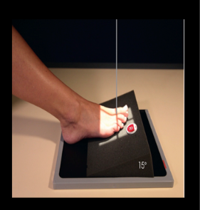
PA PROJ. TOES

AP OBLIQUE PROJ. (MEDIAL ROT.) TOES

AP PROJ. LAT ROT. TOES

PA OBLIQUE (MEDIAL ROT) TOES
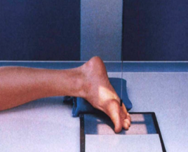
LATERAL PROJ (LATEROMEDIAL) TOES

LATERAL PROJ. (MEDIOLATERAL) TOES

TANGENTIAL PROJ. LEWIS METHOD
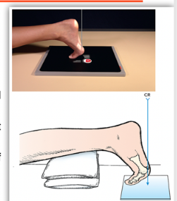
TANGENTIAL PROJ. HOLLY METHOD

TANGENTIAL PROJ. CAUSTION METHOD

AP AXIAL FOOT

AP PROJ. FOOT
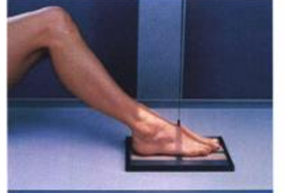


AP OBLIQUE PROJ. MEDIAL ROT. FOOT

ap oblique projection lateral rotation





PA OBLIQUE PROJECTION: GRASHEY METHOD

AP OBLIQUE LAT. ROT
PA OBLIQUE FOOT MED. ROT IS THE SAME AS _______
LATERAL PROJ. MEDIOLATERAL (FOOT)

LATERAL PROJ. LATEROMEDIAL (FOOT)
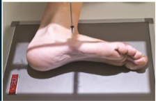





1.) AP/AP AXIAL
2.) AP OBLIQUE (MEDIAL & LATERAL ROT.)
3.) PA OBLIQUE: GRASHEY METHOD (MEDIAL & LAT. ROT.)
4.) LATERAL PROJ (MEDIOLATERAL, LATEROMEDIAL)
5.) LATERAL (WEIGHT BEARING METHOD)
6.) AP AXIAL (WEIGHT BEARING METHOD)
7.) AP AXIAL (WEIGHT BEARING COMPOSITE METHOD)
what are the projections of the foot

WHAT ARE THE PROJECTIONS/ METHODS FOR CONGENITAL CLUBFOOT
EQUINUS, ADDUCTION, SUPINATION
Talipes equinovarus
]• 3 Deviations
(equinus)
Plantar flexion and inversion of the calcaneus
adduction
Medial displacement of the forefoot
supination
Elevation of the medial border of the foot
Davis and Hatte
stated that even slight rotation of the foot can result in marked alteration in the radiographically projected relation of the ossification centers
PP: Supine; hips & knees flexed; foot flat on IR; ankles slightly extended; legs are vertical
RP: Tarsals
CR: 15o posteriorly
SS: Degree of forefoot adduction & calcaneus inversion (equinus)
AP PROJECTION: ____ METHOD (CC)
PP:
RP:
CR:
SS:
AP PROJECTION; KITE METHOD; (CC)
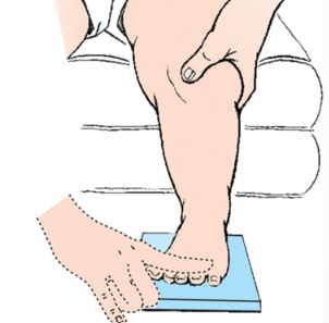
PP: Lateral recumbent; uppermost limb flexed & draw forward
RP: Midtarsal area
CR: Perpendicular
SS: Anterior talar subluxation Degree of plantar flexion (equinus)
LATERAL PROJECTION (MEDIOLATERAL) (KITE METHOD) (CC)
PP: _____; uppermost limb flexed & draw ____
RP: _____
CR: ____
SS: __& __


PP: Erect & Bending forward position; plantar surface against IR
RP: Lower leg
CR: 40o anteriorly
SS: Suroplantar projection or an axial view of congenital clubfoot
Freiberger-Hersh-Harrison:
CR 35o, 45o & 55o for demonstration of sustentaculum talar joint fusion
DORSOPLANTAR AXIAL PROJECTION (KANDEL METHOD) (CC)
PP:___ &______; plantar surface against IR
RP: _____
CR: ___
SS:
_____-____-Harrison:
CR __o, __o & _o for demonstration of__ joint fusion
AP PROJECTION; KITE METHOD (CC)


DORSOPLANTAR AXIAL PROJECTION (KANDEL METHOD) (CC)

PP: Supine/Seated; leg fully extended; dorsiflex foot w/ strip of gauze; foot ┴ to IR
RP: 3rd MT base
CR: 40o cephalad to long axis of foot
SS: Calcaneus & subtalar joint
AXIAL PROJECTION (PLANTODORSAL) (CALCANEUS)
PP: ; leg fully extended; foot w/ strip of gauze; foot ┴ to IR
RP:
CR:
SS: &
AXIAL PROJECTION (PLANTODORSAL) (CALCANEUS)

P: Prone; ankle elevated; dorsiflex ankle; foot ┴ to IR; IR vertical
RP: Dorsal surface of ankle joint
CR: 40o caudad
SS: Calcaneus, subtalar joint & sustentaculum tali
AXIAL PROJECTION (DORSOPLANTAR) (CALCANEUS)
PP:
RP:
CR:
SS:
AXIAL PROJECTION (DORSOPLANTAR) (CALCANEUS)

PP: Supine/ LATERAL RECUMB; patient turn toward affected side; plantar surface // to IR
RP: 1 in distal to medial malleolus (at subtalar joint)
CR: ┴
SS: Calcaneus & ankle joint; sinus tarsi
LATERAL PROJECTION: MEDIOLATERAL (CALCANEUS)
PP: _____/ _______; patient turn toward ___ (A/UN) side; plantar surface __ to IR
RP:
CR:
SS:
LATERAL PROJECTION, MEDIOLATERAL (CALCANEUS)

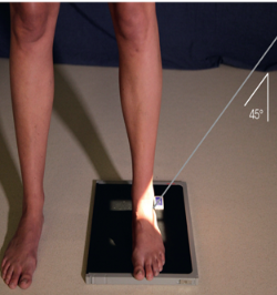
PP: Upright; posterior surface of heel at edge of IR; unaffected forward foot one step (to prevent superimposition of leg shadow)
RP: Level of 5th MT base
CR: 45o anteriorly
SS: Calcaneotalar coalition
AXIAL PROJECTION: DORSOPLANTAR (WEIGHT BEARING COALITION METHOD) (LILIENFIELD METHOD)
PP: __; posterior surface of heel at ___ of IR; ___ forward foot one step (to prevent superimposition of leg shadow)
RP:
CR:
SS:


PP: Upright; leg perpendicular to IR; calcaneus center to IR
RP: Lateral malleolus
CR: 45o caudad (medially)
SS: the calcaneal tuberosity and is useful in diagnosing stress of the calcaneus or fractures tuberosity
LATEROMEDIAL OBLIQUE PROJ. ( WEIGHT BEARING METHOD) (CALCANEUS)
PP:
RP:
CR:
SS:
LATEROMEDIAL OBLIQUE PROJ. ( WEIGHT BEARING METHOD) (CALCANEUS)
the calcaneal tuberosity and is useful in diagnosing stress of the calcaneus or fractures tuberosity

1.) AXIAL PROJ. (PLANTODORSAL)
2.) AXIAL PROJ. (DORSOPLANTAR)
3.) LATERAL PROJ. (MEDIOLATERAL)
4.) AXIAL PROJ.; DORSOPLANTAR ( WEIGHT BEARING COALITION METHOD) (LILIENFIEL METHOD)
5.) LATEROMEDIAL OBLIQUE PROJECTION (WEIGHT BEARING METHOD)
WHAT ARE THE PROJECTIONS/ METHODS FOR THE CALCANEUS
PP: Semisupine; foot & leg rotated 45o medially; knee flexed
RP: 1 in. distal & 1 in. anterior to lateral malleolus
CR: ┴
SS: Anterior subtalar articulation
LATEROMEDIAL OBLIQUE PROJECTION (ISHERWOOD METHOD)
Medial Rotation Foot
PP: ____ foot & leg rotated ___o medially; knee flexed
RP:
CR:
SS:
LATEROMEDIAL OBLIQUE PROJECTION (ISHERWOOD METHOD)

PP: Seated or semi-lateral recumbent (more comfortable); leg, foot & ankle rotated 30o medially; dorsiflex foot; 30o foam wedge
RP: 1 in. distal & 1 in. anterior to lateral malleolus
CR: 10o cephalad
SS: Middle subtalar articular surface & “end on” projection of sinus tarsi
AP AXIAL OBLIQUE PROJECTION; ISHERWOOD METHOD
MEDIAL ROTATION ANKLE
PP:
RP:
CR:
SS: &
AP AXIAL OBLIQUE; ISHERWOOD METHOD
MEDIAL ROT OF ANKLE
