chapter 35: disorders of bladder and lower urinary tract
 Call Kai
 Learn
 Practice Test
 Spaced Repetition
 Match
 Flashcards
 Knowt Play
1/25

There's no tags or description
Looks like no tags are added yet.
26 Terms
urinary bladder
hollow, muscular, distensible organ located in pelvic cavity, behind pubic symphysis
bladder wall consists of 3 main layers:
mucosa and submucosa: inner lining made of transitional epithelium (urothelium)
muscular layer: smooth muscle called detrusor
serosa: outer covering
trigone is a triangular area on:
internal floor of bladder, formed by:
two ureteric openings
one internal urethral opening
two main sphincters of urinary bladder:
internal urethral sphincter: located at bladder neck, surrounding beginning of urethra
made of smooth muscle, continuous w/ detrusor muscle — involuntary
controlled by sympathetic nervous system
external urethral sphincter: located around the urethra in deep perineal region
made of skeletal muscle - under voluntary control
innervated by pudendal nerve (S2-S4)
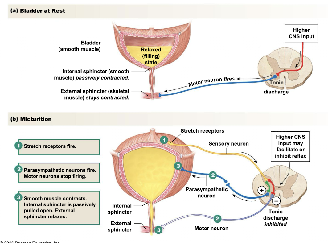
micturition
process of urination is micturition
stretch receptors in bladder walls send signal to spinal cord
smooth muscle in bladder wall contracts
internal sphincter (smooth muscle) pulled open
external sphincter (skeletal muscle) btwn bladder and urethra relaxes
in micturition, centers in brainstem and cerebral cortex override:
basic reflexes under parasympathetic ctrl
pudendal nerve sends impulses to external urethral sphincter
sphincter contracts voluntarily - prevents urine from escaping
bladder obstruction
bladder and urethral blockages can result from sev factors:
facal impaction or severe constipation: hard stool accumulates in rectum and may press against bladder or urethra
bladder tumor or metastases: tumors in bladder or cancer that has spread from other organs may block normal flow of urine
congenital obstruction of external urethral meatus
BPH: enlargement of prostate gland compresses urethra, making urination difficult - common in older men

early stages of bladder obstruction
urinary obstruction results in compensatory changes, for early:
bladder hypertrophy: becomes less flexible, making it difficult to expand and fill w/ urine
hypersensitivity to stretch - ??
decreased ability to suppress urination
later stages of bladder obstruction
bladder wall continues to thicken
detrusor contraction becomes too short: incomplete emptying and retention → frequency → urgency
long term obstruction leads to structural changes in bladder:
trabeculea: detrusor muscle becomes hypertrophied, forming
cellulae: small pockets dev btwn thickened muscle bundles
diverticula: non-muscular pouches dev outward through weak areas btwn muscle bundles
incontinence
inability to voluntarily control urination, resulting in involuntary leakage of urine
enuresis
involuntary urination by child age older than 4 years
often related to developmental delay, sleep pattern, psychosocial aspect
retention
inability to empty bladder completely, causing urine to remain in bladder
spinal cord injury at sacral lvl can block micturition reflex
may follow anesthesia (general or spinal)
neurogenic bladder
bladder dysfunction caused by dmg or disruption of CNS, ANS, or peripheral nerves responsible for controlling bladder function, resulting in loss of norm bladder ctrl
neurogenic bladder commonly presents in 2 forms:
spastic - failure to store urine
flaccid - inability to empty urine
dyssnergia
common, w/ both detrusor and external urethral sphincter contracting at same time
normalyl detrusor muscle contracts while external urethral sphincter relaxes
spastic bladder
results from dmg to spinal cord above T12 lvl or dmg to brain - stroke, tumors, MS
higher brain centers cannot ctrl reflex
bladder cannot relax to fill urine
contracts involuntarily, causing involuntarily, causing frequent, interrupted, or incomplete urination
flaccid bladder
results from dmg to spinal cord below T12 or dmg to peripheral nerves controlling bladder - peripheral neuropathies
micturition reflex is inhibited
bladder cannot contract to empty - retention and overlow incontinence
treatment goals for neurogenic bladder
prevent bladder overdistention: follow an individualized urination (or catheterization) schedule
prevent UTIs: ensure bladder empties properly to reduce urine retention and bacterial growth
prevent potentially life-threatening renal dmg: prevent urine from backing up into kidneys, which can cause hydroneprhosis
reduce undesirable social and psychological effects of disorder: reduce anxiety and social isolation associated w/ loss of bladder ctrl
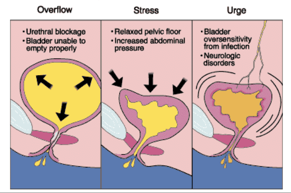
3 common types of urinary incontinence:
overactive bladder/urge incontinence
stress incontinence
overflow incontinence
overactive bladder/urge incontinence
involuntary contraction of bladder during filling
person feels sudden, strong urge to urinate followed by involuntary urine leakage
stress incontinence (common in women)
increased intra-abdominal pressure (coughing, sneezing, lifting) forces urine through sphincter
result of pelvic floor muscle dysfunction - weakening due to pregnancies, childbirth
overflow incontinence
retention of urine may be accompanied by overflow incontinence
intravesical pressure exceeds urethral pressure - involuntary loss of urine
types of urinary incontinence picture
several non-urological conditions that increase risk of bladder probs in older adults, such as:
“DIAPPERS”
dementia, infection, atrophic vaginitis, pharmaceuticals, psychological, endocrine, restricted mobility, stool impaction
bladder cancer
most bladder tumors are malignant and commonly arise from transitional epithelium of bladder
often develops as multiple tumors
diagnosed by urine cytology and biopsy
early signs: ???
tumor is invasive through wall to adjacent structures
metastasizes to pelvic lymph nodes, liver, and bone
predisposing factors of bladder cancer
working w/ chemicals in labs and industry
cig smoking
recurrent infections
heavy intake of analgesics
picture of bladder cancer
