unit 2 a&p
 Call Kai
 Learn
Learn Practice Test
 Spaced Repetition
 Match
Match Flashcards
 Knowt Play
1/369

There's no tags or description
Looks like no tags are added yet.
Name | Mastery | Learn | Test | Matching | Spaced | Call with Kai | Chat |
|---|
No analytics yet
Send a link to your students to track their progress
370 Terms
What are the major characteristics of cartilage?
Avascular
Usually no nerves within the cartilage matrix
Cells occupy lacunae
Nutrients and wastes move largely by diffusion through the matrix
avascular means…
no blood vessels penetrate the cartilage matrix.
What surrounds cartilage, and what are its layers?
The perichondrium surrounds cartilage and has:
Outer fibrous layer: dense irregular CT with type I collagen
Inner chondrogenic layer: contains chondrogenic/progenitor cells
Blood vessels are present in the perichondrium, not the cartilage matrix.

What is the progression of cartilage cells from immature to mature?
Chondrogenic cells → chondroblasts → mature chondrocytes

Chondrogenic cells
progenitor cells near inner perichondrium

Chondroblasts
matrix-secreting cells beneath perichondrium.

Mature chondrocytes
cells located within lacunae
What are lacunae and isogenous groups in cartilage?
Lacuna: space that houses a chondrocyte
Isogenous group: cluster of about 2–4 chondrocytes
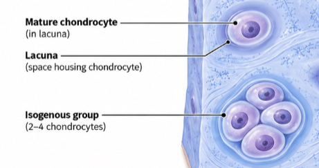
What are the major components of the extracellular matrix of hyaline cartilage?
Type II collagen fibers forming a fine network
Proteoglycan aggregates containing chondroitin and keratan sulfate chains
Hyaluronic acid forms the backbone of proteoglycan aggregates
Abundant water, which keeps the matrix hydrated and gel-like


Compare territorial vs. interterritorial matrix in hyaline cartilage.
Territorial matrix: directly around lacunae, richer in proteoglycans, stains darker/basophilic
Interterritorial matrix: farther from lacunae, contains more collagen, appears lighter and more uniform
Cartilage contains no blood vessels within its matrix. How do its cells receive nutrients and eliminate wastes?
By diffusion through the cartilage matrix, with nutrients ultimately supplied from blood vessels outside the matrix, such as those in the perichondrium.
What is interstitial growth of cartilage?
Growth from within the cartilage as chondrocytes divide and form isogenous groups.
How does interstitial growth increase the size of cartilage?
Chondrocytes divide and form isogenous groups.
The daughter cells secrete new matrix between themselves.
The new matrix separates the cells and expands the cartilage internally.
What is appositional growth of cartilage?
Growth at the surface of existing cartilage from chondrogenic cells in the inner perichondrium.
How does appositional growth increase the size of cartilage?
Chondrogenic cells in the inner perichondrium become chondroblasts.
Chondroblasts deposit new cartilage matrix.
The new matrix is added to the outside/surface of existing cartilage, increasing its size.
Compare interstitial vs. appositional growth of cartilage.
Interstitial Growth | Appositional Growth |
|---|---|
Growth from within | Growth at the surface |
Involves chondrocytes | Begins with chondrogenic cells |
Chondrocytes divide → isogenous groups | Chondrogenic cells → chondroblasts |
New matrix forms between daughter cells | New matrix is deposited onto existing cartilage |
Expands cartilage internally | Adds cartilage to the outside |

Looking at a cartilage growth diagram, how can you distinguish interstitial growth from appositional growth?
Interstitial: expansion occurs within the existing cartilage as chondrocytes divide and secrete matrix.
Appositional: new layers are added to the outer surface of the cartilage.
Chondrocytes divide within existing cartilage and the daughter cells begin secreting matrix between themselves. What type of growth is occurring?
Interstitial growth, because the cartilage is expanding from within.
Chondrogenic cells in the inner perichondrium differentiate into chondroblasts and begin depositing new matrix. What type of cartilage growth is occurring?
Appositional growth, because new cartilage is being added at the surface.
Which type of cartilage growth would directly lead to the formation of isogenous groups?
Interstitial growth, because chondrocytes divide within the cartilage to produce daughter cells.

What is a mesenchymal stem/progenitor cell?
An undifferentiated embryonic connective tissue cell that is multipotent, meaning it can give rise to several cell types, including chondrocytes, osteoblasts, and fibroblasts.
What is a chondrogenic cell?
A committed progenitor cell for chondrocytes found in the inner chondrogenic layer of the perichondrium. It can differentiate into a chondroblast.
What is a chondroblast, and what does it do?
A cartilage-forming cell that:
Actively synthesizes extracellular matrix, including type II collagen and proteoglycans
Is located at the surface of growing cartilage, under the perichondrium
Eventually becomes a chondrocyte as it becomes surrounded by its own matrix
⭐ Active matrix secretion by chondroblasts results in a dense territorial matrix.
What is the difference between an early chondrocyte and a mature chondrocyte?
Early chondrocyte: becoming surrounded by its own extracellular matrix and transitioning from matrix secretion → matrix maintenance.
Mature chondrocyte: resides in a lacuna and maintains the cartilage matrix through turnover of collagen and proteoglycans.
What is the developmental sequence of cartilage cells?
Mesenchymal stem/progenitor cell → Chondrogenic cell → Chondroblast → Early chondrocyte → Mature chondrocyte
How are appositional and interstitial growth involved in cartilage development?
Appositional growth: chondrogenic cells from the inner perichondrium differentiate into chondroblasts, adding cartilage at the surface.
Interstitial growth: chondrocytes undergo mitosis within the cartilage, producing daughter cells and expanding cartilage from within.
What is an isogenous group (cell nest) and how does it form?
A group of about 2–4 daughter chondrocytes produced by mitosis of a chondrocyte.
Identify the stages of cartilage development shown in this image.
Mesenchymal stem/progenitor cell → Chondrogenic cell → Chondroblast → Early chondrocyte → Mature chondrocyte

A cartilage cell is actively secreting large amounts of type II collagen and proteoglycans. What cell is it most likely to be?
A chondroblast.
A group of four chondrocytes is seen clustered together within cartilage. How did this group form, and what type of growth does it represent?
A chondrocyte underwent mitosis, producing an isogenous group (cell nest). This represents interstitial growth.
A cell in the inner perichondrium differentiates into a chondroblast. Is this associated with appositional or interstitial growth?
Appositional growth, because chondrogenic cells in the inner perichondrium become chondroblasts that add new cartilage at the surface.

How can you identify appositional growth on a histology image of cartilage?
Look at the surface of the cartilage near the perichondrium for:
Chondroblasts lined up near the surface
Chondroblasts actively secreting new matrix
The inner chondrogenic layer of the perichondrium nearby
How can you identify interstitial growth on a histology image of cartilage?
Look within the cartilage matrix for:
Isogenous groups of about 2–4 chondrocytes
These groups indicate recent chondrocyte division (mitosis)

On a cartilage histology image, you see many chondroblasts actively secreting matrix directly beneath the perichondrium. What does this indicate?
Active appositional growth, because chondroblasts are adding new matrix to the surface of the cartilage.
Two regions of the same cartilage sample contain the same number of chondrocytes. What histologic features could show that one region is growing faster than the other?
Look for signs of active growth, not just the total number of chondrocytes:
More isogenous groups/recent chondrocyte divisions → greater interstitial growth
More active chondroblasts at the surface depositing new matrix → greater appositional growth
Evidence of greater new matrix production
What are matrix vesicles, and what is their role in cartilage calcification?
Hypertrophic chondrocytes release matrix vesicles into the surrounding extracellular matrix. These vesicles concentrate calcium (Ca²⁺) and phosphate, which allows hydroxyapatite crystals to form.
What is hydroxyapatite, and what does it do during cartilage calcification?
Hydroxyapatite is a calcium-phosphate crystal. After forming inside matrix vesicles, the crystals spread out and become deposited in the cartilage matrix, causing the matrix to calcify.
What is the sequence of cartilage calcification?
Hypertrophic chondrocytes release matrix vesicles.
Vesicles concentrate Ca²⁺ and phosphate.
Hydroxyapatite crystals form.
Crystals spread into the surrounding cartilage matrix.
The cartilage matrix becomes calcified.
What happens to chondrocytes when the surrounding cartilage matrix becomes calcified?
Chondrocytes become trapped within enlarged lacunae.
Calcification decreases diffusion through the matrix.
The chondrocytes eventually die due to decreased diffusion.
What are chondroclasts?
Large, multinucleated cells that breaks down calcified cartilage.
What is the overall sequence from normal hyaline cartilage to cartilage degradation?
Hyaline cartilage → matrix calcification → chondrocyte death → chondroclast degradation

A hypertrophic chondrocyte releases vesicles that accumulate calcium and phosphate. What will happen next?
Hydroxyapatite crystals will form, spread from the vesicles, and become deposited in the cartilage matrix, causing calcification.
Why can cartilage calcification eventually lead to chondrocyte death?
Calcification of the matrix decreases diffusion, limiting the movement of nutrients to the chondrocytes. The trapped chondrocytes eventually die.
You see a large, multinucleated cell along an area of calcified cartilage. What cell is it, and what is it doing?
A chondroclast. It is degrading the calcified cartilage.

What are osteoprogenitor cells?
Cells that are committed to the osteoblast lineage and differentiate into osteoblasts.

What are osteoblasts, and what do they produce?
Immature bone-forming cells that secrete osteoid, which is unmineralized bone matrix.
⭐ Osteoblast = builds bone.
What is osteoid?
Unmineralized bone matrix produced and secreted by osteoblasts.

What are osteocytes, and what do they do?
Mature bone cells that:
Maintain the bone matrix
Sense mechanical stress
Osteocytes form when osteoblasts become surrounded by the matrix they produced.
What is the developmental sequence of bone-forming cells?
Mesenchymal stem cell → Osteoprogenitor cell → Osteoblast → Osteocyte
Mesenchymal stem cell: undifferentiated connective tissue cell
Osteoprogenitor: committed to osteoblast lineage
Osteoblast: produces osteoid
Osteocyte: mature cell that maintains bone matrix and senses mechanical stress

A bone cell is actively secreting unmineralized bone matrix. What cell is it, and what is the material called?
An osteoblast secreting osteoid.
An osteoblast becomes completely surrounded by the bone matrix it produced. What does it become?
A mature osteocyte, which maintains the bone matrix and senses mechanical stress.
What is endochondral ossification?
The process by which cartilage is progressively calcified, degraded, and replaced by bone.
Most bones develop through endochondral ossification.
The cartilage does NOT directly transform into bone.
What are the major steps of endochondral ossification?
Start with hyaline cartilage
Chondrocytes hypertrophy and the cartilage matrix calcifies
Decreased diffusion causes chondrocyte death → calcified cartilage remnants remain
Blood vessels and osteoprogenitor cells invade, while chondroclast/osteoclast-lineage cells degrade calcified cartilage
Osteoblasts deposit osteoid, so bone gradually replaces the cartilage
What is the periosteum?
Vascular connective tissue surrounding bone that contains osteoprogenitor cells.
What happens to chondrocytes and cartilage matrix during the early stages of endochondral ossification?
Chondrocytes undergo hypertrophy (enlargement).
The surrounding cartilage matrix calcifies—the chondrocytes themselves do not calcify.
Calcification decreases diffusion.
Chondrocytes eventually die, leaving spaces surrounded by calcified cartilage remnants.
What happens during the invasion and degradation stage of endochondral ossification?
Blood vessels invade the area.
Osteoprogenitor cells from the periosteum enter.
Chondroclast/osteoclast-lineage cells resorb and degrade the calcified cartilage.
How does bone finally replace cartilage during endochondral ossification?
Osteoprogenitor cells → osteoblasts, which produce osteoid/new bone matrix.
Identify the stages of endochondral ossification in this image.
Hyaline cartilage → chondrocyte hypertrophy & matrix calcification → chondrocyte death/cartilage breakdown → vascular invasion & degradation → bone replaces cartilage

During endochondral ossification, does cartilage turn directly into bone? Explain.
No. Cartilage is first calcified and degraded, then it is replaced by new bone produced by osteoblasts.
Why do chondrocytes die after the cartilage matrix becomes calcified?
Calcification decreases diffusion through the cartilage matrix, reducing nutrient delivery to the chondrocytes and eventually causing their death.
During endochondral ossification, what cells remove calcified cartilage and what cells produce the new bone matrix?
Chondroclast/osteoclast-lineage cells → remove calcified cartilage
Osteoblasts → secrete osteoid/new bone matrix

What is the periosteum?
A connective tissue layer covering the outer surface of bone that supports bone development.
What are the two layers of the periosteum, and what does each contain/do?
Outer fibrous layer
Dense irregular connective tissue
Contains fibroblasts
Provides protection and attachment
Inner osteogenic layer
Contains bone-forming cells
Includes osteoprogenitor cells and osteoblasts
Perichondrium
surrounds most cartilage
Inner chondrogenic layer → chondrogenic cells/chondroblasts
Periosteum
surrounds bone
Inner osteogenic layer → osteoprogenitor cells/osteoblasts
Chondro
cartilage
Osteo
bone
On a histology image, you see a dense irregular connective tissue layer surrounding bone. Its inner portion contains osteoprogenitor cells and osteoblasts. What structure are you looking at?
The periosteum.
Where would you expect to find osteoblasts vs. osteocytes on this histology image?
Osteoblasts: along the surface of bone, near the inner osteogenic layer of the periosteum
Osteocytes: within the bone matrix

What is bone mineralization?
The process where osteoid (unmineralized bone matrix) becomes hardened by the deposition of hydroxyapatite crystals.
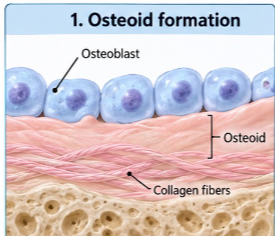
What do osteoblasts secrete to form osteoid?
Osteoblasts secrete:
Collagen fibers
Ground substance

What role do matrix vesicles play in bone mineralization?
Osteoblasts release matrix vesicles containing:
Enzymes
Calcium/phosphate-binding molecules
What are the steps of bone mineralization?
Osteoblasts produce osteoid from collagen fibers + ground substance.
Osteoblasts release matrix vesicles.
Ca²⁺ and PO₄³⁻ accumulate inside the vesicles.
Hydroxyapatite crystals form and grow.
Crystals spread into the surrounding osteoid and become embedded between collagen fibers, hardening the matrix.
Osteoid becomes mineralized bone.

How do hydroxyapatite crystals turn osteoid into mineralized bone?
The crystals:
Expand out of the matrix vesicles
Spread throughout the surrounding osteoid
Become embedded between collagen fibers
Harden the matrix, producing mineralized bone
What can happen to osteoblasts as osteoid becomes mineralized?
Some osteoblasts become trapped within the matrix they produced and mature into osteocytes located in lacunae.
Osteoblast → trapped in matrix → osteocyte
Identify the stages of bone mineralization shown in this diagram.
Osteoid formation → matrix vesicle release → Ca²⁺ + PO₄³⁻ accumulation → hydroxyapatite crystal growth → mineralized bone
An osteoblast releases matrix vesicles, and Ca²⁺ and PO₄³⁻ begin accumulating inside them. What will happen next?
The high concentrations of calcium and phosphate cause hydroxyapatite crystals to begin forming. The crystals then grow and spread into the osteoid.
What is the difference between osteoid and mineralized bone?
Osteoid: newly produced, unmineralized bone matrix containing collagen fibers and ground substance
Mineralized bone: osteoid that has been hardened by deposition of hydroxyapatite crystals
What are trabeculae?
Trabeculae are the thin, branching pieces of bone tissue that form the framework of spongy bone.
What is red bone marrow?
A soft, highly vascular tissue found in the spaces within spongy bone.
What is hematopoiesis, and where does it occur?
Hematopoiesis = production of blood cells.

What is the relationship between trabeculae and red bone marrow in spongy bone?
Trabeculae = actual bone tissue forming the branching framework
Spaces between trabeculae = contain red bone marrow
Red marrow is highly vascular and contains blood-forming hematopoietic stem cells
A histology section shows branching pieces of bone separated by large marrow-filled spaces. What type of bone is this?
Spongy bone.
Why is red bone marrow important, and where is it found?
Red bone marrow contains hematopoietic stem cells that produce blood cells. It is found in the spaces within spongy bone, between the trabeculae.
What is yellow bone marrow, and what is its main function?
Yellow bone marrow is composed mostly of adipose tissue.
Where is yellow bone marrow mainly found in adults?
Mainly in the medullary cavity of long bones, especially in the diaphysis (shaft).
What is the endosteum?
A very thin, delicate osteogenic connective tissue membrane that lines the medullary cavity.
Usually only one or a few cell layers thick
Contains osteoprogenitor cells and osteoblasts
Compare red bone marrow vs. yellow bone marrow.
🔴 Red marrow: contains hematopoietic stem cells → produces blood cells
🟡 Yellow marrow: mostly adipose tissue → stores fat/energy (found in medullary cavity of long bones)
Can yellow bone marrow convert back into red bone marrow?
Yes. If the body has a severe need for increased blood cell production, some yellow marrow can convert back toward red marrow.
You examine the medullary cavity in the diaphysis of an adult long bone and find tissue composed primarily of adipocytes. What is it, and what is its primary function?
Yellow bone marrow, and its primary function is fat/energy storage.
What is the difference between the periosteum and endosteum?
Periosteum: covers the outer surface of bone and has an outer fibrous + inner osteogenic layer.
Endosteum: very thin osteogenic membrane that lines internal bone surfaces, including the medullary cavity.
What is the bone collar?
The periosteum that forms around the diaphysis (shaft) of a developing long bone.
What is an ossification center?
A location where endochondral ossification first begins.
Primary ossification center: forms in the diaphysis
Secondary ossification centers: form later in the epiphyses
What is the epiphyseal plate (growth plate)?
A region of hyaline cartilage between the epiphysis and diaphysis that allows long bones to grow in length.
What are the major stages of long-bone development from a hyaline cartilage model?
A fetal hyaline cartilage model develops.
Cartilage begins to calcify, and a bone collar forms around the diaphysis.
The primary ossification center develops in the diaphysis.
Secondary ossification centers develop in the epiphyses.
Bone replaces most cartilage, while cartilage remains at the articular surfaces and epiphyseal plates.
The epiphyseal plates eventually ossify → epiphyseal lines.
What cartilage remains after most of the hyaline cartilage model has been replaced by bone?
Hyaline cartilage remains as:
Articular cartilage at joint surfaces
Epiphyseal plates while the bone is still growing
Identify the progression of endochondral bone formation in a developing long bone.
Hyaline cartilage model → bone collar/calcification → primary ossification center in diaphysis → secondary ossification centers in epiphyses → epiphyseal plates → epiphyseal lines
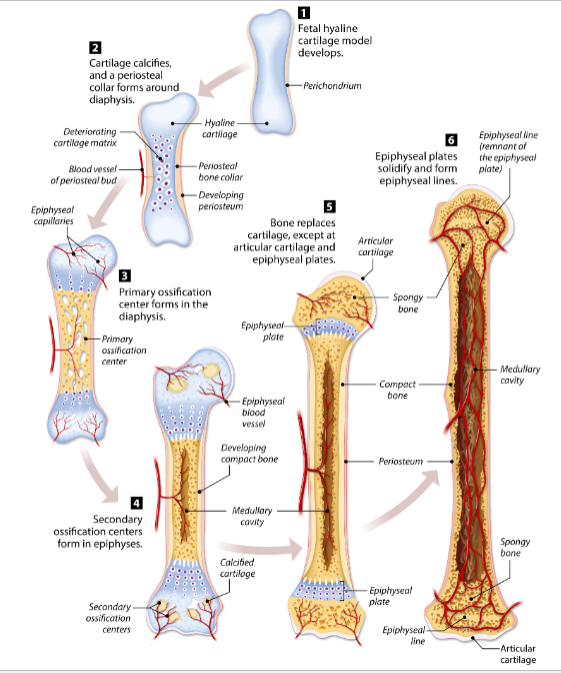
A developing long bone has an ossification center located in its shaft. Is this a primary or secondary ossification center?
Primary ossification center, because it develops in the diaphysis (shaft).
What happens to the epiphyseal plate when longitudinal bone growth ends?
The hyaline cartilage of the epiphyseal plate ossifies, leaving an epiphyseal line.
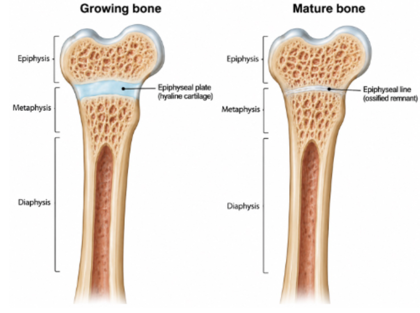
What is the epiphyseal plate (growth plate)?
A layer of hyaline cartilage located between the epiphysis and diaphysis of a growing long bone.